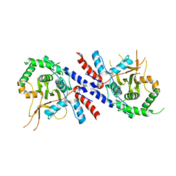
Loading
PDBj
MenuPDBj@FacebookPDBj@TwitterPDBj@YouTubewwPDB FoundationwwPDB
RCSB PDBPDBeBMRBAdv. SearchSearch help
Search by PDB author
8WCH
DownloadVisualize
BU of 8wch by Molmil
Crystal structure of SAR11_0655 bound to a co-purified ligand, L-pyroglutamate
Descriptor: PYROGLUTAMIC ACID, Probable Leu/Ile/Val-binding protein, SODIUM ION
Authors:Clifton, B.E, Laurino, P.
Deposit date:2023-09-12
Release date:2024-07-17
Method:X-RAY DIFFRACTION (1.519 Å)
Cite:Crystal structure of SAR11_0655 bound to a co-purified ligand, L-pyroglutamate
To be published
5WJP
DownloadVisualize
BU of 5wjp by Molmil
Crystal structure of the cyclohexadienyl dehydratase-like solute-binding protein SAR11_1068 from Candidatus Pelagibacter ubique.
Descriptor: Cyclohexadienyl dehydratase
Authors:Clifton, B.E, Jackson, C.J.
Deposit date:2017-07-24
Release date:2017-08-02
Last modified:2023-10-04
Method:X-RAY DIFFRACTION (1.57 Å)
Cite:Evolution of cyclohexadienyl dehydratase from an ancestral solute-binding protein.
Nat. Chem. Biol., 14, 2018
8KD0
DownloadVisualize
BU of 8kd0 by Molmil
Crystal structure of SAR11_0769 from 'Candidatus Pelagibacter ubique' HTCC1062 bound to a co-purified ligand, beta-galactopyranose
Descriptor: Probable binding protein component of ABC sugar transporter, beta-D-galactopyranose
Authors:Clifton, B.E, Laurino, P.
Deposit date:2023-08-08
Release date:2024-07-17
Method:X-RAY DIFFRACTION (1.675 Å)
Cite:Crystal structure of SAR11_0769 from 'Candidatus Pelagibacter ubique' HTCC1062 bound to a co-purified ligand, beta-galactopyranose
To Be Published
6BQE
DownloadVisualize
BU of 6bqe by Molmil
Low-resolution structure of cyclohexadienyl dehydratase from Pseudomonas aeruginosa in space group P4322.
Descriptor: ACETATE ION, Arogenate dehydratase
Authors:Clifton, B.E, Carr, P.D, Jackson, C.J.
Deposit date:2017-11-27
Release date:2017-12-13
Last modified:2018-05-30
Method:X-RAY DIFFRACTION (3.2 Å)
Cite:Evolution of cyclohexadienyl dehydratase from an ancestral solute-binding protein.
Nat. Chem. Biol., 14, 2018
5HPQ
DownloadVisualize
BU of 5hpq by Molmil
Crystal structure of cyclohexadienyl dehydratase from Pseudomonas aeruginosa bound to acetate
Descriptor: ACETATE ION, Cyclohexadienyl dehydratase
Authors:Clifton, B.E, Carr, P.D, Jackson, C.J.
Deposit date:2016-01-20
Release date:2017-01-25
Last modified:2023-09-27
Method:X-RAY DIFFRACTION (2.05 Å)
Cite:Crystal structure of cyclohexadienyl dehydratase from Pseudomonas aeruginosa bound to acetate
To Be Published
5JOS
DownloadVisualize
BU of 5jos by Molmil
Crystal structure of an ancestral cyclohexadienyl dehydratase, AncCDT-3(P188L).
Descriptor: BENZOIC ACID, CITRIC ACID, Cyclohexadienyl dehydratase, ...
Authors:Clifton, B.E, Carr, P.D, Jackson, C.J.
Deposit date:2016-05-03
Release date:2017-05-03
Last modified:2023-09-27
Method:X-RAY DIFFRACTION (2.1 Å)
Cite:To be published
To Be Published
5KKW
DownloadVisualize
BU of 5kkw by Molmil
Crystal structure of SAR11_1068 bound to a sulfobetaine (3-(1-methylpiperidinium-1-yl)propane-1-sulfonate)
Descriptor: 3-(1-methylpiperidinium-1-yl)propane-1-sulfonate, Cyclohexadienyl dehydratase, SULFATE ION
Authors:Clifton, B.E, Carr, P.D, Jackson, C.J.
Deposit date:2016-06-22
Release date:2017-07-05
Last modified:2023-09-27
Method:X-RAY DIFFRACTION (1.88 Å)
Cite:Crystal structure of SAR11_1068 bound to a sulfobetaine (3-(1-methylpiperidinium-1-yl)propane-1-sulfonate)
To Be Published
8HQQ
DownloadVisualize
BU of 8hqq by Molmil
Crystal structure of the glucose-binding protein SAR11_0769 from "Candidatus Pelagibacter ubique" HTCC1062 bound to glucose
Descriptor: Probable binding protein component of ABC sugar transporter, beta-D-glucopyranose
Authors:Clifton, B.E, Laurino, P.
Deposit date:2022-12-14
Release date:2023-12-27
Method:X-RAY DIFFRACTION (1.86 Å)
Cite:Crystal structure of the glucose-binding protein SAR11_0769 from "Candidatus Pelagibacter ubique" HTCC1062 bound to glucose
To Be Published
8HQR
DownloadVisualize
BU of 8hqr by Molmil
Crystal structure of the arginine-/lysine-binding protein SAR11_1210 from 'Candidatus Pelagibacter ubique' HTCC1062 bound to arginine
Descriptor: ABC transporter, ARGININE
Authors:Clifton, B.E, Laurino, P.
Deposit date:2022-12-14
Release date:2023-12-27
Method:X-RAY DIFFRACTION (1.32 Å)
Cite:Crystal structure of the arginine-/lysine-binding protein SAR11_1210 from 'Candidatus Pelagibacter ubique' HTCC1062 bound to arginine
To Be Published
4ZV1
DownloadVisualize
BU of 4zv1 by Molmil
An ancestral arginine-binding protein bound to arginine
Descriptor: ARGININE, AncQR
Authors:Clifton, B.E, Carr, P.D, Jackson, C.J.
Deposit date:2015-05-18
Release date:2016-02-03
Last modified:2023-09-27
Method:X-RAY DIFFRACTION (1.52 Å)
Cite:Ancestral Protein Reconstruction Yields Insights into Adaptive Evolution of Binding Specificity in Solute-Binding Proteins.
Cell Chem Biol, 23, 2016
4ZV2
DownloadVisualize
BU of 4zv2 by Molmil
An ancestral arginine-binding protein bound to glutamine
Descriptor: AncQR, GLUTAMINE
Authors:Clifton, B.E, Jackson, C.J.
Deposit date:2015-05-18
Release date:2016-02-03
Last modified:2023-09-27
Method:X-RAY DIFFRACTION (1.43 Å)
Cite:Ancestral Protein Reconstruction Yields Insights into Adaptive Evolution of Binding Specificity in Solute-Binding Proteins.
Cell Chem Biol, 23, 2016
5T0W
DownloadVisualize
BU of 5t0w by Molmil
Crystal structure of the ancestral amino acid-binding protein AncCDT-1, a precursor of cyclohexadienyl dehydratase
Descriptor: ARGININE, AncCDT-1
Authors:Clifton, B.E, Carr, P.D, Jackson, C.J.
Deposit date:2016-08-16
Release date:2017-09-06
Last modified:2023-10-04
Method:X-RAY DIFFRACTION (2.59 Å)
Cite:To be published
To Be Published
5TUJ
DownloadVisualize
BU of 5tuj by Molmil
Ancestral Cationic Amino Acid Solute Binding Protein (AncCDT-1)
Descriptor: Ancestral protein CDT-Anc1
Authors:Kaczmarski, J.A, Clifton, B.E, Carr, P.D, Jackson, C.J.
Deposit date:2016-11-06
Release date:2017-12-06
Last modified:2024-03-06
Method:X-RAY DIFFRACTION (3.352 Å)
Cite:Evolution of cyclohexadienyl dehydratase from an ancestral solute-binding protein.
Nat. Chem. Biol., 14, 2018
6WUP
DownloadVisualize
BU of 6wup by Molmil
Crystal structure of an ancestral cyclohexadienyl dehydratase, AncCDT-5
Descriptor: 4-(2-HYDROXYETHYL)-1-PIPERAZINE ETHANESULFONIC ACID, Ancestral cyclohexadienyl dehydratase, AncCDT-5, ...
Authors:Kaczmarski, J.A, Mahawaththa, M.C.
Deposit date:2020-05-05
Release date:2020-05-13
Last modified:2023-10-18
Method:X-RAY DIFFRACTION (1.49 Å)
Cite:Altered conformational sampling along an evolutionary trajectory changes the catalytic activity of an enzyme.
Nat Commun, 11, 2020
7KWW
DownloadVisualize
BU of 7kww by Molmil
X-ray Crystal Structure of PlyCB Mutant K59H
Descriptor: (4S)-2-METHYL-2,4-PENTANEDIOL, PlyCB
Authors:Williams, D.E, Broendum, S.S, Hayes, B.K, Drinkwater, N, McGowan, S.
Deposit date:2020-12-02
Release date:2021-04-07
Last modified:2023-10-18
Method:X-RAY DIFFRACTION (1.8 Å)
Cite:High avidity drives the interaction between the streptococcal C1 phage endolysin, PlyC, with the cell surface carbohydrates of Group A Streptococcus.
Mol.Microbiol., 116, 2021
7KWY
DownloadVisualize
BU of 7kwy by Molmil
X-ray Crystal Structure of PlyCB Mutant R66K
Descriptor: (4S)-2-METHYL-2,4-PENTANEDIOL, PlyCB
Authors:Williams, D.E, Broendum, S.S, Hayes, B.K, Drinkwater, N, McGowan, S.
Deposit date:2020-12-02
Release date:2021-04-07
Last modified:2023-10-18
Method:X-RAY DIFFRACTION (1.7 Å)
Cite:High avidity drives the interaction between the streptococcal C1 phage endolysin, PlyC, with the cell surface carbohydrates of Group A Streptococcus.
Mol.Microbiol., 116, 2021
7KWT
DownloadVisualize
BU of 7kwt by Molmil
X-ray Crystal Structure of PlyCB Mutant Y28H
Descriptor: PlyCB
Authors:Williams, D.E, Broendum, S.S, Hayes, B.K, Drinkwater, N, McGowan, S.
Deposit date:2020-12-02
Release date:2021-04-07
Last modified:2023-10-18
Method:X-RAY DIFFRACTION (1.79 Å)
Cite:High avidity drives the interaction between the streptococcal C1 phage endolysin, PlyC, with the cell surface carbohydrates of Group A Streptococcus.
Mol.Microbiol., 116, 2021
4N1C
DownloadVisualize
BU of 4n1c by Molmil
Structural evidence for antigen receptor evolution
Descriptor: Lysozyme C, immunoglobulin variable light chain domain
Authors:Langley, D.B, Rouet, R, Roome, B, Stock, D, Christ, D.
Deposit date:2013-10-03
Release date:2014-10-29
Last modified:2023-09-20
Method:X-RAY DIFFRACTION (1.7 Å)
Cite:Structural reconstruction of protein ancestry.
Proc. Natl. Acad. Sci. U.S.A., 114, 2017
4N1E
DownloadVisualize
BU of 4n1e by Molmil
Structural evidence for antigen receptor evolution
Descriptor: Lysozyme C, immunoglobulin variable light chain domain
Authors:Langley, D.B, Rouet, R, Stock, D, Christ, D.
Deposit date:2013-10-04
Release date:2014-10-29
Last modified:2018-04-18
Method:X-RAY DIFFRACTION (2.23 Å)
Cite:Structural reconstruction of protein ancestry.
Proc. Natl. Acad. Sci. U.S.A., 114, 2017
5V3B
DownloadVisualize
BU of 5v3b by Molmil
Human A20 OTU domain (WT) with acetamidylated C103
Descriptor: Tumor necrosis factor alpha-induced protein 3
Authors:Langley, D.B, Christ, D, Grey, S.
Deposit date:2017-03-07
Release date:2018-03-21
Last modified:2023-10-04
Method:X-RAY DIFFRACTION (3 Å)
Cite:Denisovan, modern human and mouse TNFAIP3 alleles tune A20 phosphorylation and immunity.
Nat.Immunol., 20, 2019
5V3P
DownloadVisualize
BU of 5v3p by Molmil
Human A20 OTU domain (I325N) with acetamidylated C103
Descriptor: Tumor necrosis factor alpha-induced protein 3
Authors:Langley, D.B, Christ, D, Grey, S.
Deposit date:2017-03-07
Release date:2018-03-21
Last modified:2023-10-04
Method:X-RAY DIFFRACTION (2.5 Å)
Cite:Denisovan, modern human and mouse TNFAIP3 alleles tune A20 phosphorylation and immunity.
Nat.Immunol., 20, 2019

224931

數據於2024-09-11公開中

PDB statisticsPDBj update infoContact PDBjnumon