Loading
PDBj
MenuPDBj@FacebookPDBj@TwitterPDBj@YouTubewwPDB FoundationwwPDB
RCSB PDBPDBeBMRBAdv. SearchSearch help
Search by PDB author
4EFF
DownloadVisualize
BU of 4eff by Molmil
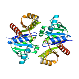
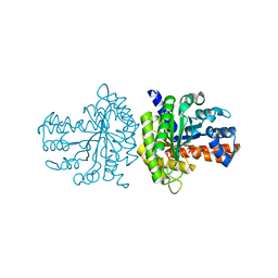
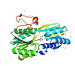
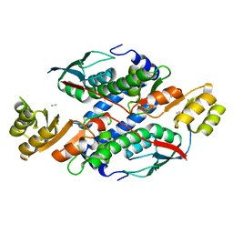
Crystal structure of aromatic-amino-acid aminotransferase from Burkholderia pseudomallei
Descriptor: Aromatic-amino-acid aminotransferase, GLYCEROL
Authors:Seattle Structural Genomics Center for Infectious Disease (SSGCID)
Deposit date:2012-03-29
Release date:2012-04-11
Last modified:2023-09-13
Method:X-RAY DIFFRACTION (1.85 Å)
Cite:Crystal structure of aromatic-amino-acid aminotransferase from Burkholderia pseudomallei
To be Published
3EMK
DownloadVisualize
BU of 3emk by Molmil
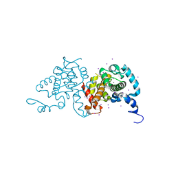
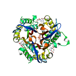
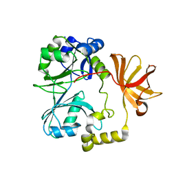
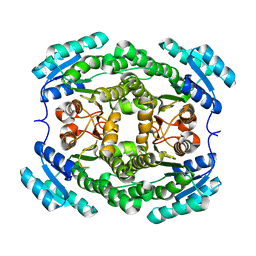
2.5A crystal structure of glucose/ribitol dehydrogenase from brucella melitensis
Descriptor: GLUCOSE/RIBITOL DEHYDROGENASE
Authors:Seattle Structural Genomics Center for Infectious Disease (SSGCID)
Deposit date:2008-09-24
Release date:2008-10-28
Last modified:2024-02-21
Method:X-RAY DIFFRACTION (2.5 Å)
Cite:2.5A crystal structure of glucose/ribitol dehydrogenase from brucella melitensis
To be Published
3K9H
DownloadVisualize
BU of 3k9h by Molmil
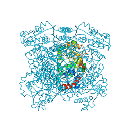
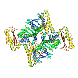
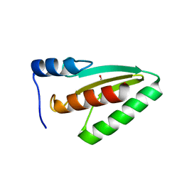
Crystal structure of a plasmid partition protein from borrelia burgdorferi at 2.25A resolution
Descriptor: PF-32 protein, SULFATE ION
Authors:Seattle Structural Genomics Center for Infectious Disease (SSGCID)
Deposit date:2009-10-15
Release date:2009-11-03
Last modified:2023-09-06
Method:X-RAY DIFFRACTION (2.25 Å)
Cite:Crystal structure of a plasmid partition protein from borrelia burgdorferi at 2.25A resolution
To be Published
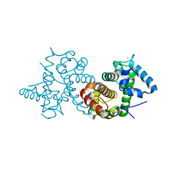
3SIA
DownloadVisualize
BU of 3sia by Molmil
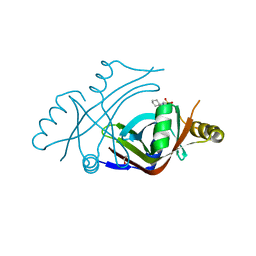
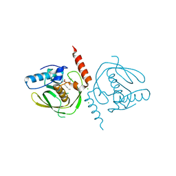
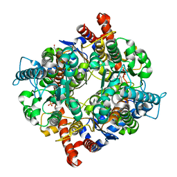
Crystal structure of URE3-binding protein, (D127A,N129A) mutant, iodide phased
Descriptor: CALCIUM ION, CHLORIDE ION, IODIDE ION, ...
Authors:Seattle Structural Genomics Center for Infectious Disease (SSGCID), Skubak, P.
Deposit date:2011-06-17
Release date:2011-06-29
Last modified:2024-02-28
Method:X-RAY DIFFRACTION (2.05 Å)
Cite:Crystal structure of URE3-binding protein
To be Published
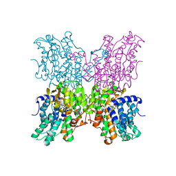
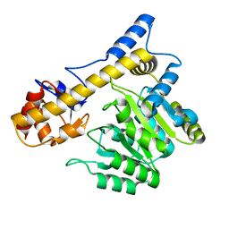
4HWG
DownloadVisualize
BU of 4hwg by Molmil
Structure of UDP-N-acetylglucosamine 2-epimerase from Rickettsia bellii
Descriptor: PHOSPHATE ION, UDP-N-acetylglucosamine 2-epimerase
Authors:Seattle Structural Genomics Center for Infectious Disease (SSGCID)
Deposit date:2012-11-07
Release date:2012-11-28
Last modified:2023-09-20
Method:X-RAY DIFFRACTION (2 Å)
Cite:Structure of UDP-N-acetylglucosamine 2-epimerase from Rickettsia bellii
TO BE PUBLISHED
3U0I
DownloadVisualize
BU of 3u0i by Molmil
Crystal Structure of a probable FAD-binding, putative uncharacterized protein from Brucella melitensis
Descriptor: 1,2-ETHANEDIOL, 2-[N-CYCLOHEXYLAMINO]ETHANE SULFONIC ACID, probable FAD-binding, ...
Authors:Seattle Structural Genomics Center for Infectious Disease (SSGCID)
Deposit date:2011-09-28
Release date:2011-10-12
Last modified:2023-09-13
Method:X-RAY DIFFRACTION (2.2 Å)
Cite:Crystal Structure of a probable FAD-binding, putative uncharacterized protein from Brucella melitensis
To be Published
3QRH
DownloadVisualize
BU of 3qrh by Molmil
Crystal structure of fructose bisphosphate aldolase from Encephalitozoon Cuniculi, bound to glyceraldehyde 3-phosphate
Descriptor: Fructose-bisphosphate aldolase, GLYCERALDEHYDE-3-PHOSPHATE
Authors:Seattle Structural Genomics Center for Infectious Disease (SSGCID)
Deposit date:2011-02-17
Release date:2011-03-23
Last modified:2024-10-16
Method:X-RAY DIFFRACTION (2 Å)
Cite:Crystal structure of fructose bisphosphate aldolase from Encephalitozoon Cuniculi, bound to glyceraldehyde 3-phosphate
TO BE PUBLISHED
3GE4
DownloadVisualize
BU of 3ge4 by Molmil
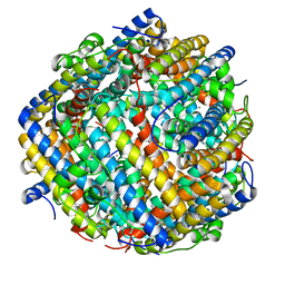
Crystal structure of ferritin:DNA-binding protein DPS from Brucella Melitensis
Descriptor: CALCIUM ION, DNA PROTECTION DURING STARVATION PROTEIN
Authors:Seattle Structural Genomics Center for Infectious Disease (SSGCID)
Deposit date:2009-02-25
Release date:2009-03-03
Last modified:2023-09-06
Method:X-RAY DIFFRACTION (1.7 Å)
Cite:Crystal structure of ferritin:DNA-binding protein DPS from Brucella Melitensis
To be Published
3S6O
DownloadVisualize
BU of 3s6o by Molmil
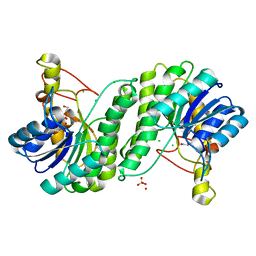
Crystal structure of a Polysaccharide deacetylase family protein from Burkholderia pseudomallei
Descriptor: 1,2-ETHANEDIOL, Polysaccharide deacetylase family protein
Authors:Seattle Structural Genomics Center for Infectious Disease (SSGCID)
Deposit date:2011-05-25
Release date:2011-06-15
Last modified:2023-09-13
Method:X-RAY DIFFRACTION (1.85 Å)
Cite:Crystal structure of a Polysaccharide deacetylase family protein from Burkholderia pseudomallei
To be Published
3SC4
DownloadVisualize
BU of 3sc4 by Molmil
Crystal structure of a Short chain dehydrogenase (A0QTM2 homolog) Mycobacterium thermoresistibile
Descriptor: 1,2-ETHANEDIOL, CHLORIDE ION, GLYCEROL, ...
Authors:Seattle Structural Genomics Center for Infectious Disease (SSGCID)
Deposit date:2011-06-06
Release date:2011-06-29
Last modified:2023-09-13
Method:X-RAY DIFFRACTION (2.5 Å)
Cite:Crystal structure of a Short chain dehydrogenase (A0QTM2 homolog) Mycobacterium thermoresistibile
To be Published
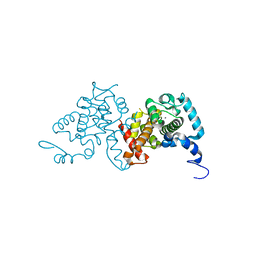
3SIB
DownloadVisualize
BU of 3sib by Molmil
Crystal structure of URE3-binding protein, wild-type
Descriptor: CALCIUM ION, SODIUM ION, URE3-BP sequence specific DNA binding protein
Authors:Seattle Structural Genomics Center for Infectious Disease (SSGCID)
Deposit date:2011-06-17
Release date:2011-06-29
Last modified:2023-09-13
Method:X-RAY DIFFRACTION (1.9 Å)
Cite:Crystal structure of URE3-binding protein
To be Published
3SJS
DownloadVisualize
BU of 3sjs by Molmil
Crystal structure of URE3-binding protein from Entamoeba histolytica, (D127A,N129A) mutant, native form
Descriptor: CALCIUM ION, URE3-BP sequence specific DNA binding protein
Authors:Seattle Structural Genomics Center for Infectious Disease (SSGCID)
Deposit date:2011-06-21
Release date:2011-07-06
Last modified:2023-09-13
Method:X-RAY DIFFRACTION (1.9 Å)
Cite:Crystal structure of URE3-binding protein from Entamoeba histolytica
to be published
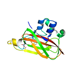
3UAM
DownloadVisualize
BU of 3uam by Molmil
Crystal structure of a chitin binding domain from Burkholderia pseudomallei
Descriptor: Chitin binding domain, GLYCEROL, NITRATE ION
Authors:Seattle Structural Genomics Center for Infectious Disease (SSGCID)
Deposit date:2011-10-21
Release date:2011-11-02
Last modified:2023-09-13
Method:X-RAY DIFFRACTION (2 Å)
Cite:Crystal structure of a chitin binding domain from Burkholderia pseudomallei
to be published
3GLQ
DownloadVisualize
BU of 3glq by Molmil
Crystal structure of S-adenosyl-L-homocysteine hydrolase from Burkholderia pseudomallei in complex with 9-beta-D-arabino-furansyl-adenine
Descriptor: 2-(6-AMINO-PURIN-9-YL)-5-HYDROXYMETHYL-TETRAHYDRO-FURAN-3,4-DIOL, Adenosylhomocysteinase, NICOTINAMIDE-ADENINE-DINUCLEOTIDE
Authors:Seattle Structural Genomics Center for Infectious Disease (SSGCID)
Deposit date:2009-03-12
Release date:2009-03-24
Last modified:2023-09-06
Method:X-RAY DIFFRACTION (2.3 Å)
Cite:Crystal structure of S-adenosyl-L-homocysteine hydrolase from Burkholderia pseudomallei in complex with 9-beta-D-arabino-furansyl-adenine
To be Published
3S99
DownloadVisualize
BU of 3s99 by Molmil
Crystal structure of a basic membrane lipoprotein from brucella melitensis, iodide soak
Descriptor: ADENINE, Basic membrane lipoprotein, IODIDE ION, ...
Authors:Seattle Structural Genomics Center for Infectious Disease (SSGCID)
Deposit date:2011-06-01
Release date:2011-06-22
Last modified:2024-04-03
Method:X-RAY DIFFRACTION (2.05 Å)
Cite:Crystal structure of a basic membrane lipoprotein from brucella melitensis, iodide soak
To be Published
3T80
DownloadVisualize
BU of 3t80 by Molmil
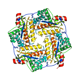
Crystal structure of 2-C-methyl-D-erythritol 2,4-cyclodiphosphate synthase from Salmonella typhimurium bound to cytidine
Descriptor: 1,2-ETHANEDIOL, 2-C-methyl-D-erythritol 2,4-cyclodiphosphate synthase, 4-AMINO-1-BETA-D-RIBOFURANOSYL-2(1H)-PYRIMIDINONE, ...
Authors:Seattle Structural Genomics Center for Infectious Disease (SSGCID), Staker, B.L, Edwards, T.E.
Deposit date:2011-07-31
Release date:2011-09-14
Last modified:2023-09-13
Method:X-RAY DIFFRACTION (2.5 Å)
Cite:Crystal structure of 2-C-methyl-D-erythritol 2,4-cyclodiphosphate synthase from Salmonella typhimurium bound to cytidine
To be Published
3INN
DownloadVisualize
BU of 3inn by Molmil
Crystal structure of pantoate-beta-alanine-ligase in complex with ATP at low occupancy at 2.1 A resolution
Descriptor: ADENOSINE-5'-TRIPHOSPHATE, Pantothenate synthetase, UNKNOWN ATOM OR ION
Authors:Seattle Structural Genomics Center for Infectious Disease (SSGCID)
Deposit date:2009-08-12
Release date:2009-10-06
Last modified:2023-09-06
Method:X-RAY DIFFRACTION (2.1 Å)
Cite:Crystal structure of pantoate-beta-alanine-ligase in complex with ATP at low occupancy at 2.1 A resolution
TO BE PUBLISHED
3U04
DownloadVisualize
BU of 3u04 by Molmil
Crystal structure of peptide deformylase from ehrlichia chaffeensis in complex with actinonin
Descriptor: ACTINONIN, CHLORIDE ION, Peptide deformylase 1, ...
Authors:Seattle Structural Genomics Center for Infectious Disease (SSGCID)
Deposit date:2011-09-28
Release date:2011-10-12
Last modified:2023-09-13
Method:X-RAY DIFFRACTION (1.7 Å)
Cite:Crystal structure of peptide deformylase from ehrlichia chaffeensis in complex with actinonin
To be Published
3GNN
DownloadVisualize
BU of 3gnn by Molmil
Crystal structure of nicotinate-nucleotide pyrophosphorylase from Burkholderi pseudomallei
Descriptor: Unknown Peptide, nicotinate-nucleotide diphosphorylase (carboxylating)
Authors:Seattle Structural Genomics Center for Infectious Disease (SSGCID)
Deposit date:2009-03-17
Release date:2009-03-31
Last modified:2024-02-21
Method:X-RAY DIFFRACTION (2.25 Å)
Cite:Crystal structure of nicotinate-nucleotide pyrophosphorylase from Burkholderi pseudomallei
To be Published
3GIR
DownloadVisualize
BU of 3gir by Molmil
Crystal structure of glycine cleavage system aminomethyltransferase T from Bartonella henselae
Descriptor: Aminomethyltransferase, SODIUM ION
Authors:Seattle Structural Genomics Center for Infectious Disease (SSGCID)
Deposit date:2009-03-06
Release date:2009-03-17
Last modified:2023-09-06
Method:X-RAY DIFFRACTION (1.6 Å)
Cite:Crystal structure of glycine cleavage system aminomethyltransferase T from Bartonella henselae
To be Published
3JST
DownloadVisualize
BU of 3jst by Molmil
Crystal structure of transcriptional coactivator/pterin dehydratase from Brucella Melitensis
Descriptor: 1,2-ETHANEDIOL, Putative pterin-4-alpha-carbinolamine dehydratase
Authors:Seattle Structural Genomics Center for Infectious Disease (SSGCID)
Deposit date:2009-09-10
Release date:2009-09-22
Last modified:2023-09-06
Method:X-RAY DIFFRACTION (2.1 Å)
Cite:Crystal structure of transcriptional coactivator/pterin dehydratase from Brucella Melitensis
To be Published
3UJH
DownloadVisualize
BU of 3ujh by Molmil
Crystal structure of substrate-bound Glucose-6-phosphate isomerase from Toxoplasma gondii
Descriptor: 1,2-ETHANEDIOL, CHLORIDE ION, GLUCOSE-6-PHOSPHATE, ...
Authors:Seattle Structural Genomics Center for Infectious Disease (SSGCID)
Deposit date:2011-11-07
Release date:2011-11-16
Last modified:2023-09-13
Method:X-RAY DIFFRACTION (2.1 Å)
Cite:Crystal structure of substrate-bound Glucose-6-phosphate isomerase from Toxoplasma gondii
To be Published
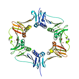
6MAN
DownloadVisualize
BU of 6man by Molmil
Crystal structure of a DnaN sliding clamp DNA polymerase III subunit beta from Rickettsia bellii RML369-C
Descriptor: 1,2-ETHANEDIOL, Beta sliding clamp, THIOCYANATE ION
Authors:Seattle Structural Genomics Center for Infectious Disease, Seattle Structural Genomics Center for Infectious Disease (SSGCID)
Deposit date:2018-08-28
Release date:2018-09-05
Last modified:2023-10-11
Method:X-RAY DIFFRACTION (2.35 Å)
Cite:Crystal structure of a DnaN sliding clamp DNA polymerase III subunit beta from Rickettsia bellii RML369-C
To Be Published
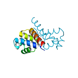
5WOQ
DownloadVisualize
BU of 5woq by Molmil
Crystal structure of an XRE family protein transcriptional regulator from Mycobacterium smegmatis
Descriptor: Transcriptional regulator ClgR
Authors:Seattle Structural Genomics Center for Infectious Disease (SSGCID)
Deposit date:2017-08-02
Release date:2017-08-16
Last modified:2023-10-04
Method:X-RAY DIFFRACTION (1.95 Å)
Cite:Crystal structure of an XRE family protein transcriptional regulator from Mycobacterium smegmatis
To Be Published
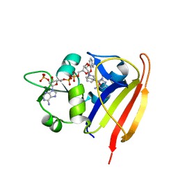
6E4E
DownloadVisualize
BU of 6e4e by Molmil
Crystal structure of dihydrofolate reductase from Staphylococcus aureus MW2 bound to NADP and p218
Descriptor: 3-(2-{3-[(2,4-diamino-6-ethylpyrimidin-5-yl)oxy]propoxy}phenyl)propanoic acid, Dihydrofolate reductase, NADP NICOTINAMIDE-ADENINE-DINUCLEOTIDE PHOSPHATE
Authors:Seattle Structural Genomics Center for Infectious Disease (SSGCID)
Deposit date:2018-07-17
Release date:2018-07-25
Last modified:2023-10-11
Method:X-RAY DIFFRACTION (1.9 Å)
Cite:Crystal structure of dihydrofolate reductase from Staphylococcus aureus MW2 bound to NADP and p218
To Be Published

226707

數據於2024-10-30公開中

PDB statisticsPDBj update infoContact PDBjnumon