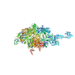
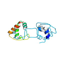
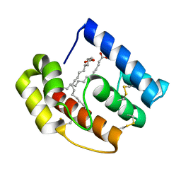
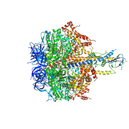
Loading
PDBj
MenuPDBj@FacebookPDBj@TwitterPDBj@YouTubewwPDB FoundationwwPDB
RCSB PDBPDBeBMRBAdv. SearchSearch help
Search by PDB author
1SKY
DownloadVisualize
BU of 1sky by Molmil
CRYSTAL STRUCTURE OF THE NUCLEOTIDE FREE ALPHA3BETA3 SUB-COMPLEX OF F1-ATPASE FROM THE THERMOPHILIC BACILLUS PS3
Descriptor: F1-ATPASE, SULFATE ION
Authors:Shirakihara, Y, Leslie, A.G.W, Abrahams, J.P, Walker, J.E, Ueda, T, Sekimoto, Y, Kambara, M, Saika, K, Kagawa, Y, Yoshida, M.
Deposit date:1997-02-26
Release date:1998-03-04
Last modified:2024-02-14
Method:X-RAY DIFFRACTION (3.2 Å)
Cite:The crystal structure of the nucleotide-free alpha 3 beta 3 subcomplex of F1-ATPase from the thermophilic Bacillus PS3 is a symmetric trimer.
Structure, 5, 1997
6OVY
DownloadVisualize
BU of 6ovy by Molmil
X-ray crystal structure of a bacterial reiterative transcription complex of pyrG promoter variant -1C
Descriptor: DNA (5'-D(*TP*AP*TP*AP*AP*TP*GP*GP*GP*TP*CP*TP*GP*AP*TP*G)-3'), DNA (5'-D(P*CP*AP*TP*CP*AP*GP*AP*GP*CP*CP*CP*CP*AP*AP*A)-3'), DNA-directed RNA polymerase subunit alpha, ...
Authors:Shin, Y, Murakami, K.S.
Deposit date:2019-05-08
Release date:2019-07-24
Last modified:2023-10-11
Method:X-RAY DIFFRACTION (2.999 Å)
Cite:Structural basis of reiterative transcription from the pyrG and pyrBI promoters by bacterial RNA polymerase.
Nucleic Acids Res., 48, 2020
6OY7
DownloadVisualize
BU of 6oy7 by Molmil
X-ray crystal structure of a bacterial reiterative transcription complex of pyrG promoter at 7 min
Descriptor: DNA (5'-D(*TP*AP*TP*AP*AP*TP*GP*GP*GP*TP*CP*TP*GP*AP*TP*GP*CP*AP*GP*G)-3'), DNA (5'-D(P*CP*CP*TP*GP*CP*AP*TP*CP*AP*GP*AP*GP*CP*CP*CP*AP*AP*AP*A)-3'), DNA-directed RNA polymerase subunit alpha, ...
Authors:Shin, Y, Murakami, K.S.
Deposit date:2019-05-14
Release date:2019-07-24
Last modified:2023-10-11
Method:X-RAY DIFFRACTION (3.04 Å)
Cite:Structural basis of reiterative transcription from the pyrG and pyrBI promoters by bacterial RNA polymerase.
Nucleic Acids Res., 48, 2020
6OY5
DownloadVisualize
BU of 6oy5 by Molmil
X-ray crystal structure of a bacterial reiterative transcription complex of pyrG promoter at 3 min
Descriptor: DNA (5'-D(*AP*TP*AP*AP*TP*GP*GP*GP*AP*GP*CP*CP*TP*CP*TP*GP*AP*TP*GP*CP*AP*G)-3'), DNA (5'-D(P*CP*CP*GP*CP*AP*TP*CP*AP*GP*AP*GP*CP*CP*CP*AP*AP*AP*AP*T)-3'), DNA-directed RNA polymerase subunit alpha, ...
Authors:Shin, Y, Murakami, K.S.
Deposit date:2019-05-14
Release date:2019-06-05
Last modified:2023-10-11
Method:X-RAY DIFFRACTION (3.1 Å)
Cite:Structural basis of reiterative transcription from the pyrG and pyrBI promoters by bacterial RNA polymerase.
Nucleic Acids Res., 48, 2020
6OVR
DownloadVisualize
BU of 6ovr by Molmil
X-ray crystal structure of a bacterial reiterative transcription complex of pyrG promoter variant -1G
Descriptor: DNA (5'-D(*TP*AP*TP*AP*AP*TP*GP*GP*GP*CP*TP*GP*AP*TP*GP*CP*AP*GP*G)-3'), DNA (5'-D(P*GP*GP*TP*GP*CP*AP*TP*CP*AP*GP*AP*GP*CP*CP*CP*GP*AP*AP*A)-3'), DNA-directed RNA polymerase subunit alpha, ...
Authors:Shin, Y, Murakami, K.S.
Deposit date:2019-05-08
Release date:2019-07-24
Last modified:2023-10-11
Method:X-RAY DIFFRACTION (2.843 Å)
Cite:Structural basis of reiterative transcription from the pyrG and pyrBI promoters by bacterial RNA polymerase.
Nucleic Acids Res., 48, 2020
6OW3
DownloadVisualize
BU of 6ow3 by Molmil
X-ray crystal structure of a bacterial reiterative transcription complex of pyrG promoter variant -1T
Descriptor: DNA (5'-D(*TP*AP*TP*AP*AP*TP*GP*GP*GP*GP*AP*TP*CP*TP*GP*AP*TP*GP*C)-3'), DNA (5'-D(P*GP*CP*AP*TP*CP*AP*GP*AP*GP*CP*CP*CP*TP*AP*AP*A)-3'), DNA-directed RNA polymerase subunit alpha, ...
Authors:Shin, Y, Murakami, K.S.
Deposit date:2019-05-09
Release date:2019-07-24
Last modified:2023-10-11
Method:X-RAY DIFFRACTION (2.766 Å)
Cite:Structural basis of reiterative transcription from the pyrG and pyrBI promoters by bacterial RNA polymerase.
Nucleic Acids Res., 48, 2020
6OY6
DownloadVisualize
BU of 6oy6 by Molmil
X-ray crystal structure of a bacterial reiterative transcription complex of pyrG promoter at 5 min
Descriptor: DNA (5'-D(*TP*AP*TP*AP*AP*TP*GP*GP*GP*GP*AP*TP*CP*TP*GP*AP*TP*GP*CP*AP*GP*G)-3'), DNA (5'-D(P*CP*CP*TP*GP*CP*AP*TP*CP*AP*GP*AP*GP*CP*CP*CP*AP*AP*AP*A)-3'), DNA-directed RNA polymerase subunit alpha, ...
Authors:Shin, Y, Murakami, K.S.
Deposit date:2019-05-14
Release date:2019-06-19
Last modified:2023-10-11
Method:X-RAY DIFFRACTION (3.096 Å)
Cite:Structural basis of reiterative transcription from the pyrG and pyrBI promoters by bacterial RNA polymerase.
Nucleic Acids Res., 48, 2020
6P70
DownloadVisualize
BU of 6p70 by Molmil
X-ray crystal structure of bacterial RNA polymerase and pyrBI promoter complex
Descriptor: DNA (5'-D(*TP*AP*TP*AP*AP*TP*CP*GP*AP*TP*CP*TP*TP*TP*GP*CP*CP*GP*GP*G)-3'), DNA (5'-D(P*TP*CP*CP*CP*GP*GP*CP*AP*AP*AP*TP*TP*GP*TP*CP*CP*G)-3'), DNA-directed RNA polymerase subunit alpha, ...
Authors:Shin, Y, Murakami, K.S.
Deposit date:2019-06-04
Release date:2019-06-19
Last modified:2023-10-11
Method:X-RAY DIFFRACTION (3.052 Å)
Cite:Structural basis of reiterative transcription from the pyrG and pyrBI promoters by bacterial RNA polymerase.
Nucleic Acids Res., 48, 2020
7XMA
DownloadVisualize
BU of 7xma by Molmil
Crystal structure of Bovine heart cytochrome c oxidase, apo structure with DMSO
Descriptor: (1R)-2-{[{[(2S)-2,3-DIHYDROXYPROPYL]OXY}(HYDROXY)PHOSPHORYL]OXY}-1-[(PALMITOYLOXY)METHYL]ETHYL (11E)-OCTADEC-11-ENOATE, (1S)-2-{[(2-AMINOETHOXY)(HYDROXY)PHOSPHORYL]OXY}-1-[(STEAROYLOXY)METHYL]ETHYL (5E,8E,11E,14E)-ICOSA-5,8,11,14-TETRAENOATE, (7R,17E,20E)-4-HYDROXY-N,N,N-TRIMETHYL-9-OXO-7-[(PALMITOYLOXY)METHYL]-3,5,8-TRIOXA-4-PHOSPHAHEXACOSA-17,20-DIEN-1-AMINIUM 4-OXIDE, ...
Authors:Nishida, Y, Shinzawa-Itoh, K, Mizuno, N, Kumasaka, T, Yoshikawa, S, Tsukihara, T, Takashima, S, Shintani, Y.
Deposit date:2022-04-25
Release date:2022-12-21
Last modified:2023-11-29
Method:X-RAY DIFFRACTION (2.2 Å)
Cite:Identifying antibiotics based on structural differences in the conserved allostery from mitochondrial heme-copper oxidases.
Nat Commun, 13, 2022
7XMB
DownloadVisualize
BU of 7xmb by Molmil
Crystal structure of Bovine heart cytochrome c oxidase, the structure complexed with an allosteric inhibitor T113
Descriptor: (1R)-2-{[{[(2S)-2,3-DIHYDROXYPROPYL]OXY}(HYDROXY)PHOSPHORYL]OXY}-1-[(PALMITOYLOXY)METHYL]ETHYL (11E)-OCTADEC-11-ENOATE, (1S)-2-{[(2-AMINOETHOXY)(HYDROXY)PHOSPHORYL]OXY}-1-[(STEAROYLOXY)METHYL]ETHYL (5E,8E,11E,14E)-ICOSA-5,8,11,14-TETRAENOATE, (7R,17E,20E)-4-HYDROXY-N,N,N-TRIMETHYL-9-OXO-7-[(PALMITOYLOXY)METHYL]-3,5,8-TRIOXA-4-PHOSPHAHEXACOSA-17,20-DIEN-1-AMINIUM 4-OXIDE, ...
Authors:Nishida, Y, Shinzawa-Itoh, K, Mizuno, N, Kumasaka, T, Yoshikawa, S, Tsukihara, T, Shintani, Y, Takashima, S.
Deposit date:2022-04-25
Release date:2022-12-21
Last modified:2023-11-29
Method:X-RAY DIFFRACTION (2.2 Å)
Cite:Identifying antibiotics based on structural differences in the conserved allostery from mitochondrial heme-copper oxidases.
Nat Commun, 13, 2022
7XMC
DownloadVisualize
BU of 7xmc by Molmil
Cryo-EM structure of Cytochrome bo3 from Escherichia coli, apo structure with DMSO
Descriptor: 1,2-dioleoyl-sn-glycero-3-phosphoethanolamine, COPPER (II) ION, Cytochrome bo(3) ubiquinol oxidase subunit 1, ...
Authors:Nishida, Y, Shigematsu, H, Iwamoto, T, Takashima, S, Shintani, Y.
Deposit date:2022-04-25
Release date:2022-12-21
Method:ELECTRON MICROSCOPY (3.09 Å)
Cite:Identifying antibiotics based on structural differences in the conserved allostery from mitochondrial heme-copper oxidases.
Nat Commun, 13, 2022
7XMD
DownloadVisualize
BU of 7xmd by Molmil
Cryo-EM structure of Cytochrome bo3 from Escherichia coli, the structure complexed with an allosteric inhibitor N4
Descriptor: 1,2-dioleoyl-sn-glycero-3-phosphoethanolamine, COPPER (II) ION, Cytochrome bo(3) ubiquinol oxidase subunit 1, ...
Authors:Nishida, Y, Shigematsu, H, Iwamoto, T, Takashima, S, Shintani, Y.
Deposit date:2022-04-25
Release date:2022-12-21
Method:ELECTRON MICROSCOPY (2.99 Å)
Cite:Identifying antibiotics based on structural differences in the conserved allostery from mitochondrial heme-copper oxidases.
Nat Commun, 13, 2022
4ZID
DownloadVisualize
BU of 4zid by Molmil
Dimeric Hydrogenobacter thermophilus cytochrome c552 obtained from Escherichia coli
Descriptor: Cytochrome c-552, HEME C
Authors:Hayashi, Y, Yamanaka, M, Nagao, S, Komori, H, Higuchi, Y, Hirota, S.
Deposit date:2015-04-28
Release date:2016-02-10
Last modified:2023-11-08
Method:X-RAY DIFFRACTION (1.8 Å)
Cite:Domain swapping oligomerization of thermostable c-type cytochrome in E. coli cells
Sci Rep, 6, 2016
7R8I
DownloadVisualize
BU of 7r8i by Molmil
Crystal structure of Pseudooceanicola lipolyticus Argonaute bound to 5' OH guide DNA in the presence of Mg2+
Descriptor: Argonaute, DNA (5'-D(*TP*TP*AP*CP*TP*GP*CP*AP*CP*AP*GP*GP*TP*GP*AP*CP*GP*A), MAGNESIUM ION
Authors:Shin, Y, Murakami, K.S.
Deposit date:2021-06-26
Release date:2022-11-09
Last modified:2024-05-22
Method:X-RAY DIFFRACTION (2.4 Å)
Cite:Crystal structure of Pseudooceanicola lipolyticus Argonaute bound to 5' OH guide DNA in the presence of Mg2+
To be published
3IE4
DownloadVisualize
BU of 3ie4 by Molmil
b-glucan binding domain of Drosophila GNBP3 defines a novel family of pattern recognition receptor
Descriptor: 1,2-ETHANEDIOL, Gram-Negative Binding Protein 3, ZINC ION
Authors:Mishima, Y, Coste, F, Kellenberger, C, Roussel, A.
Deposit date:2009-07-22
Release date:2009-08-18
Last modified:2024-03-20
Method:X-RAY DIFFRACTION (1.45 Å)
Cite:The N-terminal domain of drosophila gram-negative binding protein 3 (GNBP3) defines a novel family of fungal pattern recognition receptors
To be Published
6AJM
DownloadVisualize
BU of 6ajm by Molmil
Crystal structure of apo AtaTR
Descriptor: DUF1778 domain-containing protein, N-acetyltransferase, TRIETHYLENE GLYCOL
Authors:Yashiro, Y, Yamashita, S, Tomita, K.
Deposit date:2018-08-28
Release date:2019-01-02
Last modified:2023-11-22
Method:X-RAY DIFFRACTION (2.604 Å)
Cite:Crystal Structure of the Enterohemorrhagic Escherichia coli AtaT-AtaR Toxin-Antitoxin Complex.
Structure, 27, 2019
6AJN
DownloadVisualize
BU of 6ajn by Molmil
Crystal structure of AtaTR bound with AcCoA
Descriptor: ACETYL COENZYME *A, DUF1778 domain-containing protein, N-acetyltransferase
Authors:Yashiro, Y, Yamashita, S, Tomita, K.
Deposit date:2018-08-28
Release date:2019-01-02
Last modified:2023-11-22
Method:X-RAY DIFFRACTION (3.302 Å)
Cite:Crystal Structure of the Enterohemorrhagic Escherichia coli AtaT-AtaR Toxin-Antitoxin Complex.
Structure, 27, 2019
3AF5
DownloadVisualize
BU of 3af5 by Molmil
The crystal structure of an archaeal CPSF subunit, PH1404 from Pyrococcus horikoshii
Descriptor: ACETIC ACID, Putative uncharacterized protein PH1404, SULFATE ION, ...
Authors:Nishida, Y, Ishikawa, H, Nakagawa, N, Masui, R, Kuramitsu, S.
Deposit date:2010-02-23
Release date:2010-04-21
Last modified:2024-03-13
Method:X-RAY DIFFRACTION (2.6 Å)
Cite:Crystal structure of an archaeal cleavage and polyadenylation specificity factor subunit from Pyrococcus horikoshii
Proteins, 78, 2010
3AF6
DownloadVisualize
BU of 3af6 by Molmil
The crystal structure of an archaeal CPSF subunit, PH1404 from Pyrococcus horikoshii complexed with RNA-analog
Descriptor: 5'-R(*(SSU)P*(SSU)P*(SSU)P*(SSU)P*(SSU)P*(SSU))-3', Putative uncharacterized protein PH1404, SULFATE ION, ...
Authors:Nishida, Y, Ishikawa, H, Nakagawa, N, Masui, R, Kuramitsu, S.
Deposit date:2010-02-24
Release date:2010-04-21
Last modified:2023-11-01
Method:X-RAY DIFFRACTION (2.6 Å)
Cite:Crystal structure of an archaeal cleavage and polyadenylation specificity factor subunit from Pyrococcus horikoshii
Proteins, 78, 2010
1JDC
DownloadVisualize
BU of 1jdc by Molmil
MUTANT (E219Q) MALTOTETRAOSE-FORMING EXO-AMYLASE COCRYSTALLIZED WITH MALTOTETRAOSE (CRYSTAL TYPE 1)
Descriptor: 1,4-ALPHA MALTOTETRAHYDROLASE, CALCIUM ION, alpha-D-glucopyranose-(1-4)-alpha-D-glucopyranose-(1-4)-alpha-D-glucopyranose-(1-4)-alpha-D-glucopyranose
Authors:Yoshioka, Y, Hasegawa, K, Matsuura, Y, Katsube, Y, Kubota, M.
Deposit date:1997-06-16
Release date:1997-10-15
Last modified:2021-11-03
Method:X-RAY DIFFRACTION (1.9 Å)
Cite:Crystal structures of a mutant maltotetraose-forming exo-amylase cocrystallized with maltopentaose.
J.Mol.Biol., 271, 1997
1JDD
DownloadVisualize
BU of 1jdd by Molmil
MUTANT (E219Q) MALTOTETRAOSE-FORMING EXO-AMYLASE COCRYSTALLIZED WITH MALTOTETRAOSE (CRYSTAL TYPE 2)
Descriptor: 1,4-ALPHA MALTOTETRAHYDROLASE, CALCIUM ION, alpha-D-glucopyranose-(1-4)-alpha-D-glucopyranose-(1-4)-alpha-D-glucopyranose-(1-4)-alpha-D-glucopyranose
Authors:Yoshioka, Y, Hasegawa, K, Matsuura, Y, Katsube, Y, Kubota, M.
Deposit date:1997-06-16
Release date:1997-10-15
Last modified:2021-11-03
Method:X-RAY DIFFRACTION (1.9 Å)
Cite:Crystal structures of a mutant maltotetraose-forming exo-amylase cocrystallized with maltopentaose.
J.Mol.Biol., 271, 1997
3KDN
DownloadVisualize
BU of 3kdn by Molmil
Crystal structure of Type III Rubisco SP4 mutant complexed with 2-CABP
Descriptor: 2-CARBOXYARABINITOL-1,5-DIPHOSPHATE, MAGNESIUM ION, Ribulose bisphosphate carboxylase
Authors:Nishitani, Y, Fujihashi, M, Doi, T, Yoshida, S, Atomi, H, Imanaka, T, Miki, K.
Deposit date:2009-10-23
Release date:2010-10-06
Last modified:2023-11-22
Method:X-RAY DIFFRACTION (2.09 Å)
Cite:Structure-based catalytic optimization of a type III Rubisco from a hyperthermophile
J.Biol.Chem., 285, 2010
5DIC
DownloadVisualize
BU of 5dic by Molmil
Fatty acid binding protein OBP56a from the oral disk of the blowfly Phormia regina
Descriptor: Odorant-binding protein, PALMITIC ACID
Authors:Ishida, Y, Leal, W.S, Wilson, D.K.
Deposit date:2015-08-31
Release date:2016-10-05
Method:X-RAY DIFFRACTION (1.185 Å)
Cite:In preparation
To Be Published
3KDO
DownloadVisualize
BU of 3kdo by Molmil
Crystal structure of Type III Rubisco SP6 mutant complexed with 2-CABP
Descriptor: 2-CARBOXYARABINITOL-1,5-DIPHOSPHATE, MAGNESIUM ION, Ribulose bisphosphate carboxylase
Authors:Nishitani, Y, Fujihashi, M, Doi, T, Yoshida, S, Atomi, H, Imanaka, T, Miki, K.
Deposit date:2009-10-23
Release date:2010-10-06
Last modified:2023-11-22
Method:X-RAY DIFFRACTION (2.36 Å)
Cite:Structure-based catalytic optimization of a type III Rubisco from a hyperthermophile
J.Biol.Chem., 285, 2010
1IYZ
DownloadVisualize
BU of 1iyz by Molmil
Crystal Structures of the Quinone Oxidoreductase from Thermus thermophilus HB8 and Its Complex with NADPH
Descriptor: NADPH DIHYDRO-NICOTINAMIDE-ADENINE-DINUCLEOTIDE PHOSPHATE, QUINONE OXIDOREDUCTASE
Authors:Shimomura, Y, Kakuta, Y, Fukuyama, K, RIKEN Structural Genomics/Proteomics Initiative (RSGI)
Deposit date:2002-09-17
Release date:2003-07-15
Last modified:2023-12-27
Method:X-RAY DIFFRACTION (2.8 Å)
Cite:Crystal Structures of the Quinone Oxidoreductase from Thermus thermophilus HB8 and Its Complex with NADPH: Implication for NADPH and Substrate Recognition
J.Bacteriol., 185, 2003

221716

數據於2024-06-26公開中

PDB statisticsPDBj update infoContact PDBjnumon