Loading
PDBj
MenuPDBj@FacebookPDBj@X(formerly Twitter)PDBj@BlueSkyPDBj@YouTubewwPDB FoundationwwPDBDonate
RCSB PDBPDBeBMRBAdv. SearchSearch help
Search by PDB author
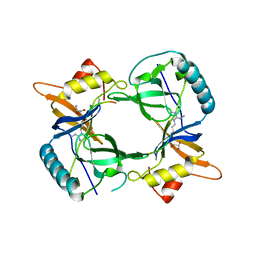
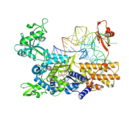
3ZGZ
DownloadVisualize
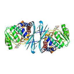
BU of 3zgz by Molmil
Ternary complex of E. coli leucyl-tRNA synthetase, tRNA(leu) and toxic moiety from agrocin 84 (TM84) in aminoacylation-like conformation
Descriptor: LEUCINE--TRNA LIGASE, MAGNESIUM ION, TRNA-LEU UAA ISOACCEPTOR, ...
Authors:Chopra, S, Palencia, A, Virus, C, Tripathy, A, Temple, B.R, Velazquez-Campoy, A, Cusack, S, Reader, J.S.
Deposit date:2012-12-19
Release date:2013-01-30
Last modified:2023-12-20
Method:X-RAY DIFFRACTION (2.4 Å)
Cite:Plant Tumour Biocontrol Agent Employs a tRNA-Dependent Mechanism to Inhibit Leucyl-tRNA Synthetase
Nat.Commun., 4, 2013
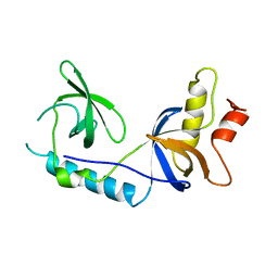
4B9W
DownloadVisualize
BU of 4b9w by Molmil
Structure of extended Tudor domain TD3 from mouse TDRD1 in complex with MILI peptide containing dimethylarginine 45.
Descriptor: GLYCEROL, PIWI-LIKE PROTEIN 2, TUDOR DOMAIN-CONTAINING PROTEIN 1
Authors:Mathioudakis, N, Palencia, A, Kadlec, J, Round, A, Tripsianes, K, Sattler, M, Pillai, R.S, Cusack, S.
Deposit date:2012-09-08
Release date:2012-10-17
Last modified:2024-11-13
Method:X-RAY DIFFRACTION (2.1 Å)
Cite:The multiple Tudor domain-containing protein TDRD1 is a molecular scaffold for mouse Piwi proteins and piRNA biogenesis factors.
Rna, 18, 2012
4B9X
DownloadVisualize
BU of 4b9x by Molmil
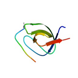
Structure of extended Tudor domain TD3 from mouse TDRD1
Descriptor: TUDOR DOMAIN-CONTAINING PROTEIN 1
Authors:Mathioudakis, N, Palencia, A, Kadlec, J, Round, A, Tripsianes, K, Sattler, M, Pillai, R.S, Cusack, S.
Deposit date:2012-09-08
Release date:2012-10-17
Last modified:2023-12-20
Method:X-RAY DIFFRACTION (2.8 Å)
Cite:The Multiple Tudor Domain-Containing Protein Tdrd1 is a Molecular Scaffold for Mouse Piwi Proteins and Pirna Biogenesis Factors.
RNA, 18, 2012
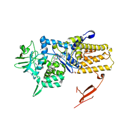
3ZIU
DownloadVisualize
BU of 3ziu by Molmil
Crystal structure of Mycoplasma mobile Leucyl-tRNA Synthetase with Leu-AMS in the active site
Descriptor: 5'-O-(L-leucylsulfamoyl)adenosine, GLYCEROL, LEUCYL-TRNA SYNTHETASE
Authors:Li, L, Palencia, A, Lukk, T, Li, Z, Luthey-Schulten, Z.A, Cusack, S, Martinis, S.A, Boniecki, M.T.
Deposit date:2013-01-10
Release date:2013-02-27
Last modified:2023-12-20
Method:X-RAY DIFFRACTION (2.07 Å)
Cite:Leucyl-tRNA Synthetase Editing Domain Functions as a Molecular Rheostat to Control Codon Ambiguity in Mycoplasma Pathogens.
Proc.Natl.Acad.Sci.USA, 110, 2013
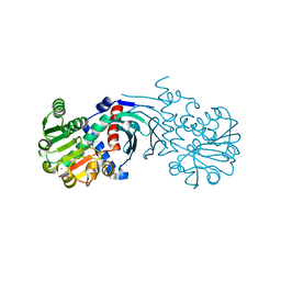
4BV8
DownloadVisualize
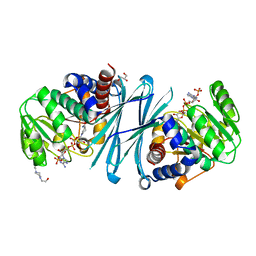
BU of 4bv8 by Molmil
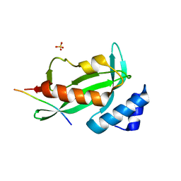
Crystal structure of the apo form of mouse Mu-crystallin.
Descriptor: GLYCEROL, POTASSIUM ION, THIOMORPHOLINE-CARBOXYLATE DEHYDROGENASE
Authors:Borel, F, Hachi, I, Palencia, A, Gaillard, M.C, Ferrer, J.L.
Deposit date:2013-06-25
Release date:2014-02-05
Last modified:2023-12-20
Method:X-RAY DIFFRACTION (2.3 Å)
Cite:Crystal Structure of Mouse Mu-Crystallin Complexed with Nadph and the T3 Thyroid Hormone
FEBS J., 281, 2014
4BVA
DownloadVisualize
BU of 4bva by Molmil
Crystal structure of the NADPH-T3 form of mouse Mu-crystallin.
Descriptor: 3,5,3'TRIIODOTHYRONINE, NADPH DIHYDRO-NICOTINAMIDE-ADENINE-DINUCLEOTIDE PHOSPHATE, POTASSIUM ION, ...
Authors:Borel, F, Hachi, I, Palencia, A, Gaillard, M.C, Ferrer, J.L.
Deposit date:2013-06-25
Release date:2014-02-05
Last modified:2023-12-20
Method:X-RAY DIFFRACTION (1.75 Å)
Cite:Crystal Structure of Mouse Mu-Crystallin Complexed with Nadph and the T3 Thyroid Hormone
FEBS J., 281, 2014
4BV9
DownloadVisualize
BU of 4bv9 by Molmil
Crystal structure of the NADPH form of mouse Mu-crystallin.
Descriptor: 4-(2-HYDROXYETHYL)-1-PIPERAZINE ETHANESULFONIC ACID, GLYCEROL, NADPH DIHYDRO-NICOTINAMIDE-ADENINE-DINUCLEOTIDE PHOSPHATE, ...
Authors:Borel, F, Hachi, I, Palencia, A, Gaillard, M.C, Ferrer, J.L.
Deposit date:2013-06-25
Release date:2014-02-05
Last modified:2024-11-06
Method:X-RAY DIFFRACTION (2.193 Å)
Cite:Crystal Structure of Mouse Mu-Crystallin Complexed with Nadph and the T3 Thyroid Hormone
FEBS J., 281, 2014
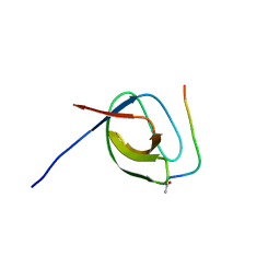
3EG0
DownloadVisualize
BU of 3eg0 by Molmil
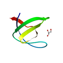
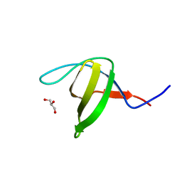
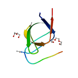
Crystal structure of the N114T mutant of ABL-SH3 domain
Descriptor: GLYCEROL, Proto-oncogene tyrosine-protein kinase ABL1
Authors:Camara-Artigas, A.
Deposit date:2008-09-10
Release date:2009-09-15
Last modified:2023-08-30
Method:X-RAY DIFFRACTION (2.3 Å)
Cite:Role of interfacial water molecules in proline-rich ligand recognition by the Src homology 3 domain of Abl.
J.Biol.Chem., 285, 2010
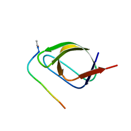
3EG2
DownloadVisualize
BU of 3eg2 by Molmil
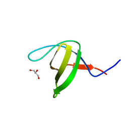
Crystal structure of the N114Q mutant of ABL-SH3 domain
Descriptor: GLYCEROL, Proto-oncogene tyrosine-protein kinase ABL1
Authors:Camara-Artigas, A.
Deposit date:2008-09-10
Release date:2009-09-15
Last modified:2023-08-30
Method:X-RAY DIFFRACTION (1.8 Å)
Cite:Role of interfacial water molecules in proline-rich ligand recognition by the Src homology 3 domain of Abl.
J.Biol.Chem., 285, 2010
3EG1
DownloadVisualize
BU of 3eg1 by Molmil
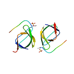
Crystal structure of the N114Q mutant of ABL-SH3 domain complexed with a designed high-affinity peptide ligand: implications for SH3-ligand interactions
Descriptor: Proto-oncogene tyrosine-protein kinase ABL1, SULFATE ION, p41 peptide
Authors:Camara-Artigas, A.
Deposit date:2008-09-10
Release date:2009-09-15
Last modified:2024-10-16
Method:X-RAY DIFFRACTION (1.85 Å)
Cite:Role of interfacial water molecules in proline-rich ligand recognition by the Src homology 3 domain of Abl.
J.Biol.Chem., 285, 2010
3EGU
DownloadVisualize
BU of 3egu by Molmil
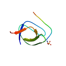
Crystal structure of the N114A mutant of ABL-SH3 domain
Descriptor: GLYCEROL, Proto-oncogene tyrosine-protein kinase ABL1, SULFATE ION
Authors:Camara-Artigas, A.
Deposit date:2008-09-11
Release date:2009-09-15
Last modified:2024-02-21
Method:X-RAY DIFFRACTION (2.25 Å)
Cite:Role of interfacial water molecules in proline-rich ligand recognition by the Src homology 3 domain of Abl.
J.Biol.Chem., 285, 2010
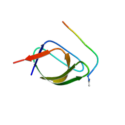
3EG3
DownloadVisualize
BU of 3eg3 by Molmil
Crystal structure of the N114A mutant of ABL-SH3 domain
Descriptor: GLYCEROL, Proto-oncogene tyrosine-protein kinase ABL1
Authors:Camara-Artigas, A.
Deposit date:2008-09-10
Release date:2009-09-15
Last modified:2023-08-30
Method:X-RAY DIFFRACTION (1.4 Å)
Cite:Role of interfacial water molecules in proline-rich ligand recognition by the Src homology 3 domain of Abl.
J.Biol.Chem., 285, 2010
2O88
DownloadVisualize
BU of 2o88 by Molmil
Crystal structure of the N114A mutant of ABL-SH3 domain complexed with a designed high-affinity peptide ligand: implications for SH3-ligand interactions
Descriptor: P41 peptide, Proto-oncogene tyrosine-protein kinase ABL1, SULFATE ION
Authors:Camara-Artigas, A.
Deposit date:2006-12-12
Release date:2007-05-01
Last modified:2024-10-30
Method:X-RAY DIFFRACTION (1.75 Å)
Cite:Crystallization by capillary counter-diffusion and structure determination of the N114A mutant of the SH3 domain of Abl tyrosine kinase complexed with a high-affinity peptide ligand.
Acta Crystallogr.,Sect.D, 63, 2007
4EJE
DownloadVisualize
BU of 4eje by Molmil
Structure Of The Tsg101 UEV Domain In Complex With an Ebola PTAP late Domain Peptide
Descriptor: Matrix protein VP40, SULFATE ION, Tumor susceptibility gene 101 protein
Authors:Camara-Artigas, A.
Deposit date:2012-04-06
Release date:2013-04-10
Last modified:2023-09-13
Method:X-RAY DIFFRACTION (2.2 Å)
Cite:To be published
To be Published
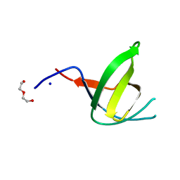
4JJB
DownloadVisualize
BU of 4jjb by Molmil
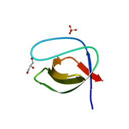
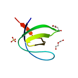
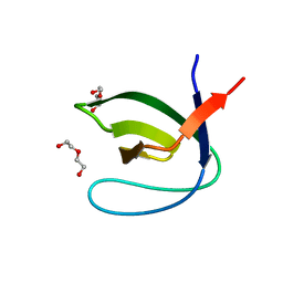
Crystal structure of the Abl-SH3 domain at pH3
Descriptor: DI(HYDROXYETHYL)ETHER, SULFATE ION, Tyrosine-protein kinase ABL1
Authors:Camara-Artigas, A, Martin-Garcia, J.M.
Deposit date:2013-03-07
Release date:2014-03-12
Last modified:2023-11-08
Method:X-RAY DIFFRACTION (1.65 Å)
Cite:Crystal structure of the Abl-SH3 domain at pH3
To be Published
4J9H
DownloadVisualize
BU of 4j9h by Molmil
Crystal structure of the ABL-SH3 domain complexed with the designed high-affinity peptide ligand P7 at pH 8
Descriptor: P7, SULFATE ION, Tyrosine-protein kinase ABL1
Authors:Camara-Artigas, A.
Deposit date:2013-02-16
Release date:2014-01-29
Last modified:2024-11-20
Method:X-RAY DIFFRACTION (1.7 Å)
Cite:Crystal structure of the ABL-SH3 domain complexed with the designed high-affinity peptide ligand P7
To be Published
4J9C
DownloadVisualize
BU of 4j9c by Molmil
Crystal structure of the Abl-SH3 domain H59Q-N96T mutant complexed with the designed high-affinity peptide ligand P17
Descriptor: DI(HYDROXYETHYL)ETHER, P17, TRIETHYLENE GLYCOL, ...
Authors:Camara-Artigas, A.
Deposit date:2013-02-16
Release date:2014-01-29
Last modified:2024-11-27
Method:X-RAY DIFFRACTION (1.051 Å)
Cite:Crystal structure of the Abl-SH3 domain H59Q-N96T mutant complexed with the designed high-affinity peptide ligand P17
To be Published
4J9I
DownloadVisualize
BU of 4j9i by Molmil
Crystal structure of the ABL-SH3 domain complexed with the designed high-affinity peptide ligand P17
Descriptor: GLYCEROL, P17, Tyrosine-protein kinase ABL1
Authors:Camara-Artigas, A.
Deposit date:2013-02-16
Release date:2014-01-29
Last modified:2024-11-06
Method:X-RAY DIFFRACTION (2.2 Å)
Cite:Crystal structure of the ABL-SH3 domain complexed with the designed high-affinity peptide ligand P17
To be Published
4J9F
DownloadVisualize
BU of 4j9f by Molmil
Crystal structure of the Abl-SH3 domain complexed with the high affinity peptide P0
Descriptor: GLYCEROL, P0, SULFATE ION, ...
Authors:Camara-Artigas, A.
Deposit date:2013-02-16
Release date:2014-01-29
Last modified:2024-11-27
Method:X-RAY DIFFRACTION (1.094 Å)
Cite:Crystal structure of the Abl-SH3 domain complexed with the high affinity peptide P0
To be Published
4J9E
DownloadVisualize
BU of 4j9e by Molmil
Crystal structure of the N114A mutant of the Abl-SH3 domain complexed with the high affinity peptide P17
Descriptor: P17, SULFATE ION, Tyrosine-protein kinase ABL1
Authors:Camara-Artigas, A.
Deposit date:2013-02-16
Release date:2014-01-29
Last modified:2024-11-20
Method:X-RAY DIFFRACTION (1.4 Å)
Cite:Crystal structure of the N114A mutant of the Abl-SH3 domain complexed with the high affinity peptide P17
To be Published
4JJD
DownloadVisualize
BU of 4jjd by Molmil
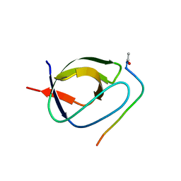
Crystal structure of the N114A Abl-SH3 domain mutant at pH4
Descriptor: DI(HYDROXYETHYL)ETHER, SODIUM ION, Tyrosine-protein kinase ABL1
Authors:Camara-Artigas, A, Martin-Garcia, J.M.
Deposit date:2013-03-07
Release date:2014-03-12
Last modified:2023-11-08
Method:X-RAY DIFFRACTION (1.6 Å)
Cite:Crystal structure of the Abl-SH3 domain at pH5
To be Published
4J9B
DownloadVisualize
BU of 4j9b by Molmil
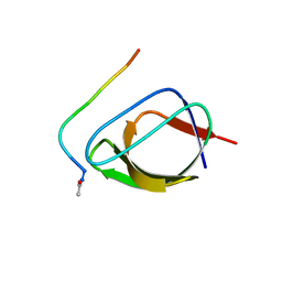
Crystal structure of the Abl-SH3 domain H59Q-N96T mutant
Descriptor: DI(HYDROXYETHYL)ETHER, Tyrosine-protein kinase ABL1
Authors:Camara-Artigas, A.
Deposit date:2013-02-16
Release date:2014-01-29
Last modified:2023-09-20
Method:X-RAY DIFFRACTION (1.702 Å)
Cite:Crystal structure of the Abl-SH3 domain H59Q-N96T mutant
To be Published
4J9D
DownloadVisualize
BU of 4j9d by Molmil
Crystal structure of the N114A mutant of the Abl-SH3 domain complexed with the high affinity peptide P0
Descriptor: P0, SULFATE ION, Tyrosine-protein kinase ABL1
Authors:Camara-Artigas, A.
Deposit date:2013-02-16
Release date:2014-01-29
Last modified:2024-11-06
Method:X-RAY DIFFRACTION (1.5 Å)
Cite:Crystal structure of the N114A mutant of the Abl-SH3 domain complexed with the high affinity peptide P0
To be Published
4JJC
DownloadVisualize
BU of 4jjc by Molmil
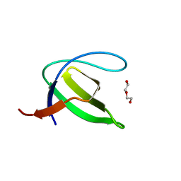
Crystal structure of the Abl-SH3 domain at pH5
Descriptor: DI(HYDROXYETHYL)ETHER, Tyrosine-protein kinase ABL1
Authors:Camara-Artigas, A, Martin-Garcia, J.M.
Deposit date:2013-03-07
Release date:2014-03-12
Last modified:2023-11-08
Method:X-RAY DIFFRACTION (1.6 Å)
Cite:Crystal structure of the Abl-SH3 domain at pH5
To be Published
4J9G
DownloadVisualize
BU of 4j9g by Molmil
Crystal structure of the ABL-SH3 domain complexed with the designed high-affinity peptide ligand P7 at pH7
Descriptor: GLYCEROL, P7, SULFATE ION, ...
Authors:Camara-Artigas, A.
Deposit date:2013-02-16
Release date:2014-01-29
Last modified:2024-11-27
Method:X-RAY DIFFRACTION (1.8 Å)
Cite:Crystal structure of the ABL-SH3 domain complexed with the designed high-affinity peptide ligand P7
To be Published

238895

數據於2025-07-16公開中

PDB statisticsPDBj update infoContact PDBjnumon