6AR7
 
 | |
6B1P
 | |
6AO8
 | |
6APH
 | |
6AQY
 | |
6AN0
 | |
6ANZ
 | |
6AQG
 | |
6AY1
 | |
6AO7
 | |
6BAC
 | |
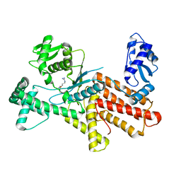
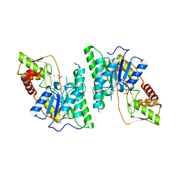
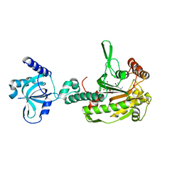
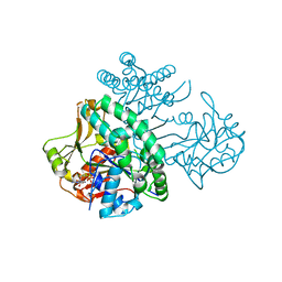
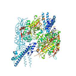
6AMY
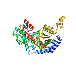
 | | Crystal structure of a 2,3,4,5-tetrahydropyridine-2,6-dicarboxylate N-succinyltransferase from Acinetobacter baumannii | | Descriptor: | 2,3,4,5-tetrahydropyridine-2,6-dicarboxylate N-succinyltransferase | | Authors: | Seattle Structural Genomics Center for Infectious Disease, Edwards, T.E, Seattle Structural Genomics Center for Infectious Disease (SSGCID) | | Deposit date: | 2017-08-11 | | Release date: | 2017-09-06 | | Last modified: | 2023-10-04 | | Method: | X-RAY DIFFRACTION (2.85 Å) | | Cite: | Crystal structure of a 2,3,4,5-tetrahydropyridine-2,6-dicarboxylate N-succinyltransferase from Acinetobacter baumannii
To Be Published
|
|
6AQH
 | |
6B8S
 | |
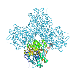
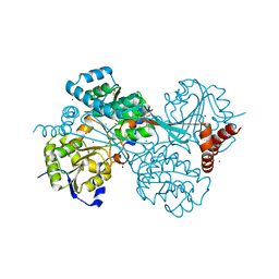
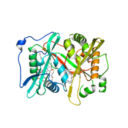
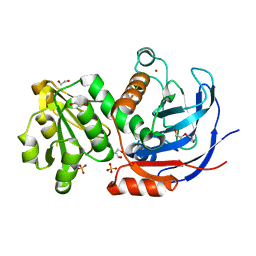
6AMZ
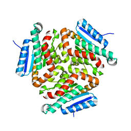
 | | Crystal structure of a domain swapped 2,3,4,5-tetrahydropyridine-2,6-dicarboxylate N-succinyltransferase from Acinetobacter baumannii | | Descriptor: | (4R)-2-METHYLPENTANE-2,4-DIOL, 2,3,4,5-tetrahydropyridine-2,6-dicarboxylate N-succinyltransferase, CALCIUM ION | | Authors: | Edwards, T.E, Seattle Structural Genomics Center for Infectious Disease (SSGCID) | | Deposit date: | 2017-08-11 | | Release date: | 2017-08-23 | | Last modified: | 2023-10-04 | | Method: | X-RAY DIFFRACTION (2.05 Å) | | Cite: | Crystal structure of a domain swapped 2,3,4,5-tetrahydropyridine-2,6-dicarboxylate N-succinyltransferase from Acinetobacter baumannii
To Be Published
|
|
6B1L
 | |
6C25
 | |
6C49
 | |
6C86
 | |
6BQZ
 | |
6BZC
 | |
6C46
 | |
6C7C
 | |
6C87
 | |
6BLJ
 | |


