Loading
PDBj
MenuPDBj@FacebookPDBj@TwitterPDBj@YouTubewwPDB FoundationwwPDB
RCSB PDBPDBeBMRBAdv. SearchSearch help
Search by PDB author
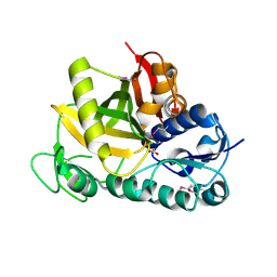
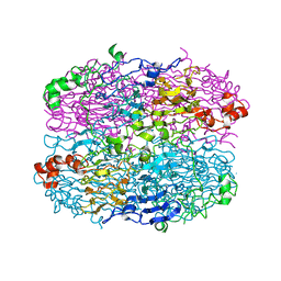
2QVH
DownloadVisualize
BU of 2qvh by Molmil
Crystal structure of O-succinylbenzoate synthase complexed with O-succinyl benzoate (OSB)
Descriptor: 2-SUCCINYLBENZOATE, MAGNESIUM ION, O-succinylbenzoate-CoA synthase
Authors:Sugadev, R, Burley, S.K, Swaminathan, S, New York SGX Research Center for Structural Genomics (NYSGXRC)
Deposit date:2007-08-08
Release date:2007-08-28
Last modified:2024-11-06
Method:X-RAY DIFFRACTION (1.76 Å)
Cite:Crystal structure of O-succinylbenzoate synthase complexed with O-succinyl benzoate.
To be Published
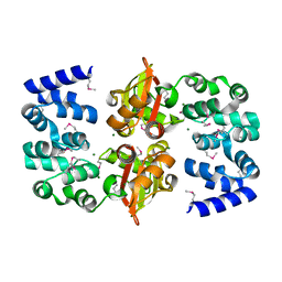
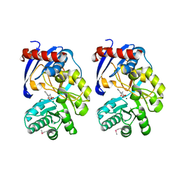
2RDY
DownloadVisualize
BU of 2rdy by Molmil
Crystal structure of a putative glycoside hydrolase family protein from Bacillus halodurans
Descriptor: BH0842 protein
Authors:Sugadev, R, Burley, S.K, Swaminathan, S, New York SGX Research Center for Structural Genomics (NYSGXRC)
Deposit date:2007-09-25
Release date:2007-10-09
Last modified:2024-11-06
Method:X-RAY DIFFRACTION (2.03 Å)
Cite:Crystal structure of a putative glycoside hydrolase family protein from Bacillus halodurans.
To be Published
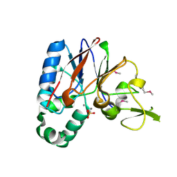
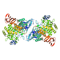
3DF7
DownloadVisualize
BU of 3df7 by Molmil
Crystal structure of a putative ATP-grasp superfamily protein from Archaeoglobus fulgidus
Descriptor: ACETATE ION, Putative ATP-grasp superfamily protein
Authors:Sugadev, R, Burley, S.K, Swaminathan, S, New York SGX Research Center for Structural Genomics (NYSGXRC)
Deposit date:2008-06-11
Release date:2008-08-05
Last modified:2024-10-30
Method:X-RAY DIFFRACTION (1.87 Å)
Cite:Crystal structure of a putative ATP-grasp superfamily protein from Archaeoglobus fulgidus
To be Published
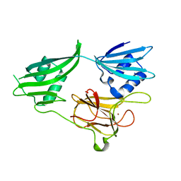
2OUX
DownloadVisualize
BU of 2oux by Molmil
Crystal structure of the soluble part of a magnesium transporter
Descriptor: MAGNESIUM ION, Magnesium transporter
Authors:Sugadev, R, Burley, S.K, Swaminathan, S, New York SGX Research Center for Structural Genomics (NYSGXRC)
Deposit date:2007-02-12
Release date:2007-03-20
Last modified:2024-10-30
Method:X-RAY DIFFRACTION (2.16 Å)
Cite:Structural studies on cytosolic domain of magnesium transporter MgtE from Enterococcus faecalis.
Proteins, 78, 2010
2QJC
DownloadVisualize
BU of 2qjc by Molmil
Crystal structure of a putative diadenosine tetraphosphatase
Descriptor: Diadenosine tetraphosphatase, putative, MANGANESE (II) ION, ...
Authors:Sugadev, R, Burley, S.K, Swaminathan, S, New York SGX Research Center for Structural Genomics (NYSGXRC)
Deposit date:2007-07-06
Release date:2007-07-24
Last modified:2021-02-03
Method:X-RAY DIFFRACTION (2.05 Å)
Cite:Structural genomics of protein phosphatases.
J.Struct.Funct.Genom., 8, 2007
2GU1
DownloadVisualize
BU of 2gu1 by Molmil
Crystal structure of a zinc containing peptidase from vibrio cholerae
Descriptor: SODIUM ION, ZINC ION, Zinc peptidase
Authors:Sugadev, R, Kumaran, D, Swaminathan, S, Burley, S.K, New York SGX Research Center for Structural Genomics (NYSGXRC)
Deposit date:2006-04-28
Release date:2006-07-04
Last modified:2024-02-14
Method:X-RAY DIFFRACTION (1.9 Å)
Cite:Crystal structure of a putative lysostaphin peptidase from Vibrio cholerae.
Proteins, 72, 2008
1XGF
DownloadVisualize
BU of 1xgf by Molmil
Backbone Structure of COCOSIN, an 11S storage protein from cocos nucifera
Descriptor: cocosin
Authors:Balasundaresan, D, Ponnuswamy, M.N.
Deposit date:2004-09-17
Release date:2005-11-29
Last modified:2023-08-23
Method:X-RAY DIFFRACTION (2.61 Å)
Cite:CRYSTAL STRUCTURE OF COCOSIN FROM COCOS NUCIFERA, A BACKBONE MODEL
TO BE PUBLISHED
<12


227111

數據於2024-11-06公開中

PDB statisticsPDBj update infoContact PDBjnumon