6W14
 
 | |
6UM4
 | |
6WBD
 | |
6WFM
 | |
6W04
 | |
6VS4
 | |
6UJD
 | |
6WCI
 | |
6WGY
 | |
6V45
 | |
6UYH
 | |
6VU9
 | |
6WNG
 | |
6TZ7
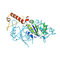
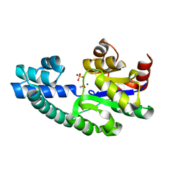
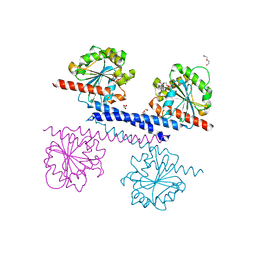
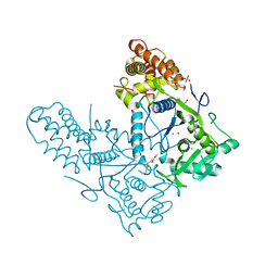
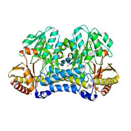
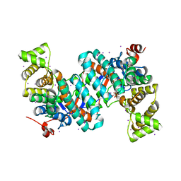
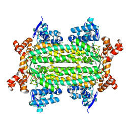
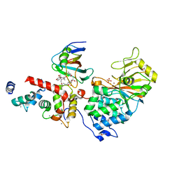 | | Crystal Structure of Aspergillus fumigatus Calcineurin A, Calcineurin B, FKBP12 and FK506 (Tacrolimus) | | Descriptor: | 1,2-ETHANEDIOL, 8-DEETHYL-8-[BUT-3-ENYL]-ASCOMYCIN, CALCIUM ION, ... | | Authors: | Fox III, D, Horanyi, P.S. | | Deposit date: | 2019-08-10 | | Release date: | 2019-09-18 | | Last modified: | 2023-10-11 | | Method: | X-RAY DIFFRACTION (2.5 Å) | | Cite: | Harnessing calcineurin-FK506-FKBP12 crystal structures from invasive fungal pathogens to develop antifungal agents.
Nat Commun, 10, 2019
|
|


