Loading
PDBj
MenuPDBj@FacebookPDBj@TwitterPDBj@YouTubewwPDB FoundationwwPDB
RCSB PDBPDBeBMRBAdv. SearchSearch help
Search by PDB author
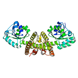
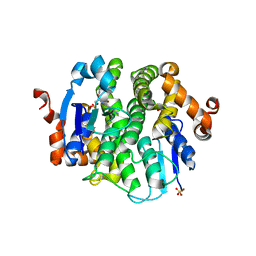
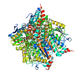
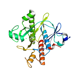
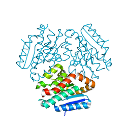
3I3F
DownloadVisualize
BU of 3i3f by Molmil
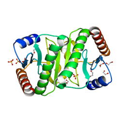
Hypothetical protein from Giardia lamblia GL50803_14299
Descriptor: BUTANOIC ACID, Hypothetical protein, PENTANOIC ACID
Authors:Seattle Structural Genomics Center for Infectious Disease (SSGCID)
Deposit date:2009-06-30
Release date:2009-09-01
Last modified:2011-07-13
Method:X-RAY DIFFRACTION (1.35 Å)
Cite:Hypothetical protein from Giardia lamblia GL50803_14299
To be Published
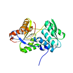
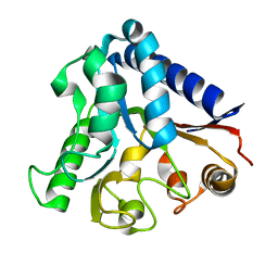
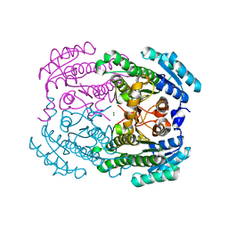
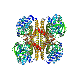
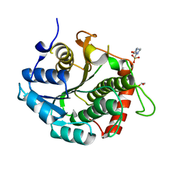
3I3R
DownloadVisualize
BU of 3i3r by Molmil
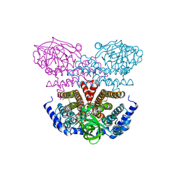
X-ray structure dihydrofolate reductase/thymidylate synthase from babesia bovis at 2.35A resolution
Descriptor: CHLORIDE ION, Dihydrofolate reductase/thymidylate synthase
Authors:Seattle Structural Genomics Center for Infectious Disease (SSGCID), Seattle Structural Genomics Center for Infectious Disease (SSGCID)
Deposit date:2009-06-30
Release date:2009-08-18
Last modified:2023-09-06
Method:X-RAY DIFFRACTION (2.35 Å)
Cite:Inhibitor-bound complexes of dihydrofolate reductase-thymidylate synthase from Babesia bovis.
Acta Crystallogr.,Sect.F, 67, 2011
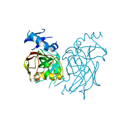
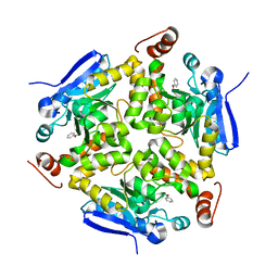
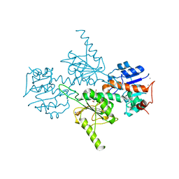
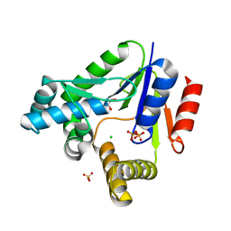
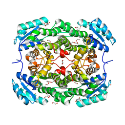
3HWI
DownloadVisualize
BU of 3hwi by Molmil
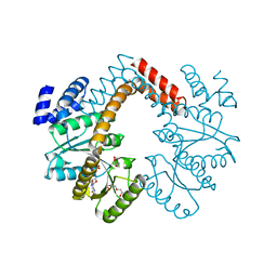
Crystal structure of probable thiosulfate sulfurtransferase Cysa2 (Rhodanese-like protein) from Mycobacterium tuberculosis
Descriptor: Thiosulfate sulfurtransferase
Authors:Seattle Structural Genomics Center for Infectious Disease (SSGCID)
Deposit date:2009-06-17
Release date:2009-06-30
Last modified:2023-09-06
Method:X-RAY DIFFRACTION (2.29 Å)
Cite:Increasing the structural coverage of tuberculosis drug targets.
Tuberculosis (Edinb), 95, 2015
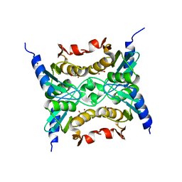
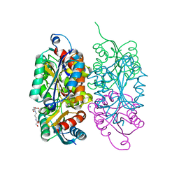
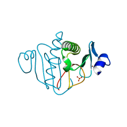
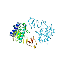
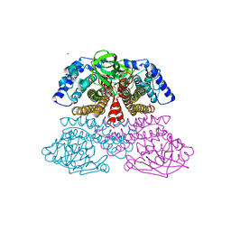
3HWK
DownloadVisualize
BU of 3hwk by Molmil
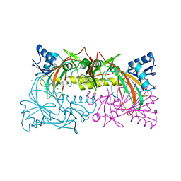
Crystal structure of methylcitrate synthase from Mycobacterium tuberculosis
Descriptor: Methylcitrate synthase, SUCCINIC ACID
Authors:Seattle Structural Genomics Center for Infectious Disease (SSGCID)
Deposit date:2009-06-17
Release date:2009-06-30
Last modified:2023-09-06
Method:X-RAY DIFFRACTION (2.3 Å)
Cite:Increasing the structural coverage of tuberculosis drug targets.
Tuberculosis (Edinb), 95, 2015
3H7F
DownloadVisualize
BU of 3h7f by Molmil
Crystal structure of serine hydroxymethyltransferase from Mycobacterium tuberculosis
Descriptor: MAGNESIUM ION, Serine hydroxymethyltransferase 1
Authors:Seattle Structural Genomics Center for Infectious Disease (SSGCID)
Deposit date:2009-04-24
Release date:2009-05-05
Last modified:2023-11-22
Method:X-RAY DIFFRACTION (1.5 Å)
Cite:Increasing the structural coverage of tuberculosis drug targets.
Tuberculosis (Edinb), 95, 2015
3GP3
DownloadVisualize
BU of 3gp3 by Molmil
Crystal structure of phosphoglyceromutase from Burkholderia pseudomallei with 2-phosphoserine
Descriptor: 2,3-bisphosphoglycerate-dependent phosphoglycerate mutase, PHOSPHITE ION, PHOSPHOSERINE, ...
Authors:Seattle Structural Genomics Center for Infectious Disease (SSGCID)
Deposit date:2009-03-20
Release date:2009-03-31
Last modified:2024-02-21
Method:X-RAY DIFFRACTION (1.5 Å)
Cite:An ensemble of structures of Burkholderia pseudomallei 2,3-bisphosphoglycerate-dependent phosphoglycerate mutase.
Acta Crystallogr.,Sect.F, 67, 2011
3GWE
DownloadVisualize
BU of 3gwe by Molmil
2.1 Angstrom crystal structure of 3-oxoacyl-(acyl-carrier-protein) synthase III
Descriptor: 3-oxoacyl-(Acyl-carrier-protein) synthase III
Authors:Seattle Structural Genomics Center for Infectious Disease (SSGCID)
Deposit date:2009-03-31
Release date:2009-04-28
Last modified:2023-09-06
Method:X-RAY DIFFRACTION (2.1 Å)
Cite:Combining functional and structural genomics to sample the essential Burkholderia structome.
Plos One, 8, 2013
3JS4
DownloadVisualize
BU of 3js4 by Molmil
Crystal structure of iron superoxide dismutase from Anaplasma phagocytophilum
Descriptor: FE (III) ION, SODIUM ION, Superoxide dismutase
Authors:Seattle Structural Genomics Center for Infectious Disease (SSGCID)
Deposit date:2009-09-09
Release date:2009-09-22
Last modified:2023-09-06
Method:X-RAY DIFFRACTION (1.95 Å)
Cite:Crystal structure of Iron superoxide dismutase from Anaplasma phagocytophilum
To be Published
3JS9
DownloadVisualize
BU of 3js9 by Molmil
Crystal structure of nucleoside diphosphate kinase family protein from Babesia bovis
Descriptor: Nucleoside diphosphate kinase family protein, SULFATE ION
Authors:Seattle Structural Genomics Center for Infectious Disease (SSGCID)
Deposit date:2009-09-09
Release date:2009-09-22
Last modified:2023-09-06
Method:X-RAY DIFFRACTION (2.5 Å)
Cite:Crystal structure of nucleoside diphosphate kinase family protein from Babesia bovis
To be Published
3K2H
DownloadVisualize
BU of 3k2h by Molmil
Co-crystal structure of dihydrofolate reductase/thymidylate synthase from Babesia bovis with dUMP, Pemetrexed and NADP
Descriptor: 1,2-ETHANEDIOL, 2'-DEOXYURIDINE 5'-MONOPHOSPHATE, 2-{4-[2-(2-AMINO-4-OXO-4,7-DIHYDRO-3H-PYRROLO[2,3-D]PYRIMIDIN-5-YL)-ETHYL]-BENZOYLAMINO}-PENTANEDIOIC ACID, ...
Authors:Seattle Structural Genomics Center for Infectious Disease (SSGCID)
Deposit date:2009-09-30
Release date:2009-10-13
Last modified:2023-09-06
Method:X-RAY DIFFRACTION (2.2 Å)
Cite:Inhibitor-bound complexes of dihydrofolate reductase-thymidylate synthase from Babesia bovis.
Acta Crystallogr.,Sect.F, 67, 2011
3GVC
DownloadVisualize
BU of 3gvc by Molmil
Crystal structure of probable short-chain dehydrogenase-reductase from Mycobacterium tuberculosis
Descriptor: Probable short-chain type dehydrogenase/reductase
Authors:Seattle Structural Genomics Center for Infectious Disease (SSGCID)
Deposit date:2009-03-30
Release date:2009-04-07
Last modified:2023-09-06
Method:X-RAY DIFFRACTION (2.45 Å)
Cite:Increasing the structural coverage of tuberculosis drug targets.
Tuberculosis (Edinb), 95, 2015
3GK0
DownloadVisualize
BU of 3gk0 by Molmil
Crystal structure of pyridoxal phosphate biosynthetic protein from Burkholderia pseudomallei
Descriptor: 1-DEOXY-D-XYLULOSE-5-PHOSPHATE, PHOSPHATE ION, Pyridoxine 5'-phosphate synthase
Authors:Seattle Structural Genomics Center for Infectious Disease (SSGCID)
Deposit date:2009-03-09
Release date:2009-03-17
Last modified:2023-09-06
Method:X-RAY DIFFRACTION (2.28 Å)
Cite:Combining functional and structural genomics to sample the essential Burkholderia structome.
Plos One, 8, 2013
3JS5
DownloadVisualize
BU of 3js5 by Molmil
Crystal structure of protein tyrosine phosphatase from Entamoeba histolytica with Hepes in the active site. High resolution, alternative crystal form with 1 molecule in asymmetric unit
Descriptor: 4-(2-HYDROXYETHYL)-1-PIPERAZINE ETHANESULFONIC ACID, Protein tyrosine phosphatase, SODIUM ION
Authors:Seattle Structural Genomics Center for Infectious Disease (SSGCID)
Deposit date:2009-09-09
Release date:2009-09-22
Last modified:2023-09-06
Method:X-RAY DIFFRACTION (1.94 Å)
Cite:Crystal structure and putative substrate identification for the Entamoeba histolytica low molecular weight tyrosine phosphatase.
Mol.Biochem.Parasitol., 193, 2014
3I4T
DownloadVisualize
BU of 3i4t by Molmil
Crystal structure of putative diphthine synthase from Entamoeba histolytica
Descriptor: diphthine synthase
Authors:Seattle Structural Genomics Center for Infectious Disease (SSGCID)
Deposit date:2009-07-02
Release date:2009-07-28
Last modified:2023-09-06
Method:X-RAY DIFFRACTION (2.49 Å)
Cite:Crystal structure of putative diphthine synthase from Entamoeba histolytica
To be Published
3I0P
DownloadVisualize
BU of 3i0p by Molmil
Crystal structure of malate dehydrogenase from Entamoeba histolytica
Descriptor: GLYCEROL, Malate dehydrogenase, NICOTINAMIDE-ADENINE-DINUCLEOTIDE
Authors:Seattle Structural Genomics Center for Infectious Disease (SSGCID)
Deposit date:2009-06-25
Release date:2009-07-14
Last modified:2023-09-06
Method:X-RAY DIFFRACTION (2.6 Å)
Cite:Crystal structure of malate dehydrogenase from Entamoeba histolytica
To be Published
3KC6
DownloadVisualize
BU of 3kc6 by Molmil
Crystal structure of the large c-terminal domain of polymerase basic protein 2 from influenza virus a/viet nam/1203/2004 (h5n1)
Descriptor: 1,2-ETHANEDIOL, Polymerase PB2
Authors:Seattle Structural Genomics Center for Infectious Disease (SSGCID)
Deposit date:2009-10-20
Release date:2010-03-09
Last modified:2023-09-06
Method:X-RAY DIFFRACTION (2.05 Å)
Cite:Biological and structural characterization of a host-adapting amino acid in influenza virus.
Plos Pathog., 6, 2010
3KHW
DownloadVisualize
BU of 3khw by Molmil
Crystal structure of the large c-terminal domain of polymerase basic protein 2 from influenza virus a/mexico/indre4487/2009(h1n1)
Descriptor: GLYCEROL, Polymerase PB2
Authors:Seattle Structural Genomics Center for Infectious Disease (SSGCID)
Deposit date:2009-10-31
Release date:2010-03-09
Last modified:2023-09-06
Method:X-RAY DIFFRACTION (2.1 Å)
Cite:Biological and structural characterization of a host-adapting amino acid in influenza virus.
Plos Pathog., 6, 2010
3LAA
DownloadVisualize
BU of 3laa by Molmil
Crystal structure of the trimeric autotransporter adhesin head domain BpaA from Burkholderia pseudomallei
Descriptor: Haemagglutinin family protein
Authors:Seattle Structural Genomics Center for Infectious Disease (SSGCID)
Deposit date:2010-01-06
Release date:2010-05-12
Last modified:2023-09-06
Method:X-RAY DIFFRACTION (1.35 Å)
Cite:Structure of a Burkholderia pseudomallei trimeric autotransporter adhesin head.
Plos One, 5, 2010
3LG6
DownloadVisualize
BU of 3lg6 by Molmil
Crystal structure of putative glutathione transferase from Coccidioides immitis
Descriptor: SULFATE ION, putative glutathione transferase
Authors:Seattle Structural Genomics Center for Infectious Disease (SSGCID)
Deposit date:2010-01-19
Release date:2010-01-26
Last modified:2023-09-06
Method:X-RAY DIFFRACTION (2.2 Å)
Cite:Structures of a putative zeta-class glutathione S-transferase from the pathogenic fungus Coccidioides immitis.
Acta Crystallogr.,Sect.F, 67, 2011
3LNT
DownloadVisualize
BU of 3lnt by Molmil
Crystal structure of phosphoglyceromutase from Burkholderia Pseudomallei 1710B with bound malonic acid
Descriptor: 1,2-ETHANEDIOL, 2,3-bisphosphoglycerate-dependent phosphoglycerate mutase, MALONATE ION, ...
Authors:Seattle Structural Genomics Center for Infectious Disease (SSGCID)
Deposit date:2010-02-03
Release date:2010-02-09
Last modified:2024-02-21
Method:X-RAY DIFFRACTION (2.1 Å)
Cite:An ensemble of structures of Burkholderia pseudomallei 2,3-bisphosphoglycerate-dependent phosphoglycerate mutase.
Acta Crystallogr.,Sect.F, 67, 2011
3LA9
DownloadVisualize
BU of 3la9 by Molmil
Crystal structure of the trimeric autotransporter adhesin head domain BpaA from Burkholderia pseudomallei, iodide phased
Descriptor: Haemagglutinin family protein, IODIDE ION
Authors:Seattle Structural Genomics Center for Infectious Disease (SSGCID)
Deposit date:2010-01-06
Release date:2010-05-12
Last modified:2024-02-21
Method:X-RAY DIFFRACTION (2.05 Å)
Cite:Structure of a Burkholderia pseudomallei trimeric autotransporter adhesin head.
Plos One, 5, 2010
3LR3
DownloadVisualize
BU of 3lr3 by Molmil
Periplasmic domain of the risS sensor protein from Burkholderia pesuromallei, low pH native structure
Descriptor: Sensor protein
Authors:Seattle Structural Genomics Center for Infectious Disease (SSGCID)
Deposit date:2010-02-10
Release date:2010-06-16
Last modified:2023-09-06
Method:X-RAY DIFFRACTION (2.1 Å)
Cite:Structural basis for pH sensing by the periplasmic domain of the risS histidine kinase from Burkholderia pseudomallei
To be Published
3LUZ
DownloadVisualize
BU of 3luz by Molmil
Crystal structure of extragenic suppressor protein suhB from Bartonella henselae, via combined iodide SAD molecular replacement
Descriptor: Extragenic suppressor protein suhB, IODIDE ION, MAGNESIUM ION
Authors:Seattle Structural Genomics Center for Infectious Disease (SSGCID)
Deposit date:2010-02-18
Release date:2010-03-02
Last modified:2023-09-06
Method:X-RAY DIFFRACTION (2.05 Å)
Cite:SAD phasing using iodide ions in a high-throughput structural genomics environment.
J Struct Funct Genomics, 12, 2011
3LR0
DownloadVisualize
BU of 3lr0 by Molmil
Periplasmic domain of the risS sensor protein from Burkholderia pseudomallei, iodide phased at low pH
Descriptor: IODIDE ION, Sensor protein
Authors:Seattle Structural Genomics Center for Infectious Disease (SSGCID)
Deposit date:2010-02-10
Release date:2010-06-16
Last modified:2024-02-21
Method:X-RAY DIFFRACTION (1.9 Å)
Cite:Structural basis for pH sensing by the periplasmic domain of the risS histidine kinase from Bukholderia pseudomallei
To be Published
3LR4
DownloadVisualize
BU of 3lr4 by Molmil
Periplasmic domain of the risS sensor protein from Burkholderia pseudomallei, barium phased at low pH
Descriptor: BARIUM ION, CHLORIDE ION, Sensor protein
Authors:Seattle Structural Genomics Center for Infectious Disease (SSGCID)
Deposit date:2010-02-10
Release date:2010-06-16
Last modified:2024-02-21
Method:X-RAY DIFFRACTION (1.9 Å)
Cite:Structural basis for pH sensing by the periplasmic domain of the risS histidine kinase from Burkholderia pseudomallei
To be Published

221051

數據於2024-06-12公開中

PDB statisticsPDBj update infoContact PDBjnumon