Loading
PDBj
MenuPDBj@FacebookPDBj@TwitterPDBj@YouTubewwPDB FoundationwwPDB
RCSB PDBPDBeBMRBAdv. SearchSearch help
Search by PDB author
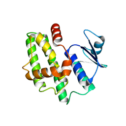
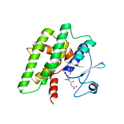
7DW1
DownloadVisualize
BU of 7dw1 by Molmil
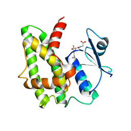
Crystal structure of a glutathione S-transferase SbGSTU6 from Salix babylonica in complex with glutathione
Descriptor: GLUTATHIONE, Glutathione S-transferase
Authors:Zhuge, X.L, Yang, H.L.
Deposit date:2021-01-15
Release date:2021-07-21
Last modified:2023-11-29
Method:X-RAY DIFFRACTION (2.27 Å)
Cite:Crystal structure of a glutathione S-transferase SbGSTU6 from Salix babylonica in complex with glutathione
To Be Published
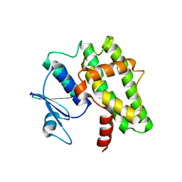
7DWD
DownloadVisualize
BU of 7dwd by Molmil
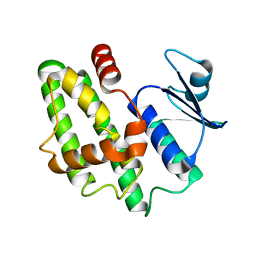
Crystal structure of a glutathione S-transferase SbGSTU7 from Salix babylonica
Descriptor: Glutathione S-transferase
Authors:Zhuge, X.L, Yang, H.L.
Deposit date:2021-01-17
Release date:2021-08-04
Last modified:2023-11-29
Method:X-RAY DIFFRACTION (1.58 Å)
Cite:Crystal structure of a glutathione S-transferase SbGSTU7 from Salix babylonica
To Be Published
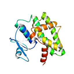
7DWF
DownloadVisualize
BU of 7dwf by Molmil
Crystal structure of a glutathione S-transferase mutant SbGSTU7(T53I) from Salix babylonica
Descriptor: Glutathione S-transferase
Authors:Zhuge, X.L, Yang, H.L.
Deposit date:2021-01-17
Release date:2021-08-04
Last modified:2023-11-29
Method:X-RAY DIFFRACTION (2.21 Å)
Cite:Crystal structure of a glutathione S-transferase mutant SbGSTU7(T53I) from Salix babylonica
To Be Published
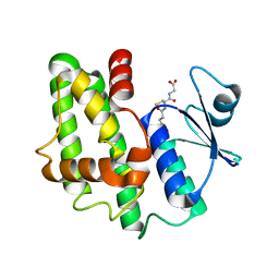
7DW2
DownloadVisualize
BU of 7dw2 by Molmil
Crystal structure of a glutathione S-transferase SbGSTU6 from Salix babylonica
Descriptor: Glutathione S-transferase
Authors:Zhuge, X.L, Yang, H.L.
Deposit date:2021-01-15
Release date:2021-08-04
Last modified:2023-11-29
Method:X-RAY DIFFRACTION (1.74 Å)
Cite:Crystal structure of a glutathione S-transferase SbGSTU6 from Salix babylonica
To Be Published
7DW3
DownloadVisualize
BU of 7dw3 by Molmil
Crystal structure of a glutathione S-transferase mutant SbGSTU6(I55T) from Salix babylonica
Descriptor: Glutathione S-transferase
Authors:Zhuge, X.L, Yang, H.L.
Deposit date:2021-01-15
Release date:2021-08-04
Last modified:2023-11-29
Method:X-RAY DIFFRACTION (1.93 Å)
Cite:Crystal structure of a glutathione S-transferase SbGSTU6 from Salix babylonica in complex with glutathione
To Be Published
7DWG
DownloadVisualize
BU of 7dwg by Molmil
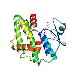
Crystal structure of a glutathione S-transferase mutant SbGSTU7(T53I) from Salix babylonica in complex with glutathione
Descriptor: GLUTATHIONE, Glutathione S-transferase
Authors:Zhuge, X.L, Yang, H.L.
Deposit date:2021-01-17
Release date:2021-08-04
Last modified:2023-11-29
Method:X-RAY DIFFRACTION (1.67 Å)
Cite:Crystal structure of a glutathione S-transferase mutant SbGSTU7(T53I) from Salix babylonica in complex with glutathione
To Be Published
7DW4
DownloadVisualize
BU of 7dw4 by Molmil
Crystal structure of a glutathione S-transferase mutant SbGSTU6(I55T) from Salix babylonica in complex with glutathione
Descriptor: GLUTATHIONE, Glutathione S-transferase
Authors:Zhuge, X.L, Yang, H.L.
Deposit date:2021-01-15
Release date:2021-08-04
Last modified:2023-11-29
Method:X-RAY DIFFRACTION (1.57 Å)
Cite:Crystal structure of a glutathione S-transferase mutant SbGSTU6(I55T) from Salix babylonica in complex with glutathione
To Be Published
7DWE
DownloadVisualize
BU of 7dwe by Molmil
Crystal structure of a glutathione S-transferase SbGSTU7 from Salix babylonica in complex with glutathione
Descriptor: GLUTATHIONE, Glutathione S-transferase
Authors:Zhuge, X.L, Yang, H.L.
Deposit date:2021-01-17
Release date:2021-08-04
Last modified:2023-11-29
Method:X-RAY DIFFRACTION (1.77 Å)
Cite:Crystal structure of a glutathione S-transferase SbGSTU7 from Salix babylonica in complex with glutathione
To Be Published
8E7B
DownloadVisualize
BU of 8e7b by Molmil
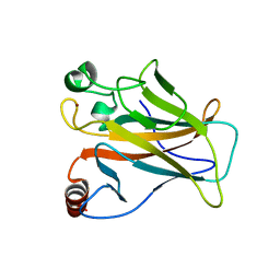
Crystal structure of the p53 (Y107H) core domain monoclinic P form
Descriptor: Cellular tumor antigen p53, ZINC ION
Authors:Lovell, S, Liu, L, Battaile, K.P, Miller, S, Karanicolas, J.
Deposit date:2022-08-23
Release date:2023-05-17
Last modified:2023-10-25
Method:X-RAY DIFFRACTION (2.5 Å)
Cite:An African-Specific Variant of TP53 Reveals PADI4 as a Regulator of p53-Mediated Tumor Suppression.
Cancer Discov, 13, 2023
8E7A
DownloadVisualize
BU of 8e7a by Molmil
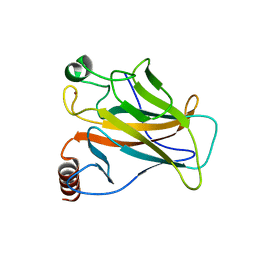
Crystal structure of the p53 (Y107H) core domain orthorhombic P form
Descriptor: Cellular tumor antigen p53, ZINC ION
Authors:Lovell, S, Liu, L, Battaile, K.P, Miller, S, Karanicolas, J.
Deposit date:2022-08-23
Release date:2023-05-17
Last modified:2023-10-25
Method:X-RAY DIFFRACTION (1.3 Å)
Cite:An African-Specific Variant of TP53 Reveals PADI4 as a Regulator of p53-Mediated Tumor Suppression.
Cancer Discov, 13, 2023

227111

数据于2024-11-06公开中

PDB statisticsPDBj update infoContact PDBjnumon