Loading
PDBj
MenuPDBj@FacebookPDBj@TwitterPDBj@YouTubewwPDB FoundationwwPDB
RCSB PDBPDBeBMRBAdv. SearchSearch help
Search by PDB author
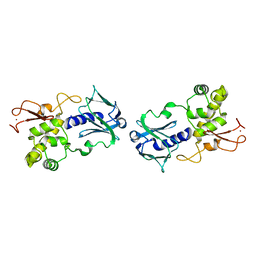
1VDM
DownloadVisualize
BU of 1vdm by Molmil
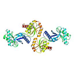
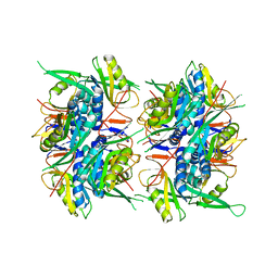
Crystal structure of purine phosphoribosyltransferase from Pyrococcus horikoshii Ot3
Descriptor: purine phosphoribosyltransferase
Authors:Sugahara, M, Kunishima, N, RIKEN Structural Genomics/Proteomics Initiative (RSGI)
Deposit date:2004-03-23
Release date:2005-05-24
Last modified:2023-12-27
Method:X-RAY DIFFRACTION (2.5 Å)
Cite:Crystal structure of purine phosphoribosyltransferase from Pyrococcus horikoshii Ot3
To be Published
1EE8
DownloadVisualize
BU of 1ee8 by Molmil
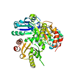
CRYSTAL STRUCTURE OF MUTM (FPG) PROTEIN FROM THERMUS THERMOPHILUS HB8
Descriptor: MUTM (FPG) PROTEIN, ZINC ION
Authors:Sugahara, M, Mikawa, T, Kumasaka, T, Yamamoto, M, Kato, R, Fukuyama, K, Inoue, Y, Kuramitsu, S, RIKEN Structural Genomics/Proteomics Initiative (RSGI)
Deposit date:2000-01-31
Release date:2001-01-31
Last modified:2024-02-07
Method:X-RAY DIFFRACTION (1.9 Å)
Cite:Crystal structure of a repair enzyme of oxidatively damaged DNA, MutM (Fpg), from an extreme thermophile, Thermus thermophilus HB8.
EMBO J., 19, 2000
6K2R
DownloadVisualize
BU of 6k2r by Molmil
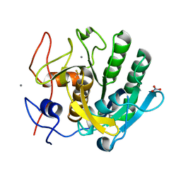
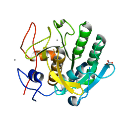
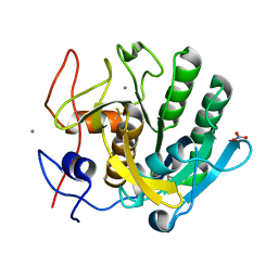
Crystal structure of proteinase K from Engyodontium album
Descriptor: CALCIUM ION, NITRATE ION, Proteinase K
Authors:Sugahara, M, Motomura, K, Numata, K.
Deposit date:2019-05-15
Release date:2020-05-20
Last modified:2023-09-06
Method:X-RAY DIFFRACTION (1.6 Å)
Cite:Crystal structure of proteinase K from Engyodontium album
To Be Published
6K2X
DownloadVisualize
BU of 6k2x by Molmil
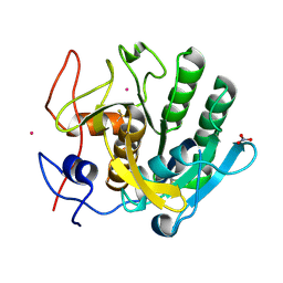
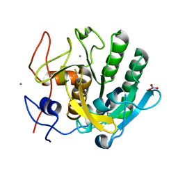
Crystal structure of proteinase K from Engyodontium album
Descriptor: NITRATE ION, PRASEODYMIUM ION, Proteinase K
Authors:Sugahara, M, Motomura, K, Numata, K.
Deposit date:2019-05-15
Release date:2020-05-20
Method:X-RAY DIFFRACTION (1.65 Å)
Cite:Crystal structure of proteinase K from Engyodontium album
To Be Published
4YM8
DownloadVisualize
BU of 4ym8 by Molmil
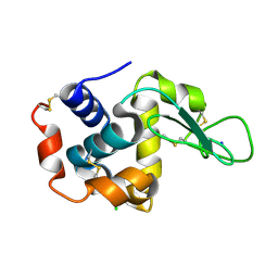
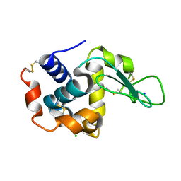
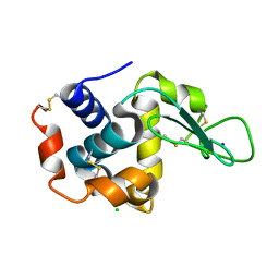
Crystal structure of hen egg-white lysozyme
Descriptor: CHLORIDE ION, Lysozyme C, SODIUM ION
Authors:Sugahara, M, Nakane, T, Suzuki, M, Nango, E.
Deposit date:2015-03-06
Release date:2015-12-23
Last modified:2018-01-24
Method:X-RAY DIFFRACTION (2.1 Å)
Cite:Crystal structure of hen egg-white lysozyme
To Be Published
1J3B
DownloadVisualize
BU of 1j3b by Molmil
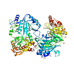
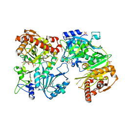
Crystal structure of ATP-dependent phosphoenolpyruvate carboxykinase from Thermus thermophilus HB8
Descriptor: ATP-dependent phosphoenolpyruvate carboxykinase, CALCIUM ION, GLYCEROL, ...
Authors:Sugahara, M, Miyano, M, Kunishima, N, RIKEN Structural Genomics/Proteomics Initiative (RSGI)
Deposit date:2003-01-21
Release date:2003-02-11
Last modified:2023-10-25
Method:X-RAY DIFFRACTION (2 Å)
Cite:Structure of ATP-dependent phosphoenolpyruvate carboxykinase from Thermus thermophilus HB8 showing the structural basis of induced fit and thermostability.
Acta Crystallogr.,Sect.D, 61, 2005
1XKV
DownloadVisualize
BU of 1xkv by Molmil
Crystal Structure Of ATP-Dependent Phosphoenolpyruvate Carboxykinase From Thermus thermophilus HB8
Descriptor: ADENOSINE-5'-TRIPHOSPHATE, ATP-dependent phosphoenolpyruvate carboxykinase, CALCIUM ION, ...
Authors:Sugahara, M, Kunishima, N, RIKEN Structural Genomics/Proteomics Initiative (RSGI)
Deposit date:2004-09-29
Release date:2005-10-11
Last modified:2023-10-25
Method:X-RAY DIFFRACTION (2.2 Å)
Cite:Structure of ATP-dependent phosphoenolpyruvate carboxykinase from Thermus thermophilus HB8 showing the structural basis of induced fit and thermostability.
Acta Crystallogr.,Sect.D, 61, 2005
1UJN
DownloadVisualize
BU of 1ujn by Molmil
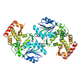
Crystal structure of dehydroquinate synthase from Thermus thermophilus HB8
Descriptor: dehydroquinate synthase
Authors:Sugahara, M, Yokoyama, S, Kuramitsu, S, Miyano, M, Kunishima, N, RIKEN Structural Genomics/Proteomics Initiative (RSGI)
Deposit date:2003-08-06
Release date:2003-09-02
Last modified:2023-10-25
Method:X-RAY DIFFRACTION (1.8 Å)
Cite:Crystal structure of dehydroquinate synthase from Thermus thermophilus HB8 showing functional importance of the dimeric state.
Proteins, 58, 2005
1VBK
DownloadVisualize
BU of 1vbk by Molmil
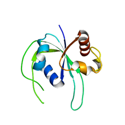
Crystal structure of PH1313 from Pyrococcus horikoshii Ot3
Descriptor: (4R)-2-METHYLPENTANE-2,4-DIOL, hypothetical protein PH1313
Authors:Sugahara, M, Kunishima, N, RIKEN Structural Genomics/Proteomics Initiative (RSGI)
Deposit date:2004-02-27
Release date:2005-04-19
Last modified:2023-12-27
Method:X-RAY DIFFRACTION (1.9 Å)
Cite:Purification, crystallization and preliminary crystallographic analysis of the putative thiamine-biosynthesis protein PH1313 from Pyrococcus horikoshii OT3
Acta Crystallogr.,Sect.F, 63, 2007
2E8H
DownloadVisualize
BU of 2e8h by Molmil
Crystal structure of PH0725 from Pyrococcus horikoshii OT3
Descriptor: Probable diphthine synthase, S-ADENOSYL-L-HOMOCYSTEINE, SODIUM ION
Authors:Sugahara, M, Taketa, M, Tanaka, Y, Matsuura, Y, Kunishima, N, RIKEN Structural Genomics/Proteomics Initiative (RSGI)
Deposit date:2007-01-19
Release date:2007-07-24
Last modified:2023-10-25
Method:X-RAY DIFFRACTION (2.1 Å)
Cite:Crystal structure of PH0725 from Pyrococcus horikoshii OT3
To be Published
6K2P
DownloadVisualize
BU of 6k2p by Molmil
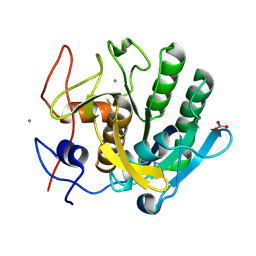
Crystal structure of proteinase K from Engyodontium album
Descriptor: CALCIUM ION, NITRATE ION, Proteinase K
Authors:Sugahara, M, Motomura, K, Numata, K.
Deposit date:2019-05-15
Release date:2020-05-20
Last modified:2023-09-06
Method:X-RAY DIFFRACTION (1.6 Å)
Cite:Crystal structure of proteinase K from Engyodontium album
To Be Published
6K2W
DownloadVisualize
BU of 6k2w by Molmil
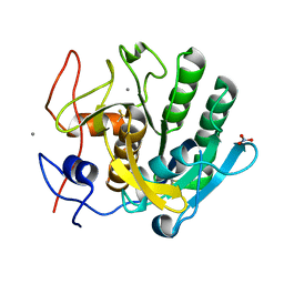
Crystal structure of proteinase K from Engyodontium album
Descriptor: CALCIUM ION, NITRATE ION, Proteinase K
Authors:Sugahara, M, Motomura, K, Numata, K.
Deposit date:2019-05-15
Release date:2020-05-20
Method:X-RAY DIFFRACTION (1.7 Å)
Cite:Crystal structure of proteinase K from Engyodontium album
To Be Published
6K2S
DownloadVisualize
BU of 6k2s by Molmil
Crystal structure of proteinase K from Engyodontium album
Descriptor: CALCIUM ION, NITRATE ION, Proteinase K
Authors:Sugahara, M, Motomura, K, Numata, K.
Deposit date:2019-05-15
Release date:2020-05-20
Last modified:2023-09-06
Method:X-RAY DIFFRACTION (1.6 Å)
Cite:Crystal structure of proteinase K from Engyodontium album
To Be Published
6K2T
DownloadVisualize
BU of 6k2t by Molmil
Crystal structure of proteinase K from Engyodontium album
Descriptor: CALCIUM ION, NITRATE ION, Proteinase K
Authors:Sugahara, M, Motomura, K, Numata, K.
Deposit date:2019-05-15
Release date:2020-05-20
Last modified:2023-09-06
Method:X-RAY DIFFRACTION (1.6 Å)
Cite:Crystal structure of proteinase K from Engyodontium album
To Be Published
6K2V
DownloadVisualize
BU of 6k2v by Molmil
Crystal structure of proteinase K from Engyodontium album
Descriptor: CALCIUM ION, NITRATE ION, Proteinase K
Authors:Sugahara, M, Motomura, K, Numata, K.
Deposit date:2019-05-15
Release date:2020-05-20
Last modified:2023-09-06
Method:X-RAY DIFFRACTION (1.6 Å)
Cite:Crystal structure of proteinase K from Engyodontium album
To Be Published
3WUM
DownloadVisualize
BU of 3wum by Molmil
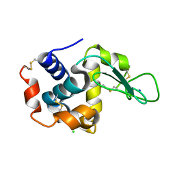
Crystal structure of hen egg-white lysozyme
Descriptor: CHLORIDE ION, Lysozyme C, SODIUM ION
Authors:Sugahara, M, Nango, E, Suzuki, M.
Deposit date:2014-04-28
Release date:2014-11-05
Last modified:2018-01-24
Method:X-RAY DIFFRACTION (2 Å)
Cite:Grease matrix as a versatile carrier of proteins for serial crystallography
Nat.Methods, 12, 2015
3WXU
DownloadVisualize
BU of 3wxu by Molmil
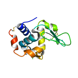
Crystal structure of hen egg-white lysozyme
Descriptor: CHLORIDE ION, Lysozyme C, SODIUM ION
Authors:Sugahara, M, Nango, E, Suzuki, M.
Deposit date:2014-08-08
Release date:2014-11-05
Last modified:2018-01-24
Method:X-RAY DIFFRACTION (2 Å)
Cite:Grease matrix as a versatile carrier of proteins for serial crystallography
Nat.Methods, 12, 2015
3WUL
DownloadVisualize
BU of 3wul by Molmil
Crystal structure of hen egg-white lysozyme
Descriptor: CHLORIDE ION, Lysozyme C, SODIUM ION
Authors:Sugahara, M, Nango, E, Suzuki, M.
Deposit date:2014-04-28
Release date:2014-11-05
Last modified:2018-01-24
Method:X-RAY DIFFRACTION (2 Å)
Cite:Grease matrix as a versatile carrier of proteins for serial crystallography
Nat.Methods, 12, 2015
3WXT
DownloadVisualize
BU of 3wxt by Molmil
Crystal structure of hen egg-white lysozyme
Descriptor: CHLORIDE ION, Lysozyme C, SODIUM ION
Authors:Sugahara, M, Nango, E, Suzuki, M.
Deposit date:2014-08-08
Release date:2014-11-05
Last modified:2018-01-24
Method:X-RAY DIFFRACTION (2 Å)
Cite:Grease matrix as a versatile carrier of proteins for serial crystallography
Nat.Methods, 12, 2015
2ZBN
DownloadVisualize
BU of 2zbn by Molmil
Crystal structure of PH1033 from Pyrococcus horikoshii OT3
Descriptor: UPF0310 protein PH1033
Authors:Sugahara, M, Kunishima, N, RIKEN Structural Genomics/Proteomics Initiative (RSGI)
Deposit date:2007-10-26
Release date:2008-04-29
Last modified:2023-11-01
Method:X-RAY DIFFRACTION (2 Å)
Cite:Nucleant-mediated protein crystallization with the application of microporous synthetic zeolites.
Acta Crystallogr.,Sect.D, 64, 2008
3WIF
DownloadVisualize
BU of 3wif by Molmil
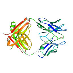
Crystal structure of anti-prostaglandin E2 Fab fragment 9Cl-PGF2beta complex
Descriptor: (Z)-7-[(1R,2R,3R,5R)-5-chloranyl-3-oxidanyl-2-[(E,3S)-3-oxidanyloct-1-enyl]cyclopentyl]hept-5-enoic acid, mAb Fab H fragment, mAb Fab L fragment
Authors:Sugahara, M, Ago, H, Saino, H, Miyano, M.
Deposit date:2013-09-12
Release date:2014-09-17
Last modified:2023-11-08
Method:X-RAY DIFFRACTION (1.7 Å)
Cite:Crystal structure of anti-Prostaglandin E2 Fab fragment with Prostaglandin E2
To be Published
3WE6
DownloadVisualize
BU of 3we6 by Molmil
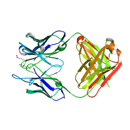
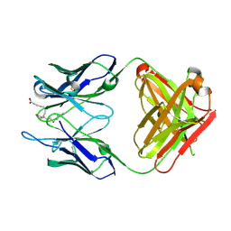
Crystal structure of anti-Prostaglandin E2 Fab fragment
Descriptor: ISOPROPYL ALCOHOL, mAb Fab H fragment, mAb Fab L fragment
Authors:Sugahara, M, Ago, H, Saino, H, Miyano, M.
Deposit date:2013-07-01
Release date:2014-07-02
Last modified:2023-11-08
Method:X-RAY DIFFRACTION (2.02 Å)
Cite:Crystal structure of anti-Prostaglandin E2 Fab fragment with Prostaglandin E2
To be Published
3WFH
DownloadVisualize
BU of 3wfh by Molmil
Crystal structure of anti-Prostaglandin E2 Fab fragment PGE2 complex
Descriptor: (Z)-7-[(1R,2R,3R)-3-hydroxy-2-[(E,3S)-3-hydroxyoct-1-enyl]-5-oxo-cyclopentyl]hept-5-enoic acid, mAb Fab H fragment, mAb Fab L fragment
Authors:Sugahara, M, Ago, H, Saino, H, Miyano, M.
Deposit date:2013-07-19
Release date:2014-07-23
Last modified:2023-11-08
Method:X-RAY DIFFRACTION (1.9 Å)
Cite:Crystal structure of anti-Prostaglandin E2 Fab fragment with Prostaglandin E2
To be Published
3AKR
DownloadVisualize
BU of 3akr by Molmil
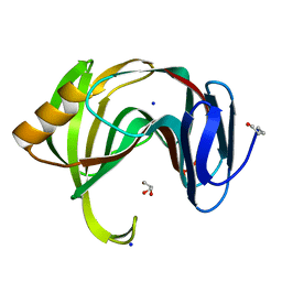
Crystal structure of xylanase from Trichoderma longibrachiatum
Descriptor: GLYCEROL, SODIUM ION, xylanase
Authors:Sugahara, M, Kunishima, N.
Deposit date:2010-07-15
Release date:2011-06-01
Last modified:2023-11-01
Method:X-RAY DIFFRACTION (1.1 Å)
Cite:Packing Space Expansion of Protein Crystallization Screening with Synthetic Zeolite as a Heteroepitaxic Nucleant
Cryst.Growth Des., 11, 2011
2HD9
DownloadVisualize
BU of 2hd9 by Molmil
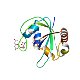
Crystal structure of PH1033 from Pyrococcus horikoshii OT3
Descriptor: CALCIUM ION, CITRIC ACID, GLYCEROL, ...
Authors:Sugahara, M, Kunishima, N, RIKEN Structural Genomics/Proteomics Initiative (RSGI)
Deposit date:2006-06-20
Release date:2006-12-20
Last modified:2023-10-25
Method:X-RAY DIFFRACTION (1.35 Å)
Cite:Nucleant-mediated protein crystallization with the application of microporous synthetic zeolites.
Acta Crystallogr.,Sect.D, 64, 2008

224004

数据于2024-08-21公开中

PDB statisticsPDBj update infoContact PDBjnumon