Loading
PDBj
MenuPDBj@FacebookPDBj@TwitterPDBj@YouTubewwPDB FoundationwwPDB
RCSB PDBPDBeBMRBAdv. SearchSearch help
Search by PDB author
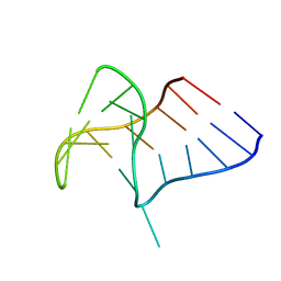
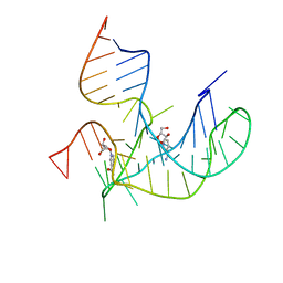
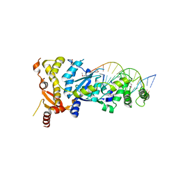
6GRC
DownloadVisualize
BU of 6grc by Molmil
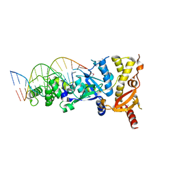
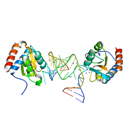
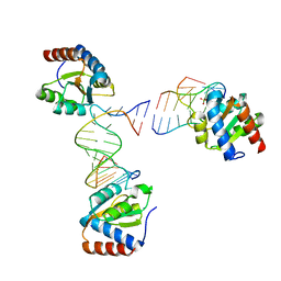
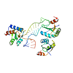
eukaryotic junction-resolving enzyme GEN-1 binding with Sodium
Descriptor: DNA (5'-D(*TP*AP*CP*CP*CP*AP*CP*CP*AP*CP*CP*GP*CP*TP*CP*A)-3'), DNA (5'-D(*TP*GP*AP*GP*CP*GP*GP*TP*GP*GP*TP*TP*GP*GP*T)-3'), MAGNESIUM ION, ...
Authors:Lilley, D.M.J, Liu, Y, Freeman, D.J.
Deposit date:2018-06-11
Release date:2018-09-26
Last modified:2019-02-13
Method:X-RAY DIFFRACTION (2.452 Å)
Cite:A monovalent ion in the DNA binding interface of the eukaryotic junction-resolving enzyme GEN1.
Nucleic Acids Res., 46, 2018
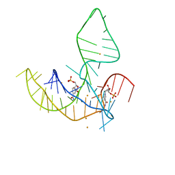
6GRD
DownloadVisualize
BU of 6grd by Molmil
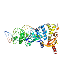
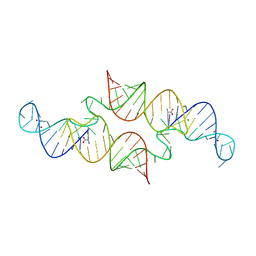
eukaryotic junction-resolving enzyme GEN-1 binding with Cesium
Descriptor: CESIUM ION, DNA (5'-D(*TP*AP*CP*CP*CP*AP*CP*CP*AP*CP*CP*GP*CP*TP*CP*A)-3'), DNA (5'-D(*TP*GP*AP*GP*CP*GP*GP*TP*GP*GP*TP*TP*GP*GP*T)-3'), ...
Authors:Lilley, D.M.J, Liu, Y, Freeman, D.J.
Deposit date:2018-06-11
Release date:2018-09-26
Last modified:2018-11-28
Method:X-RAY DIFFRACTION (2.66 Å)
Cite:A monovalent ion in the DNA binding interface of the eukaryotic junction-resolving enzyme GEN1.
Nucleic Acids Res., 46, 2018
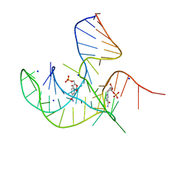
6GRB
DownloadVisualize
BU of 6grb by Molmil
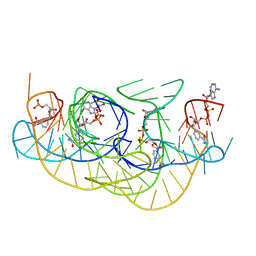
eukaryotic junction-resolving enzyme GEN-1 binding with Potassium
Descriptor: DNA (5'-D(*TP*AP*CP*CP*CP*AP*CP*CP*AP*CP*CP*GP*CP*TP*CP*A)-3'), DNA (5'-D(*TP*GP*AP*GP*CP*GP*GP*TP*GP*GP*TP*TP*GP*GP*T)-3'), MAGNESIUM ION, ...
Authors:Lilley, D.M.J, Liu, Y, Freeman, D.J.
Deposit date:2018-06-11
Release date:2018-09-26
Last modified:2024-01-17
Method:X-RAY DIFFRACTION (2.4 Å)
Cite:A monovalent ion in the DNA binding interface of the eukaryotic junction-resolving enzyme GEN1.
Nucleic Acids Res., 46, 2018
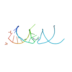
6R47
DownloadVisualize
BU of 6r47 by Molmil
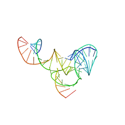
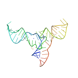
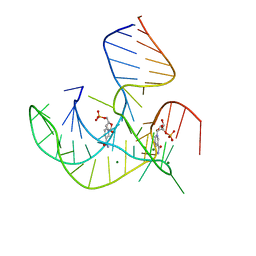
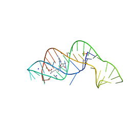
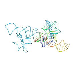
The structure of pistol ribozyme bound to magnesium
Descriptor: MAGNESIUM ION, RNA, RNA (50-MER)
Authors:Lilley, D.M.J, Wilson, T.M, Liu, Y.
Deposit date:2019-03-22
Release date:2019-07-03
Last modified:2024-05-15
Method:X-RAY DIFFRACTION (3.1 Å)
Cite:Comparison of the Structures and Mechanisms of the Pistol and Hammerhead Ribozymes.
J.Am.Chem.Soc., 141, 2019
8ITS
DownloadVisualize
BU of 8its by Molmil
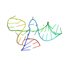
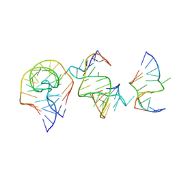
Crystal structure of DUF-3268 k-junction
Descriptor: MAGNESIUM ION, RNA (46-MER)
Authors:Li, M, Lilley, D.M.J, Huang, L.
Deposit date:2023-03-22
Release date:2024-03-27
Method:X-RAY DIFFRACTION (1.94 Å)
Cite:Crystal structure of DUF-3268 k-junction
To Be Published
7V9E
DownloadVisualize
BU of 7v9e by Molmil
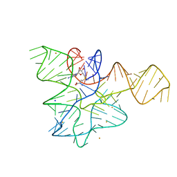
Crystal structure of a methyl transferase ribozyme
Descriptor: BARIUM ION, GUANINE, RNA (68-MER), ...
Authors:Deng, J, Lilley, D.M.J, Huang, L.
Deposit date:2021-08-25
Release date:2022-03-23
Last modified:2024-05-29
Method:X-RAY DIFFRACTION (2.3 Å)
Cite:Structure and mechanism of a methyltransferase ribozyme.
Nat.Chem.Biol., 18, 2022
7EAG
DownloadVisualize
BU of 7eag by Molmil
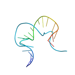
Crystal structure of the RAGATH-18 k-turn
Descriptor: RNA (5'-R(*GP*UP*CP*UP*AP*UP*GP*AP*AP*GP*GP*CP*UP*GP*GP*AP*GP*AP*C)-3')
Authors:Huang, L, Lilley, D.M.J.
Deposit date:2021-03-07
Release date:2021-06-02
Last modified:2023-11-29
Method:X-RAY DIFFRACTION (2.5 Å)
Cite:Structure and folding of four putative kink turns identified in structured RNA species in a test of structural prediction rules.
Nucleic Acids Res., 49, 2021
7EAF
DownloadVisualize
BU of 7eaf by Molmil
Crystal structure of SAM-I riboswitch with the Actinomyces-1 k-turn
Descriptor: BARIUM ION, RNA (94-MER), S-ADENOSYLMETHIONINE, ...
Authors:Huang, L, Lilley, D.M.J.
Deposit date:2021-03-07
Release date:2021-06-02
Last modified:2023-11-29
Method:X-RAY DIFFRACTION (2.85 Å)
Cite:Structure and folding of four putative kink turns identified in structured RNA species in a test of structural prediction rules.
Nucleic Acids Res., 49, 2021
6Q8U
DownloadVisualize
BU of 6q8u by Molmil
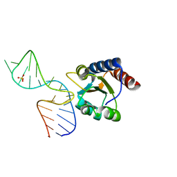
Structure of the standard kink turn HmKt-7 variant A2bm6A bound with AfL7Ae protein
Descriptor: 50S ribosomal protein L7Ae, RNA (5'-R(*CP*GP*GP*CP*GP*AP*AP*GP*(6MZ)P*AP*CP*CP*GP*GP*GP*GP*AP*GP*CP*CP*G)-3'), SODIUM ION
Authors:Huang, L, Lilley, D.M.J.
Deposit date:2018-12-16
Release date:2019-07-03
Last modified:2024-01-24
Method:X-RAY DIFFRACTION (1.99 Å)
Cite:Effect of methylation of adenine N6on kink turn structure depends on location.
Rna Biol., 16, 2019
6Q8V
DownloadVisualize
BU of 6q8v by Molmil
Structure of the standard kink turn HmKt-7 variant A2bm6A.
Descriptor: RNA (5'-R(*GP*GP*CP*GP*AP*AP*GP*(6MZ)P*AP*CP*CP*GP*GP*GP*GP*AP*GP*CP*C)-3')
Authors:Huang, L, Lilley, D.M.J.
Deposit date:2018-12-16
Release date:2019-07-03
Last modified:2024-01-24
Method:X-RAY DIFFRACTION (2.09 Å)
Cite:Effect of methylation of adenine N6on kink turn structure depends on location.
Rna Biol., 16, 2019
6QN3
DownloadVisualize
BU of 6qn3 by Molmil
Structure of the Glutamine II Riboswitch
Descriptor: BROMIDE ION, GLUTAMINE, MAGNESIUM ION, ...
Authors:Huang, L, Lilley, D.M.J.
Deposit date:2019-02-08
Release date:2019-06-12
Last modified:2024-05-15
Method:X-RAY DIFFRACTION (2.3 Å)
Cite:Structure and ligand binding of the glutamine-II riboswitch.
Nucleic Acids Res., 47, 2019
8HBA
DownloadVisualize
BU of 8hba by Molmil
Crystal structure of NAD-II riboswitch (single strand) with NAD
Descriptor: BETA-NICOTINAMIDE RIBOSE MONOPHOSPHATE, NICOTINAMIDE-ADENINE-DINUCLEOTIDE, RNA (55-MER)
Authors:Peng, X, Lilley, D.M.J, Huang, L.
Deposit date:2022-10-27
Release date:2023-03-22
Last modified:2023-11-29
Method:X-RAY DIFFRACTION (2.64 Å)
Cite:Crystal structures of the NAD+-II riboswitch reveal two distinct ligand-binding pockets.
Nucleic Acids Res., 51, 2023
8HB1
DownloadVisualize
BU of 8hb1 by Molmil
Crystal structure of NAD-II riboswitch (two strands) with NMN
Descriptor: BETA-NICOTINAMIDE RIBOSE MONOPHOSPHATE, MAGNESIUM ION, RNA (30-MER), ...
Authors:Peng, X, Lilley, D.M.J, Huang, L.
Deposit date:2022-10-27
Release date:2023-03-22
Last modified:2024-05-29
Method:X-RAY DIFFRACTION (2.23 Å)
Cite:Crystal structures of the NAD+-II riboswitch reveal two distinct ligand-binding pockets.
Nucleic Acids Res., 51, 2023
8HB3
DownloadVisualize
BU of 8hb3 by Molmil
Crystal structure of NAD-II riboswitch (two strands) with NR
Descriptor: Nicotinamide riboside, RNA (31-MER), RNA (5'-R(*AP*GP*AP*GP*CP*GP*UP*UP*GP*CP*GP*UP*CP*CP*GP*AP*AP*AP*GP*UP*(CBV)P*GP*CP*C)-3')
Authors:Peng, X, Lilley, D.M.J, Huang, L.
Deposit date:2022-10-27
Release date:2023-03-22
Last modified:2023-11-29
Method:X-RAY DIFFRACTION (2.87 Å)
Cite:Crystal structures of the NAD+-II riboswitch reveal two distinct ligand-binding pockets.
Nucleic Acids Res., 51, 2023
8HB8
DownloadVisualize
BU of 8hb8 by Molmil
Crystal structure of NAD-II riboswitch (single strand) with NMN
Descriptor: BARIUM ION, BETA-NICOTINAMIDE RIBOSE MONOPHOSPHATE, RNA (55-MER)
Authors:Peng, X, Lilley, D.M.J, Huang, L.
Deposit date:2022-10-27
Release date:2023-03-22
Last modified:2023-11-29
Method:X-RAY DIFFRACTION (2.3 Å)
Cite:Crystal structures of the NAD+-II riboswitch reveal two distinct ligand-binding pockets.
Nucleic Acids Res., 51, 2023
8I3Z
DownloadVisualize
BU of 8i3z by Molmil
Crystal structure of NAD-II riboswitch (two strands) with NMN at 1.67 angstrom
Descriptor: BETA-NICOTINAMIDE RIBOSE MONOPHOSPHATE, RNA (31-MER), RNA (5'-R(*AP*GP*AP*GP*CP*GP*UP*UP*GP*CP*GP*UP*CP*CP*GP*AP*AP*AP*GP*UP*(CBV)P*GP*CP*C)-3'), ...
Authors:Peng, X, Lilley, D.M.J, Huang, L.
Deposit date:2023-01-18
Release date:2023-03-22
Last modified:2024-05-29
Method:X-RAY DIFFRACTION (1.67 Å)
Cite:Crystal structures of the NAD+-II riboswitch reveal two distinct ligand-binding pockets.
Nucleic Acids Res., 51, 2023
6HBT
DownloadVisualize
BU of 6hbt by Molmil
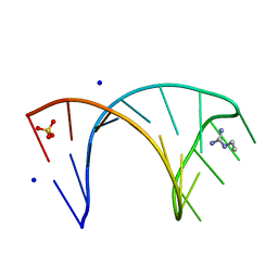
The structure of the G. violaceus guanidine II riboswitch P2 stem-loop with arcaine
Descriptor: 1-(4-carbamimidamidobutyl)guanidine, RNA (5'-R(*GP*GP*UP*GP*GP*GP*GP*AP*CP*GP*AP*CP*CP*CP*CP*AP*(CBV)P*C)-3'), SULFATE ION
Authors:Huang, L, Lilley, D.M.J.
Deposit date:2018-08-13
Release date:2019-02-20
Last modified:2024-05-15
Method:X-RAY DIFFRACTION (1.66 Å)
Cite:Structure-guided design of a high-affinity ligand for a riboswitch.
Rna, 25, 2019
6HBX
DownloadVisualize
BU of 6hbx by Molmil
The structure of the G. violaceus guanidine II riboswitch P2 stem-loop with ethylguanidine
Descriptor: N-ETHYLGUANIDINE, RNA (5'-R(*GP*GP*UP*GP*GP*GP*GP*AP*CP*GP*AP*CP*CP*CP*CP*AP*(CBV)P*C)-3'), SODIUM ION, ...
Authors:Huang, L, Lilley, D.M.J.
Deposit date:2018-08-13
Release date:2019-02-20
Last modified:2024-05-15
Method:X-RAY DIFFRACTION (1.54 Å)
Cite:Structure-guided design of a high-affinity ligand for a riboswitch.
Rna, 25, 2019
6HC5
DownloadVisualize
BU of 6hc5 by Molmil
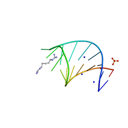
The structure of the G. violaceus guanidine II riboswitch P2 stem-loop with audouine
Descriptor: 1-(5-carbamimidamidopentyl)guanidine, RNA (5'-R(*GP*GP*UP*GP*GP*GP*GP*AP*CP*GP*AP*CP*CP*CP*CP*AP*(CBV)P*C)-3'), SODIUM ION, ...
Authors:Huang, L, Lilley, D.M.J.
Deposit date:2018-08-14
Release date:2019-02-20
Last modified:2024-05-15
Method:X-RAY DIFFRACTION (1.413 Å)
Cite:Structure-guided design of a high-affinity ligand for a riboswitch.
Rna, 25, 2019
6HCT
DownloadVisualize
BU of 6hct by Molmil
Crystal structure of Archeoglobus fulgidus L7Ae bound to its cognate UTR k-turn
Descriptor: 50S ribosomal protein L7Ae, RNA (5'-R(*GP*CP*CP*GP*AP*UP*GP*AP*AP*UP*G)-3'), RNA (5'-R(*GP*CP*CP*GP*AP*UP*GP*AP*AP*UP*GP*CP*AP*UP*GP*AP*AP*GP*C)-3'), ...
Authors:Huang, L, Lilley, D.M.J.
Deposit date:2018-08-16
Release date:2018-10-31
Last modified:2024-01-17
Method:X-RAY DIFFRACTION (3.091 Å)
Cite:The role of RNA structure in translational regulation by L7Ae protein in archaea.
RNA, 25, 2019
6FZ0
DownloadVisualize
BU of 6fz0 by Molmil
Crystal structure of the metY SAM V riboswitch
Descriptor: MAGNESIUM ION, S-ADENOSYLMETHIONINE, SODIUM ION, ...
Authors:Huang, L, Lilley, D.M.J.
Deposit date:2018-03-13
Release date:2018-07-04
Last modified:2024-05-08
Method:X-RAY DIFFRACTION (2.499 Å)
Cite:Structure and ligand binding of the SAM-V riboswitch.
Nucleic Acids Res., 46, 2018
4C40
DownloadVisualize
BU of 4c40 by Molmil
The molecular recognition of kink turn structure by the L7Ae class of proteins
Descriptor: 5'-(*GP*GP*CP*GP*AP*AP*GP*AP*AP*CP*CP*GP*GP*GP *GP*AP*GP*CP*C)-3'
Authors:Huang, L, Lilley, D.M.J.
Deposit date:2013-08-28
Release date:2013-11-06
Last modified:2023-12-20
Method:X-RAY DIFFRACTION (2.2 Å)
Cite:The Molecular Recognition of Kink-Turn Structure by the L7Ae Class of Proteins.
RNA, 19, 2013
4C4W
DownloadVisualize
BU of 4c4w by Molmil
Structure of a rare, non-standard sequence k-turn bound by L7Ae protein
Descriptor: 50S RIBOSOMAL PROTEIN L7AE, DIHYDROGENPHOSPHATE ION, TSKT-23, ...
Authors:Huang, L, Lilley, D.M.J.
Deposit date:2013-09-09
Release date:2014-02-12
Last modified:2023-12-20
Method:X-RAY DIFFRACTION (2.95 Å)
Cite:Structure of a Rare, Non-Standard Sequence K-Turn Bound by L7Ae Protein
Nucleic Acids Res., 42, 2014
4BW0
DownloadVisualize
BU of 4bw0 by Molmil
The molecular recognition of kink turn structure by the L7Ae class of proteins
Descriptor: 50S RIBOSOMAL PROTEIN L7AE, HMKT-7, SULFATE ION
Authors:Huang, L, Lilley, D.M.J.
Deposit date:2013-06-29
Release date:2013-11-06
Last modified:2023-12-20
Method:X-RAY DIFFRACTION (2.33 Å)
Cite:The Molecular Recognition of Kink-Turn Structure by the L7Ae Class of Proteins.
RNA, 19, 2013
4B5R
DownloadVisualize
BU of 4b5r by Molmil
SAM-I riboswitch bearing the H. marismortui K-t-7
Descriptor: BARIUM ION, POTASSIUM ION, S-ADENOSYLMETHIONINE, ...
Authors:Daldrop, P, Lilley, D.M.J.
Deposit date:2012-08-07
Release date:2013-01-30
Last modified:2023-12-20
Method:X-RAY DIFFRACTION (2.95 Å)
Cite:The Plasticity of a Structural Motif in RNA: Structural Polymorphism of a Kink Turn as a Function of its Environment.
RNA, 19, 2013


1234>

226707

数据于2024-10-30公开中

PDB statisticsPDBj update infoContact PDBjnumon