Loading
PDBj
MenuPDBj@FacebookPDBj@X(formerly Twitter)PDBj@BlueSkyPDBj@YouTubewwPDB FoundationwwPDB
RCSB PDBPDBeBMRBAdv. SearchSearch help
Search by PDB author
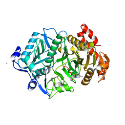
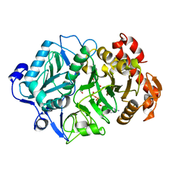
2PXZ
DownloadVisualize
BU of 2pxz by Molmil
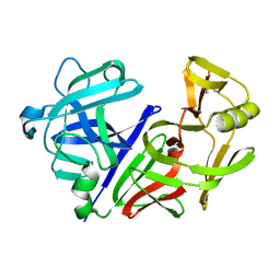
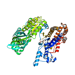
E. coli phosphoenolpyruvate carboxykinase (PEPCK) complexed with ATP, Mg2+, Mn2+, carbon dioxide and oxaloacetate
Descriptor: ADENOSINE-5'-TRIPHOSPHATE, CARBON DIOXIDE, MAGNESIUM ION, ...
Authors:Delbaere, L.T.J, Cotelesage, J.J.H, Goldie, H.
Deposit date:2007-05-14
Release date:2008-04-22
Last modified:2024-02-21
Method:X-RAY DIFFRACTION (2.23 Å)
Cite:Crystal structure of E. coli phosphoenolpyruvate carboxykinase (PEPCK) complexed with ATP, Mg2+, Mn2+, carbon dioxide and oxaloacetate
To be Published
2PY7
DownloadVisualize
BU of 2py7 by Molmil
Crystal structure of E. coli phosphoenolpyruvate carboxykinase mutant Lys213Ser complexed with ATP-Mg2+-Mn2+
Descriptor: ADENOSINE-5'-TRIPHOSPHATE, MAGNESIUM ION, MANGANESE (II) ION, ...
Authors:Delbaere, L.T.J, Cotelesage, J.J.H, Goldie, H.
Deposit date:2007-05-15
Release date:2008-04-22
Last modified:2023-08-30
Method:X-RAY DIFFRACTION (2.2 Å)
Cite:Crystal structure of E. coli phosphoenolpyruvate carboxykinase mutant Lys213Ser
To be Published
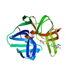
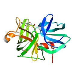
1SGC
DownloadVisualize
BU of 1sgc by Molmil
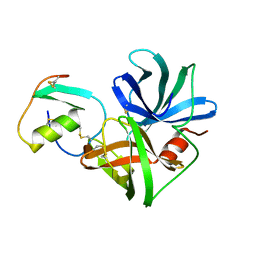
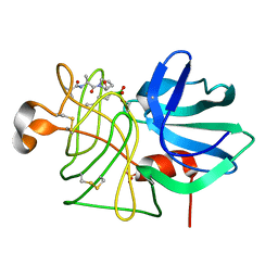
THE 1.8 ANGSTROMS STRUCTURE OF THE COMPLEX BETWEEN CHYMOSTATIN AND STREPTOMYCES GRISEUS PROTEASE A. A MODEL FOR SERINE PROTEASE CATALYTIC TETRAHEDRAL INTERMEDIATES
Descriptor: CHYMOSTATIN A, PROTEINASE A
Authors:Delbaere, L.T.J, Brayer, G.D.
Deposit date:1986-04-18
Release date:1986-07-14
Last modified:2024-06-05
Method:X-RAY DIFFRACTION (1.8 Å)
Cite:The 1.8 A structure of the complex between chymostatin and Streptomyces griseus protease A. A model for serine protease catalytic tetrahedral intermediates.
J.Mol.Biol., 183, 1985
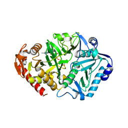
1YTM
DownloadVisualize
BU of 1ytm by Molmil
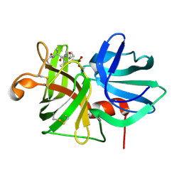
Crystal structure of phosphoenolpyruvate carboxykinase of Anaerobiospirillum succiniciproducens complexed with ATP, oxalate, magnesium and manganese ions
Descriptor: ADENOSINE-5'-TRIPHOSPHATE, MAGNESIUM ION, MANGANESE (II) ION, ...
Authors:Delbaere, L.T.J, Cotelesage, J.J.H.
Deposit date:2005-02-10
Release date:2006-01-24
Last modified:2023-08-23
Method:X-RAY DIFFRACTION (2.2 Å)
Cite:Crystal structure of Anaerobiospirillum succiniciproducens PEP carboxykinase reveals an important active site loop
Int.J.Biochem.Cell Biol., 37, 2005
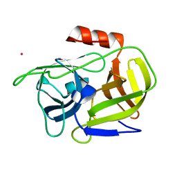
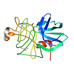
2O8L
DownloadVisualize
BU of 2o8l by Molmil
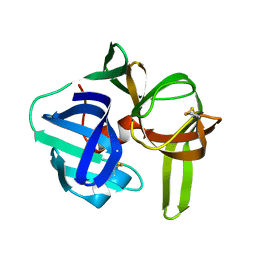
Structure of V8 protease from staphylococcus aureus
Descriptor: POTASSIUM ION, V8 protease
Authors:Delbaere, L.T.J, Prasad, L, Leduc, Y.
Deposit date:2006-12-12
Release date:2007-01-02
Last modified:2023-08-30
Method:X-RAY DIFFRACTION (1.5 Å)
Cite:The Structure of a Universally Employed Enazyme: V8 Protease from Staphylococcus Aureus
Acta Crystallogr.,Sect.D, 60, 2004
2JEL
DownloadVisualize
BU of 2jel by Molmil
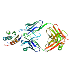
JEL42 FAB/HPR COMPLEX
Descriptor: HISTIDINE-CONTAINING PROTEIN, JEL42 FAB FRAGMENT, SULFATE ION
Authors:Prasad, L, Waygood, E.B, Lee, J.S, Delbaere, L.T.J.
Deposit date:1998-02-24
Release date:1998-05-27
Last modified:2024-11-13
Method:X-RAY DIFFRACTION (2.5 Å)
Cite:The 2.5 A resolution structure of the jel42 Fab fragment/HPr complex
J.Mol.Biol., 280, 1998
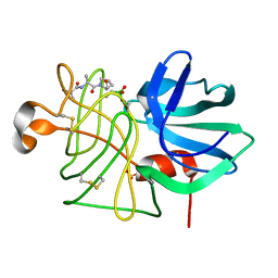
2ALP
DownloadVisualize
BU of 2alp by Molmil
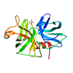
REFINED STRUCTURE OF ALPHA-LYTIC PROTEASE AT 1.7 ANGSTROMS RESOLUTION. ANALYSIS OF HYDROGEN BONDING AND SOLVENT STRUCTURE
Descriptor: ALPHA-LYTIC PROTEASE, SULFATE ION
Authors:Fujinaga, M, Delbaere, L.T.J, Brayer, G.D, James, M.N.G.
Deposit date:1985-03-07
Release date:1985-07-17
Last modified:2024-11-13
Method:X-RAY DIFFRACTION (1.7 Å)
Cite:Refined structure of alpha-lytic protease at 1.7 A resolution. Analysis of hydrogen bonding and solvent structure.
J.Mol.Biol., 184, 1985
1LIN
DownloadVisualize
BU of 1lin by Molmil
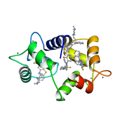
CALMODULIN COMPLEXED WITH TRIFLUOPERAZINE (1:4 COMPLEX)
Descriptor: 10-[3-(4-METHYL-PIPERAZIN-1-YL)-PROPYL]-2-TRIFLUOROMETHYL-10H-PHENOTHIAZINE, CALCIUM ION, CALMODULIN
Authors:Vandonselaar, M, Hickie, R.A, Quail, J.W, Delbaere, L.T.J.
Deposit date:1995-10-11
Release date:1996-03-08
Last modified:2024-02-14
Method:X-RAY DIFFRACTION (2 Å)
Cite:Trifluoperazine-induced conformational change in Ca(2+)-calmodulin.
Nat.Struct.Biol., 1, 1994
1JXN
DownloadVisualize
BU of 1jxn by Molmil
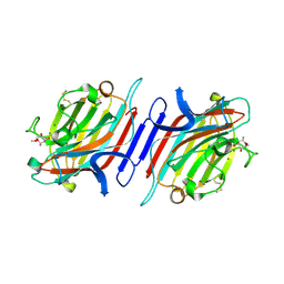
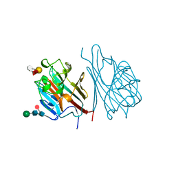
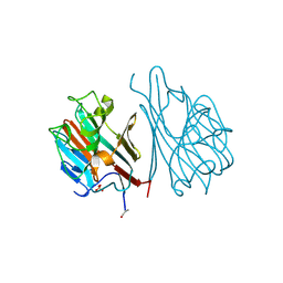
Crystal Structure of the Lectin I from Ulex europaeus in complex with the methyl glycoside of alpha-L-fucose
Descriptor: (4R)-2-METHYLPENTANE-2,4-DIOL, CALCIUM ION, MANGANESE (II) ION, ...
Authors:Audette, G.F, Olson, D.J.H, Ross, A.R.S, Quail, J.W, Delbaere, L.T.J.
Deposit date:2001-09-07
Release date:2002-12-06
Last modified:2024-10-30
Method:X-RAY DIFFRACTION (2.3 Å)
Cite:Examination of the Structural Basis for O(H) Blood Group Specificity by Ulex europaeus Lectin I
Can.J.Chem., 80, 2002
1GSL
DownloadVisualize
BU of 1gsl by Molmil
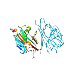
LECTIN (FOURTH ISOLATED FROM (GRIFFONIA SIMPLICIFOLIA)) COMPLEX WITH Y HUMAN BLOOD GROUP DETERMINANT
Descriptor: CALCIUM ION, GRIFFONIA SIMPLICIFOLIA LECTIN 4, MANGANESE (II) ION, ...
Authors:Delbaere, L.T.J.
Deposit date:1996-06-15
Release date:1996-12-23
Last modified:2024-10-30
Method:X-RAY DIFFRACTION (2 Å)
Cite:Structures of the lectin IV of Griffonia simplicifolia and its complex with the Lewis b human blood group determinant at 2.0 A resolution.
J.Mol.Biol., 230, 1993
5SGA
DownloadVisualize
BU of 5sga by Molmil
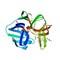
Structures of product and inhibitor complexes of Streptomyces griseus protease a at 1.8 Angstroms resolution. a model for serine protease catalysis
Descriptor: PROTEINASE A (SGPA), TETRAPEPTIDE ACE-PRO-ALA-PRO-TYR
Authors:Sielecki, A.R, James, M.N.G.
Deposit date:1990-05-29
Release date:1991-10-15
Last modified:2024-11-06
Method:X-RAY DIFFRACTION (1.8 Å)
Cite:Structures of product and inhibitor complexes of Streptomyces griseus protease A at 1.8 A resolution. A model for serine protease catalysis.
J.Mol.Biol., 144, 1980
4SGA
DownloadVisualize
BU of 4sga by Molmil
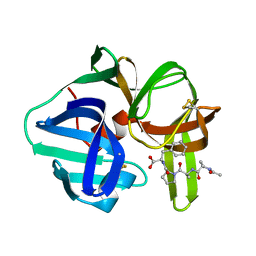
STRUCTURES OF PRODUCT AND INHIBITOR COMPLEXES OF STREPTOMYCES GRISEUS PROTEASE A AT 1.8 ANGSTROMS RESOLUTION. A MODEL FOR SERINE PROTEASE CATALYSIS
Descriptor: PROTEINASE A (SGPA), TETRAPEPTIDE ACE-PRO-ALA-PRO-PHE
Authors:Sielecki, A.R, James, M.N.G.
Deposit date:1990-05-29
Release date:1991-10-15
Last modified:2024-10-23
Method:X-RAY DIFFRACTION (1.8 Å)
Cite:Structures of product and inhibitor complexes of Streptomyces griseus protease A at 1.8 A resolution. A model for serine protease catalysis.
J.Mol.Biol., 144, 1980
3SGA
DownloadVisualize
BU of 3sga by Molmil
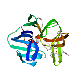
STRUCTURES OF PRODUCT AND INHIBITOR COMPLEXES OF STREPTOMYCES GRISEUS PROTEASE A AT 1.8 ANGSTROMS RESOLUTION. A MODEL FOR SERINE PROTEASE CATALYSIS
Descriptor: ACE-PRO-ALA-PRO-PHE-ALDEHYDE, PROTEINASE A (SGPA)
Authors:Sielecki, A.R, James, M.N.G.
Deposit date:1990-05-29
Release date:1991-10-15
Last modified:2024-06-05
Method:X-RAY DIFFRACTION (1.8 Å)
Cite:Structures of product and inhibitor complexes of Streptomyces griseus protease A at 1.8 A resolution. A model for serine protease catalysis.
J.Mol.Biol., 144, 1980
2SGA
DownloadVisualize
BU of 2sga by Molmil
ELECTRON DENSITY CALCULATIONS AS AN EXTENSION OF PROTEIN STRUCTURE REFINEMENT. STREPTOMYCES GRISEUS PROTEASE AT 1.5 ANGSTROMS RESOLUTION
Descriptor: PROTEINASE A
Authors:James, M.N.G, Sielecki, A.R.
Deposit date:1983-01-21
Release date:1983-04-21
Last modified:2024-06-05
Method:X-RAY DIFFRACTION (1.5 Å)
Cite:Electron density calculations as an extension of protein structure refinement. Streptomyces griseus protease A at 1.5 A resolution.
J.Mol.Biol., 182, 1985
3SGB
DownloadVisualize
BU of 3sgb by Molmil
STRUCTURE OF THE COMPLEX OF STREPTOMYCES GRISEUS PROTEASE B AND THE THIRD DOMAIN OF THE TURKEY OVOMUCOID INHIBITOR AT 1.8 ANGSTROMS RESOLUTION
Descriptor: PROTEINASE B (SGPB), TURKEY OVOMUCOID INHIBITOR (OMTKY3)
Authors:Read, R.J, Fujinaga, M, Sielecki, A.R, James, M.N.G.
Deposit date:1983-01-21
Release date:1983-07-12
Last modified:2024-10-30
Method:X-RAY DIFFRACTION (1.8 Å)
Cite:Structure of the complex of Streptomyces griseus protease B and the third domain of the turkey ovomucoid inhibitor at 1.8-A resolution.
Biochemistry, 22, 1983
3APP
DownloadVisualize
BU of 3app by Molmil
STRUCTURE AND REFINEMENT OF PENICILLOPEPSIN AT 1.8 ANGSTROMS RESOLUTION
Descriptor: PENICILLOPEPSIN
Authors:Sielecki, A.R, James, M.N.G.
Deposit date:1990-11-27
Release date:1991-01-15
Last modified:2024-11-13
Method:X-RAY DIFFRACTION (1.8 Å)
Cite:Structure and refinement of penicillopepsin at 1.8 A resolution.
J.Mol.Biol., 163, 1983
7LPR
DownloadVisualize
BU of 7lpr by Molmil
STRUCTURAL BASIS FOR BROAD SPECIFICITY IN ALPHA-LYTIC PROTEASE MUTANTS
Descriptor: ALPHA-LYTIC PROTEASE, METHOXYSUCCINYL-ALA-ALA-PRO-LEUCINE BORONIC ACID INHIBITOR, SULFATE ION
Authors:Fujishige, A, Bone, R, Agard, D.A.
Deposit date:1991-08-05
Release date:1993-01-15
Last modified:2024-06-05
Method:X-RAY DIFFRACTION (2.05 Å)
Cite:Structural basis for broad specificity in alpha-lytic protease mutants.
Biochemistry, 30, 1991
9LPR
DownloadVisualize
BU of 9lpr by Molmil
STRUCTURAL BASIS FOR BROAD SPECIFICITY IN ALPHA-LYTIC PROTEASE MUTANTS
Descriptor: ALPHA-LYTIC PROTEASE, METHOXYSUCCINYL-ALA-ALA-PRO-LEUCINE BORONIC ACID INHIBITOR, SULFATE ION
Authors:Bone, R, Agard, D.A.
Deposit date:1991-08-05
Release date:1993-01-15
Last modified:2024-10-09
Method:X-RAY DIFFRACTION (2.2 Å)
Cite:Structural basis for broad specificity in alpha-lytic protease mutants.
Biochemistry, 30, 1991
8LPR
DownloadVisualize
BU of 8lpr by Molmil
STRUCTURAL BASIS FOR BROAD SPECIFICITY IN ALPHA-LYTIC PROTEASE MUTANTS
Descriptor: ALPHA-LYTIC PROTEASE, METHOXYSUCCINYL-ALA-ALA-PRO-PHENYLALANINE BORONIC ACID INHIBITOR, SULFATE ION
Authors:Fujishige, A, Bone, R, Agard, D.A.
Deposit date:1991-08-05
Release date:1993-01-15
Last modified:2024-10-30
Method:X-RAY DIFFRACTION (2.25 Å)
Cite:Structural basis for broad specificity in alpha-lytic protease mutants.
Biochemistry, 30, 1991
5LPR
DownloadVisualize
BU of 5lpr by Molmil
STRUCTURAL BASIS FOR BROAD SPECIFICITY IN ALPHA-LYTIC PROTEASE MUTANTS
Descriptor: ALPHA-LYTIC PROTEASE, METHOXYSUCCINYL-ALA-ALA-PRO-ALANINE BORONIC ACID INHIBITOR, SULFATE ION
Authors:Fujishige, A, Bone, R, Agard, D.A.
Deposit date:1991-08-05
Release date:1993-01-15
Last modified:2024-06-05
Method:X-RAY DIFFRACTION (2.13 Å)
Cite:Structural basis for broad specificity in alpha-lytic protease mutants.
Biochemistry, 30, 1991
6LPR
DownloadVisualize
BU of 6lpr by Molmil
STRUCTURAL BASIS FOR BROAD SPECIFICITY IN ALPHA-LYTIC PROTEASE MUTANTS
Descriptor: ALPHA-LYTIC PROTEASE, METHOXYSUCCINYL-ALA-ALA-PRO-NORLEUCINE BORONIC ACID INHIBITOR, SULFATE ION
Authors:Bone, R, Agard, D.A.
Deposit date:1991-08-05
Release date:1993-01-15
Last modified:2024-06-05
Method:X-RAY DIFFRACTION (2.1 Å)
Cite:Structural basis for broad specificity in alpha-lytic protease mutants.
Biochemistry, 30, 1991
1LEC
DownloadVisualize
BU of 1lec by Molmil
STRUCTURES OF THE LECTIN IV OF GRIFFONIA SIMPLICIFOLIA AND ITS COMPLEX WITH THE LEWIS B HUMAN BLOOD GROUP DETERMINANT AT 2.0 ANGSTROMS RESOLUTION
Descriptor: CALCIUM ION, MANGANESE (II) ION, SULFATE ION, ...
Authors:Delbaere, L, Vandonselaar, M, Quail, J.
Deposit date:1992-12-17
Release date:1994-01-31
Last modified:2024-10-30
Method:X-RAY DIFFRACTION (2 Å)
Cite:Structures of the lectin IV of Griffonia simplicifolia and its complex with the Lewis b human blood group determinant at 2.0 A resolution.
J.Mol.Biol., 230, 1993
1LED
DownloadVisualize
BU of 1led by Molmil
STRUCTURES OF THE LECTIN IV OF GRIFFONIA SIMPLICIFOLIA AND ITS COMPLEX WITH THE LEWIS B HUMAN BLOOD GROUP DETERMINANT AT 2.0 ANGSTROMS RESOLUTION
Descriptor: CALCIUM ION, MANGANESE (II) ION, SULFATE ION, ...
Authors:Delbaere, L, Vandonselaar, M, Quail, J.
Deposit date:1992-12-17
Release date:1994-01-31
Last modified:2020-07-29
Method:X-RAY DIFFRACTION (2 Å)
Cite:Structures of the lectin IV of Griffonia simplicifolia and its complex with the Lewis b human blood group determinant at 2.0 A resolution.
J.Mol.Biol., 230, 1993
3MBC
DownloadVisualize
BU of 3mbc by Molmil
Crystal structure of monomeric isocitrate dehydrogenase from Corynebacterium glutamicum in complex with NADP
Descriptor: Isocitrate dehydrogenase [NADP], MAGNESIUM ION, NADP NICOTINAMIDE-ADENINE-DINUCLEOTIDE PHOSPHATE
Authors:Sidhu, N.S, Aich, S, Sheldrick, G.M, Delbaere, L.T.J.
Deposit date:2010-03-25
Release date:2011-04-06
Last modified:2023-09-06
Method:X-RAY DIFFRACTION (1.9 Å)
Cite:Structure of a highly NADP+-specific isocitrate dehydrogenase.
Acta Crystallogr.,Sect.D, 67, 2011
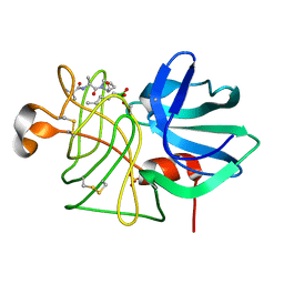
3LPR
DownloadVisualize
BU of 3lpr by Molmil
STRUCTURAL BASIS FOR BROAD SPECIFICITY IN ALPHA-LYTIC PROTEASE MUTANTS
Descriptor: ALPHA-LYTIC PROTEASE, METHOXYSUCCINYL-ALA-ALA-PRO-NORLEUCINE BORONIC ACID INHIBITOR, SULFATE ION
Authors:Bone, R, Agard, D.A.
Deposit date:1991-08-05
Release date:1993-01-15
Last modified:2024-06-05
Method:X-RAY DIFFRACTION (2.15 Å)
Cite:Structural basis for broad specificity in alpha-lytic protease mutants.
Biochemistry, 30, 1991


123>

227344

数据于2024-11-13公开中

PDB statisticsPDBj update infoContact PDBjnumon