Loading
PDBj
MenuPDBj@FacebookPDBj@X(formerly Twitter)PDBj@BlueSkyPDBj@YouTubewwPDB FoundationwwPDB
RCSB PDBPDBeBMRBAdv. SearchSearch help
Search by PDB author
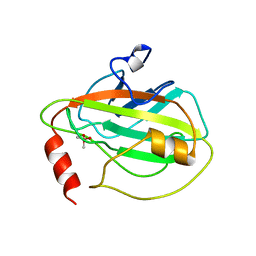
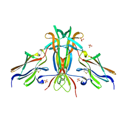
1QOU
DownloadVisualize
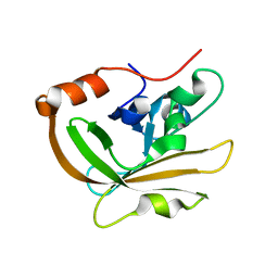
BU of 1qou by Molmil
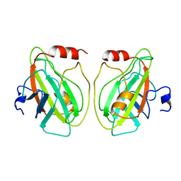
CEN (Centroradialis) protein from Antirrhinum
Descriptor: CEN
Authors:Banfield, M.J, Brady, R.L.
Deposit date:1999-11-17
Release date:2000-03-31
Last modified:2024-11-20
Method:X-RAY DIFFRACTION (1.9 Å)
Cite:The Structure of Antirrhinum Centroradialis Protein (Cen) Suggests a Role as a Kinase Regulator
J.Mol.Biol., 297, 2000
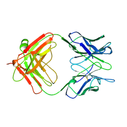
1CLO
DownloadVisualize
BU of 1clo by Molmil
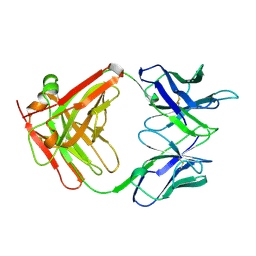
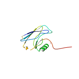
ANTI-CARCINOEMBRYONIC ANTIGEN MONOCLONAL ANTIBODY A5B7
Descriptor: A5B7 MONOCLONAL ANTIBODY
Authors:Banfield, M.J, King, D.J, Mountain, A, Brady, R.L.
Deposit date:1996-05-01
Release date:1997-05-15
Last modified:2024-10-30
Method:X-RAY DIFFRACTION (2.1 Å)
Cite:VL:VH domain rotations in engineered antibodies: crystal structures of the Fab fragments from two murine antitumor antibodies and their engineered human constructs.
Proteins, 29, 1997
1HCF
DownloadVisualize
BU of 1hcf by Molmil
Crystal structure of TrkB-d5 bound to neurotrophin-4/5
Descriptor: BDNF/NT-3 GROWTH FACTORS RECEPTOR, NEUROTROPHIN-4, SULFATE ION
Authors:Banfield, M.J, Naylor, R.L, Robertson, A.G.S, Allen, S.J, Dawbarn, D, Brady, R.L.
Deposit date:2001-05-03
Release date:2001-12-06
Last modified:2024-11-06
Method:X-RAY DIFFRACTION (2.7 Å)
Cite:Specificity in Trk-Receptor:Neurotrophin Interaction: The Crystal Structure of Trkb-D5 in Complex with Neurotrophin-4/5
Structure, 9, 2001
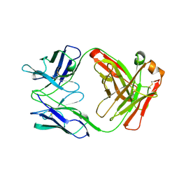
1AD0
DownloadVisualize
BU of 1ad0 by Molmil
FAB FRAGMENT OF ENGINEERED HUMAN MONOCLONAL ANTIBODY A5B7
Descriptor: ANTIBODY A5B7 (HEAVY CHAIN), ANTIBODY A5B7 (LIGHT CHAIN)
Authors:Banfield, M.J, Brady, R.L.
Deposit date:1997-02-19
Release date:1998-02-25
Last modified:2024-11-20
Method:X-RAY DIFFRACTION (2.5 Å)
Cite:VL:VH domain rotations in engineered antibodies: crystal structures of the Fab fragments from two murine antitumor antibodies and their engineered human constructs.
Proteins, 29, 1997
1AE6
DownloadVisualize
BU of 1ae6 by Molmil
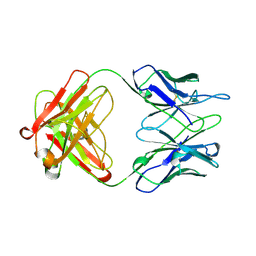
IGG-FAB FRAGMENT OF MOUSE MONOCLONAL ANTIBODY CTM01
Descriptor: IGG CTM01 FAB (HEAVY CHAIN), IGG CTM01 FAB (LIGHT CHAIN)
Authors:Banfield, M.J, Brady, R.L.
Deposit date:1997-03-06
Release date:1998-03-18
Last modified:2024-10-30
Method:X-RAY DIFFRACTION (3 Å)
Cite:VL:VH domain rotations in engineered antibodies: crystal structures of the Fab fragments from two murine antitumor antibodies and their engineered human constructs.
Proteins, 29, 1997
1AD9
DownloadVisualize
BU of 1ad9 by Molmil
IGG-FAB FRAGMENT OF ENGINEERED HUMAN MONOCLONAL ANTIBODY CTM01
Descriptor: IGG CTM01 FAB (HEAVY CHAIN), IGG CTM01 FAB (LIGHT CHAIN), SULFATE ION
Authors:Banfield, M.J, Brady, R.L.
Deposit date:1997-02-24
Release date:1998-02-25
Last modified:2024-10-23
Method:X-RAY DIFFRACTION (2.8 Å)
Cite:VL:VH domain rotations in engineered antibodies: crystal structures of the Fab fragments from two murine antitumor antibodies and their engineered human constructs.
Proteins, 29, 1997
1BD9
DownloadVisualize
BU of 1bd9 by Molmil
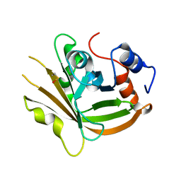
HUMAN PHOSPHATIDYLETHANOLAMINE BINDING PROTEIN
Descriptor: PHOSPHATIDYLETHANOLAMINE BINDING PROTEIN
Authors:Banfield, M.J, Barker, J.J, Perry, A.C.F, Brady, R.L.
Deposit date:1998-05-12
Release date:1998-09-16
Last modified:2024-02-07
Method:X-RAY DIFFRACTION (2.05 Å)
Cite:Function from structure? The crystal structure of human phosphatidylethanolamine-binding protein suggests a role in membrane signal transduction.
Structure, 6, 1998
1BEH
DownloadVisualize
BU of 1beh by Molmil
HUMAN PHOSPHATIDYLETHANOLAMINE BINDING PROTEIN IN COMPLEX WITH CACODYLATE
Descriptor: CACODYLATE ION, PHOSPHATIDYLETHANOLAMINE BINDING PROTEIN
Authors:Banfield, M.J, Barker, J.J, Perry, A, Brady, R.L.
Deposit date:1998-05-14
Release date:1998-09-16
Last modified:2024-05-22
Method:X-RAY DIFFRACTION (1.75 Å)
Cite:Function from structure? The crystal structure of human phosphatidylethanolamine-binding protein suggests a role in membrane signal transduction.
Structure, 6, 1998
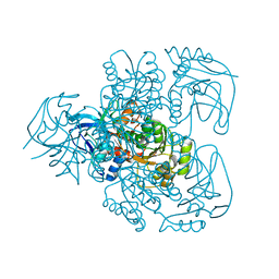
1E3J
DownloadVisualize
BU of 1e3j by Molmil
Ketose reductase (sorbitol dehydrogenase) from silverleaf whitefly
Descriptor: BORIC ACID, NADP(H)-DEPENDENT KETOSE REDUCTASE, PHOSPHATE ION, ...
Authors:Banfield, M.J, Salvucci, M.E, Baker, E.N, Smith, C.A.
Deposit date:2000-06-19
Release date:2001-02-04
Last modified:2024-05-08
Method:X-RAY DIFFRACTION (2.3 Å)
Cite:Crystal Structure of Nadp(H)-Dependent Ketose Reductase from Besimia Argentifolii at 2.3 Angstrom Resolution
J.Mol.Biol., 306, 2001
1H5Y
DownloadVisualize
BU of 1h5y by Molmil
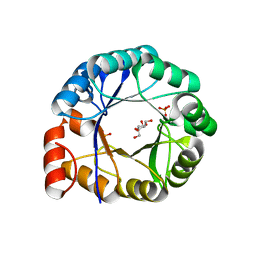
HisF protein from Pyrobaculum aerophilum
Descriptor: GLYCEROL, HISF, PHOSPHATE ION
Authors:Banfield, M.J, Lott, J.S, McCarthy, A.A, Baker, E.N.
Deposit date:2001-05-31
Release date:2001-06-01
Last modified:2023-12-13
Method:X-RAY DIFFRACTION (2 Å)
Cite:Structure of Hisf, a Histidine Biosynthetic Protein from Pyrobaculum Aerophilum
Acta Crystallogr.,Sect.D, 57, 2001
3FSV
DownloadVisualize
BU of 3fsv by Molmil
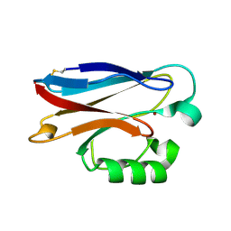
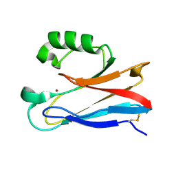
Pseudomonas aeruginosa Azurin with mutated metal-binding loop sequence (CAAAHAAAM)
Descriptor: Azurin, COPPER (II) ION
Authors:Banfield, M.J.
Deposit date:2009-01-12
Release date:2009-03-10
Last modified:2024-11-20
Method:X-RAY DIFFRACTION (2.3 Å)
Cite:Metal-binding loop length and not sequence dictates structure.
Proc.Natl.Acad.Sci.USA, 106, 2009
2HXA
DownloadVisualize
BU of 2hxa by Molmil
Crystal structure of Cu(I) Azurin with the metal-binding loop sequence "CTFPGHSALM" replaced with "CSPHQGAGM", at pH3.5
Descriptor: Azurin, COPPER (I) ION
Authors:Banfield, M.J.
Deposit date:2006-08-03
Release date:2007-01-30
Last modified:2024-11-20
Method:X-RAY DIFFRACTION (2.21 Å)
Cite:Engineering Copper Sites in Proteins: Loops Confer Native Structures and Properties to Chimeric Cupredoxins.
J.Am.Chem.Soc., 129, 2007
2HX9
DownloadVisualize
BU of 2hx9 by Molmil
Crystal structure of Cu(I) Azurin with the metal-binding loop sequence "CTFPGHSALM" replaced with "CSPHQGAGM", at pH4
Descriptor: Azurin, COPPER (I) ION
Authors:Banfield, M.J.
Deposit date:2006-08-03
Release date:2007-01-30
Last modified:2024-10-30
Method:X-RAY DIFFRACTION (1.7 Å)
Cite:Engineering Copper Sites in Proteins: Loops Confer Native Structures and Properties to Chimeric Cupredoxins.
J.Am.Chem.Soc., 129, 2007
2HX7
DownloadVisualize
BU of 2hx7 by Molmil
Crystal structure of Cu(II) Azurin with the metal-binding loop sequence "CTFPGHSALM" replaced with "CSPHQGAGM"
Descriptor: Azurin, COPPER (II) ION
Authors:Banfield, M.J.
Deposit date:2006-08-03
Release date:2007-01-30
Last modified:2024-11-20
Method:X-RAY DIFFRACTION (1.55 Å)
Cite:Engineering Copper Sites in Proteins: Loops Confer Native Structures and Properties to Chimeric Cupredoxins.
J.Am.Chem.Soc., 129, 2007
2HX8
DownloadVisualize
BU of 2hx8 by Molmil
Crystal structure of Cu(I) Azurin with the metal-binding loop sequence "CTFPGHSALM" replaced with "CSPHQGAGM", at pH5
Descriptor: Azurin, COPPER (I) ION
Authors:Banfield, M.J.
Deposit date:2006-08-03
Release date:2007-01-30
Last modified:2024-11-20
Method:X-RAY DIFFRACTION (1.6 Å)
Cite:Engineering Copper Sites in Proteins: Loops Confer Native Structures and Properties to Chimeric Cupredoxins.
J.Am.Chem.Soc., 129, 2007
3FN5
DownloadVisualize
BU of 3fn5 by Molmil
Crystal structure of sortase A (Spy1154) from Streptococcus pyogenes serotype M1 strain SF370
Descriptor: 4-(2-HYDROXYETHYL)-1-PIPERAZINE ETHANESULFONIC ACID, Sortase A
Authors:Banfield, M.J.
Deposit date:2008-12-23
Release date:2009-01-06
Last modified:2023-09-06
Method:X-RAY DIFFRACTION (1.5 Å)
Cite:Crystal Structure of Streptococcus pyogenes Sortase A: Implications for Sortase mechanism
J.Biol.Chem., 284, 2009
3FN6
DownloadVisualize
BU of 3fn6 by Molmil
Crystal structure of sortase A from Streptococcus pyogenes serotype M1 strain SF370 with the active site Cys in its sulphenic acid form
Descriptor: Sortase A
Authors:Banfield, M.J.
Deposit date:2008-12-23
Release date:2009-01-06
Last modified:2023-09-06
Method:X-RAY DIFFRACTION (1.9 Å)
Cite:Crystal Structure of Streptococcus pyogenes Sortase A: Implications for Sortase mechanism
J.Biol.Chem., 284, 2009
3FN7
DownloadVisualize
BU of 3fn7 by Molmil
Crystal structure of sortase A (Spy1154) from Streptococcus pyogenes serotype M1 strain SF370
Descriptor: Sortase A
Authors:Banfield, M.J.
Deposit date:2008-12-23
Release date:2009-01-06
Last modified:2023-09-06
Method:X-RAY DIFFRACTION (1.75 Å)
Cite:Crystal Structure of Streptococcus pyogenes Sortase A: Implications for Sortase mechanism
J.Biol.Chem., 284, 2009
3FSW
DownloadVisualize
BU of 3fsw by Molmil
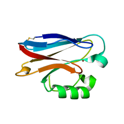
Pseudomonas aeruginosa Azurin with mutated metal-binding loop sequence (CAAAAHAAAM)
Descriptor: Azurin, COPPER (II) ION
Authors:Banfield, M.J.
Deposit date:2009-01-12
Release date:2009-03-10
Last modified:2024-10-30
Method:X-RAY DIFFRACTION (2 Å)
Cite:Metal-binding loop length and not sequence dictates structure.
Proc.Natl.Acad.Sci.USA, 106, 2009
3FSZ
DownloadVisualize
BU of 3fsz by Molmil
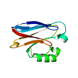
Pseudomonas aeruginosa Azurin with mutated metal-binding loop sequence (CAAAAHAAAAM)
Descriptor: Azurin, COPPER (II) ION
Authors:Banfield, M.J.
Deposit date:2009-01-12
Release date:2009-03-10
Last modified:2024-10-30
Method:X-RAY DIFFRACTION (2 Å)
Cite:Metal-binding loop length and not sequence dictates structure.
Proc.Natl.Acad.Sci.USA, 106, 2009
3FT0
DownloadVisualize
BU of 3ft0 by Molmil
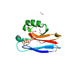
Pseudomonas aeruginosa Azurin with mutated metal-binding loop sequence (CAAAAHAAAAM), chemically reduced
Descriptor: Azurin, COPPER (I) ION
Authors:Banfield, M.J.
Deposit date:2009-01-12
Release date:2009-03-10
Last modified:2024-11-20
Method:X-RAY DIFFRACTION (1.8 Å)
Cite:Metal-binding loop length and not sequence dictates structure.
Proc.Natl.Acad.Sci.USA, 106, 2009
3FSA
DownloadVisualize
BU of 3fsa by Molmil
Pseudomonas aeruginosa Azurin with mutated metal-binding loop sequence (CAAHAAM); chemically reduced.
Descriptor: Azurin, COPPER (I) ION
Authors:Banfield, M.J.
Deposit date:2009-01-09
Release date:2009-03-10
Last modified:2024-10-16
Method:X-RAY DIFFRACTION (0.98 Å)
Cite:Metal-binding loop length and not sequence dictates structure.
Proc.Natl.Acad.Sci.USA, 106, 2009
3FS9
DownloadVisualize
BU of 3fs9 by Molmil
Pseudomonas aeruginosa Azurin with mutated metal-binding loop sequence (CAAHAAM)
Descriptor: Azurin, COPPER (II) ION
Authors:Banfield, M.J.
Deposit date:2009-01-09
Release date:2009-03-10
Last modified:2024-11-06
Method:X-RAY DIFFRACTION (1.05 Å)
Cite:Metal-binding loop length and not sequence dictates structure.
Proc.Natl.Acad.Sci.USA, 106, 2009
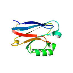
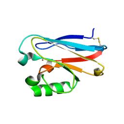
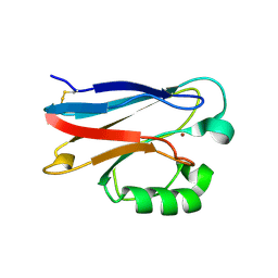
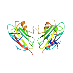
2FT6
DownloadVisualize
BU of 2ft6 by Molmil
Structure of Cu(II)azurin with the metal-binding loop sequence "CTFPGHSALM" replaced with "CTPHPM"
Descriptor: Azurin, COPPER (II) ION
Authors:Banfield, M.J.
Deposit date:2006-01-24
Release date:2006-04-11
Last modified:2024-11-06
Method:X-RAY DIFFRACTION (1.25 Å)
Cite:Basic requirements for a metal-binding site in a protein: The influence of loop shortening on the cupredoxin azurin.
Proc.Natl.Acad.Sci.Usa, 103, 2006
2FT8
DownloadVisualize
BU of 2ft8 by Molmil
Structure of Cu(I)azurin, pH8, with the metal-binding loop sequence "CTFPGHSALM" replaced with "CTPHPM"
Descriptor: Azurin, COPPER (I) ION
Authors:Banfield, M.J.
Deposit date:2006-01-24
Release date:2006-04-11
Last modified:2024-11-06
Method:X-RAY DIFFRACTION (1.55 Å)
Cite:Basic requirements for a metal-binding site in a protein: The influence of loop shortening on the cupredoxin azurin.
Proc.Natl.Acad.Sci.Usa, 103, 2006

227561

数据于2024-11-20公开中

PDB statisticsPDBj update infoContact PDBjnumon