Loading
PDBj
MenuPDBj@FacebookPDBj@TwitterPDBj@YouTubewwPDB FoundationwwPDB
RCSB PDBPDBeBMRBAdv. SearchSearch help
Search by PDB author
2A8A
DownloadVisualize
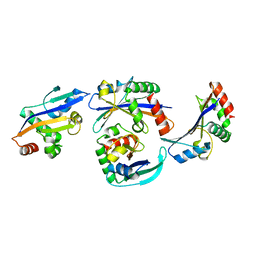
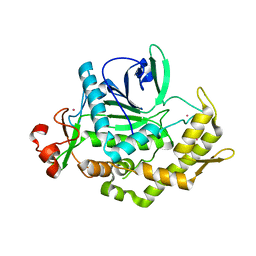
BU of 2a8a by Molmil
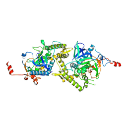
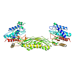
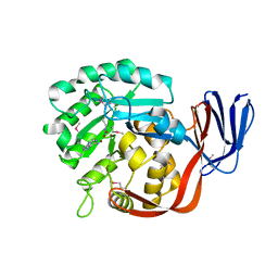
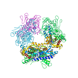
Crystal structure of Clostridium botulinum neurotoxin serotype F light chain
Descriptor: Botulinum neurotoxin type F, CADMIUM ION, ZINC ION
Authors:Agarwal, R, Binz, T, Swaminathan, S.
Deposit date:2005-07-07
Release date:2005-09-20
Last modified:2023-08-23
Method:X-RAY DIFFRACTION (2 Å)
Cite:Structural analysis of botulinum neurotoxin serotype f light chain: implications on substrate binding and inhibitor design
Biochemistry, 44, 2005
2A97
DownloadVisualize
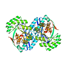
BU of 2a97 by Molmil
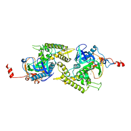
Crystal structure of catalytic domain of Clostridium botulinum neurotoxin serotype F
Descriptor: Botulinum neurotoxin type F, CADMIUM ION, ZINC ION
Authors:Agarwal, R, Binz, T, Swaminathan, S.
Deposit date:2005-07-11
Release date:2005-09-20
Last modified:2023-08-23
Method:X-RAY DIFFRACTION (1.8 Å)
Cite:Structural analysis of botulinum neurotoxin serotype f light chain: implications on substrate binding and inhibitor design
Biochemistry, 44, 2005
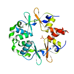
1YAV
DownloadVisualize
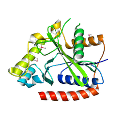
BU of 1yav by Molmil
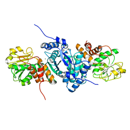
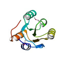
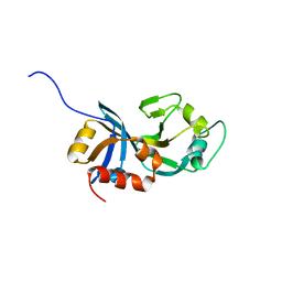
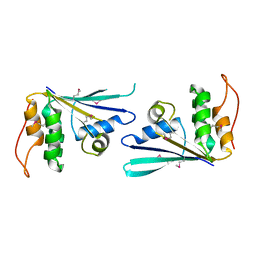
Crystal structure of CBS domain-containing protein ykuL from Bacillus subtilis
Descriptor: SULFATE ION, hypothetical protein BSU14130
Authors:Kumaran, D, Swaminathan, S, Burley, S.K, New York SGX Research Center for Structural Genomics (NYSGXRC)
Deposit date:2004-12-17
Release date:2004-12-28
Last modified:2024-02-14
Method:X-RAY DIFFRACTION (2.1 Å)
Cite:Crystal Structure of a hypothetical protein (ykul) containing CBS domains from Bacillus subtilis.
To be Published
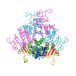
1YBD
DownloadVisualize
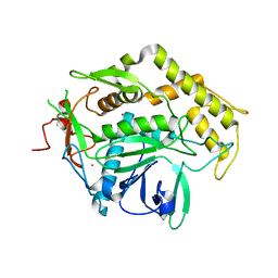
BU of 1ybd by Molmil
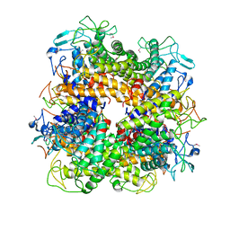
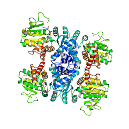
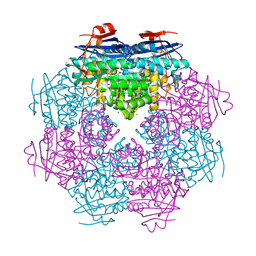
Crystal structure analysis of uridylate kinase from Neisseria meningitidis
Descriptor: FORMIC ACID, GLYCEROL, Uridylate kinase
Authors:Kumaran, D, Swaminathan, S, Burley, S.K, New York SGX Research Center for Structural Genomics (NYSGXRC)
Deposit date:2004-12-20
Release date:2005-02-15
Last modified:2021-02-03
Method:X-RAY DIFFRACTION (2.6 Å)
Cite:Crystal structure analysis of uridylate kinase from Neisseria meningitidis
To be Published
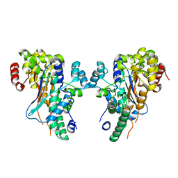
1YBE
DownloadVisualize
BU of 1ybe by Molmil
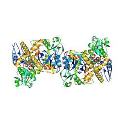
Crystal Structure of a Nicotinate phosphoribosyltransferase
Descriptor: Nicotinate phosphoribosyltransferase
Authors:Seetharaman, J, Swaminathan, S, Burley, S.K, New York SGX Research Center for Structural Genomics (NYSGXRC)
Deposit date:2004-12-20
Release date:2005-01-11
Last modified:2024-02-14
Method:X-RAY DIFFRACTION (2.5 Å)
Cite:Crystal Structure of a Nicotinate phosphoribosyltransferase
To be Published
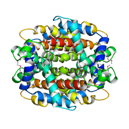
1Y9I
DownloadVisualize
BU of 1y9i by Molmil
Crystal structure of low temperature requirement C protein from Listeria monocytogenes
Descriptor: CALCIUM ION, GLYCEROL, MAGNESIUM ION, ...
Authors:Kumaran, D, Swaminathan, S, Burley, S.K, New York SGX Research Center for Structural Genomics (NYSGXRC)
Deposit date:2004-12-15
Release date:2004-12-28
Last modified:2021-02-03
Method:X-RAY DIFFRACTION (1.8 Å)
Cite:Crystal structure of phosphatidylglycerophosphatase (PGPase), a putative membrane-bound lipid phosphatase, reveals a novel binuclear metal binding site and two "proton wires".
Proteins, 64, 2006
1ZL6
DownloadVisualize
BU of 1zl6 by Molmil
Crystal structure of Tyr350Ala mutant of Clostridium botulinum neurotoxin E catalytic domain
Descriptor: SULFATE ION, ZINC ION, botulinum neurotoxin type E
Authors:Agarwal, R, Binz, T, Swaminathan, S.
Deposit date:2005-05-05
Release date:2005-06-28
Last modified:2023-08-23
Method:X-RAY DIFFRACTION (2.4 Å)
Cite:Analysis of Active Site Residues of Botulinum Neurotoxin E by Mutational, Functional, and Structural Studies: Glu335Gln Is an Apoenzyme.
Biochemistry, 44, 2005
1ZN3
DownloadVisualize
BU of 1zn3 by Molmil
Crystal structure of Glu335Ala mutant of Clostridium botulinum neurotoxin type E
Descriptor: CHLORIDE ION, ZINC ION, botulinum neurotoxin type E
Authors:Agarwal, R, Binz, T, Swaminathan, S.
Deposit date:2005-05-11
Release date:2005-07-05
Last modified:2023-08-23
Method:X-RAY DIFFRACTION (2.6 Å)
Cite:Analysis of Active Site Residues of Botulinum Neurotoxin E by Mutational, Functional, and Structural Studies: Glu335Gln Is an Apoenzyme.
Biochemistry, 44, 2005
2A9F
DownloadVisualize
BU of 2a9f by Molmil
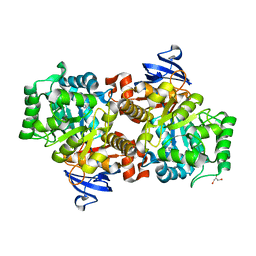
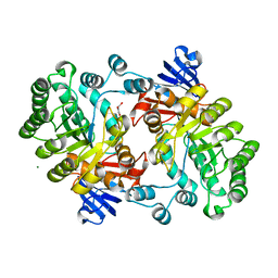
Crystal structure of a putative malic enzyme ((S)-malate:NAD+ oxidoreductase (decarboxylating))
Descriptor: MAGNESIUM ION, putative malic enzyme ((S)-malate:NAD+ oxidoreductase (decarboxylating))
Authors:Seetharaman, J, Swaminathan, S, Burley, S.K, New York SGX Research Center for Structural Genomics (NYSGXRC)
Deposit date:2005-07-11
Release date:2005-08-02
Last modified:2024-02-14
Method:X-RAY DIFFRACTION (2.5 Å)
Cite:Crystal structure of a putative malic enzyme ((S)-malate:NAD+ oxidoreductase (decarboxylating))
To be Published
2AFA
DownloadVisualize
BU of 2afa by Molmil
Crystal Structure of putative NAG isomerase from Salmonella typhimurium
Descriptor: NAG isomerase
Authors:Kumaran, D, Swaminathan, S, Burley, S.K, New York SGX Research Center for Structural Genomics (NYSGXRC)
Deposit date:2005-07-25
Release date:2005-08-16
Last modified:2021-02-03
Method:X-RAY DIFFRACTION (2.15 Å)
Cite:Crystal Structure of putative NAG isomerase from Salmonella typhimurium
To be Published
2IMO
DownloadVisualize
BU of 2imo by Molmil
Crystal structure of allantoate amidohydrolase from Escherichia coli at pH 4.6
Descriptor: Allantoate amidohydrolase
Authors:Agarwal, R, Burley, S.K, Swaminathan, S, New York SGX Research Center for Structural Genomics (NYSGXRC)
Deposit date:2006-10-04
Release date:2006-10-17
Last modified:2021-02-03
Method:X-RAY DIFFRACTION (2.8 Å)
Cite:Structural Analysis of a Ternary Complex of Allantoate Amidohydrolase from Escherichia coli Reveals its Mechanics.
J.Mol.Biol., 368, 2007
2IMG
DownloadVisualize
BU of 2img by Molmil
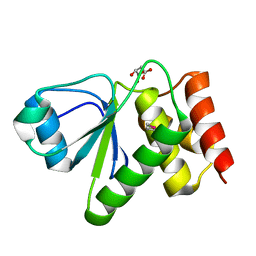
Crystal structure of dual specificity protein phosphatase 23 from Homo sapiens in complex with ligand malate ion
Descriptor: D-MALATE, Dual specificity protein phosphatase 23
Authors:Agarwal, R, Burley, S.K, Swaminathan, S, New York SGX Research Center for Structural Genomics (NYSGXRC)
Deposit date:2006-10-04
Release date:2006-10-17
Last modified:2021-02-03
Method:X-RAY DIFFRACTION (1.93 Å)
Cite:Structure of human dual specificity protein phosphatase 23, VHZ, enzyme-substrate/product complex.
J.Biol.Chem., 283, 2008
2I9U
DownloadVisualize
BU of 2i9u by Molmil
Crystal Structure of Guanine Deaminase from C. acetobutylicum with bound guanine in the active site
Descriptor: Cytosine/guanine deaminase related protein, FE (III) ION, GLYCEROL, ...
Authors:Kumaran, D, Burley, S.K, Swaminathan, S, New York SGX Research Center for Structural Genomics (NYSGXRC)
Deposit date:2006-09-06
Release date:2006-09-19
Last modified:2024-02-21
Method:X-RAY DIFFRACTION (2.05 Å)
Cite:Crystal Structure of Guanine Deaminase from C. acetobutylicum with bound guanine in the active site
To be Published
2IOJ
DownloadVisualize
BU of 2ioj by Molmil
Crystal structure of protein AF1212 from Archaeoglobus fulgidus, Pfam DRTGG
Descriptor: Hypothetical protein AF_1212
Authors:Tyagi, R, Burley, S.K, Swaminathan, S, New York SGX Research Center for Structural Genomics (NYSGXRC)
Deposit date:2006-10-10
Release date:2006-10-31
Last modified:2021-02-03
Method:X-RAY DIFFRACTION (2.15 Å)
Cite:The crystal structure of a hypothetical protein (Y1212_ARCFU) from Archeoglobus fulgidus
To be Published
2ICS
DownloadVisualize
BU of 2ics by Molmil
Crystal structure of an adenine deaminase
Descriptor: ADENINE, Adenine Deaminase, ZINC ION
Authors:Sugadev, R, Kumaran, D, Burley, S.K, Swaminathan, S, New York SGX Research Center for Structural Genomics (NYSGXRC)
Deposit date:2006-09-13
Release date:2006-10-17
Last modified:2021-02-03
Method:X-RAY DIFFRACTION (2.3 Å)
Cite:Crystal structure of an adenine deaminase
TO BE PUBLISHED
2HAF
DownloadVisualize
BU of 2haf by Molmil
Crystal structure of a putative translation repressor from Vibrio cholerae
Descriptor: Putative translation repressor
Authors:Sugadev, R, Seetharaman, J, Kumaran, D, Swaminathan, S, Burley, S.K, New York SGX Research Center for Structural Genomics (NYSGXRC)
Deposit date:2006-06-12
Release date:2006-07-04
Last modified:2024-02-14
Method:X-RAY DIFFRACTION (2.88 Å)
Cite:Crystal structure of a putative translation repressor from Vibrio cholerae
To be Published
2HAE
DownloadVisualize
BU of 2hae by Molmil
Crystal structure of a putative malic enzyme (malate oxidoreductase)
Descriptor: Malate oxidoreductase, NICOTINAMIDE-ADENINE-DINUCLEOTIDE, ZINC ION
Authors:Seetharaman, J, Swaminathan, S, Burley, S.K, New York SGX Research Center for Structural Genomics (NYSGXRC)
Deposit date:2006-06-12
Release date:2006-07-04
Last modified:2023-08-30
Method:X-RAY DIFFRACTION (3.13 Å)
Cite:Crystal structure of a putative malic enzyme (malate oxidoreductase)
To be Published
2I6E
DownloadVisualize
BU of 2i6e by Molmil
Crystal structure of protein DR0370 from Deinococcus radiodurans, Pfam DUF178
Descriptor: Hypothetical protein, SULFATE ION
Authors:Tyagi, R, Kumaran, D, Burley, S.K, Swaminathan, S, New York SGX Research Center for Structural Genomics (NYSGXRC)
Deposit date:2006-08-28
Release date:2006-09-05
Last modified:2021-02-03
Method:X-RAY DIFFRACTION (2.5 Å)
Cite:X-ray structures of two proteins belonging to Pfam DUF178 revealed unexpected structural similarity to the DUF191 Pfam family.
Bmc Struct.Biol., 7, 2007
2GUW
DownloadVisualize
BU of 2guw by Molmil
Crystal structure of AMP Nucleosidase from Salmonella typhimurium LT2
Descriptor: AMP nucleosidase
Authors:Rao, K.N, Swaminathan, S, Burley, S.K, New York SGX Research Center for Structural Genomics (NYSGXRC)
Deposit date:2006-05-01
Release date:2006-06-06
Last modified:2024-02-14
Method:X-RAY DIFFRACTION (2.64 Å)
Cite:Crystal structure of AMP Nucleosidase from Salmonella typhimurium LT2
To be Published
2GOK
DownloadVisualize
BU of 2gok by Molmil
Crystal structure of the imidazolonepropionase from Agrobacterium tumefaciens at 1.87 A resolution
Descriptor: CHLORIDE ION, FE (III) ION, GLYCEROL, ...
Authors:Tyagi, R, Kumaran, D, Swaminathan, S, Burley, S.K, New York SGX Research Center for Structural Genomics (NYSGXRC)
Deposit date:2006-04-13
Release date:2006-04-25
Last modified:2024-02-14
Method:X-RAY DIFFRACTION (1.87 Å)
Cite:X-ray structure of imidazolonepropionase from Agrobacterium tumefaciens at 1.87 A resolution.
Proteins, 69, 2007
2NWU
DownloadVisualize
BU of 2nwu by Molmil
Crystal structure of protein SSO1042 from Sulfolobus solfataricus, Pfam DUF54
Descriptor: UPF0201 protein SSO1042
Authors:Rao, K.N, Burley, S.K, Swaminathan, S, New York SGX Research Center for Structural Genomics (NYSGXRC)
Deposit date:2006-11-16
Release date:2006-12-12
Last modified:2023-12-27
Method:X-RAY DIFFRACTION (2.4 Å)
Cite:UPF201 archaeal specific family members reveal structural similarity to RNA-binding proteins but low likelihood for RNA-binding function.
Plos One, 3, 2008
2OG9
DownloadVisualize
BU of 2og9 by Molmil
Crystal Structure of mandelate racemase/muconate lactonizing enzyme from Polaromonas sp. JS666
Descriptor: CALCIUM ION, Mandelate racemase/muconate lactonizing enzyme
Authors:Kumaran, D, Burley, S.K, Swaminathan, S, New York SGX Research Center for Structural Genomics (NYSGXRC)
Deposit date:2007-01-05
Release date:2007-02-27
Last modified:2023-12-27
Method:X-RAY DIFFRACTION (1.9 Å)
Cite:Crystal Structure of mandelate racemase/muconate lactonizing enzyme from Polaromonas sp. JS666
To be Published
2OGK
DownloadVisualize
BU of 2ogk by Molmil
Crystal structure of protein AF2318 from Archaeglobus fulgidus, Pfam DUF54
Descriptor: Hypothetical protein
Authors:Rao, K.N, Burley, S.K, Swaminathan, S, New York SGX Research Center for Structural Genomics (NYSGXRC)
Deposit date:2007-01-05
Release date:2007-01-30
Last modified:2023-12-27
Method:X-RAY DIFFRACTION (3 Å)
Cite:UPF201 archaeal specific family members reveal structural similarity to RNA-binding proteins but low likelihood for RNA-binding function.
Plos One, 3, 2008
2OVL
DownloadVisualize
BU of 2ovl by Molmil
Crystal structure of a racemase from Streptomyces coelicolor A3(2)
Descriptor: Putative racemase, SODIUM ION
Authors:Rao, K.N, Burley, S.K, Swaminathan, S, New York SGX Research Center for Structural Genomics (NYSGXRC)
Deposit date:2007-02-14
Release date:2007-03-20
Last modified:2021-02-03
Method:X-RAY DIFFRACTION (2.13 Å)
Cite:Crystal structure of a racemase from Streptomyces coelicolor A3(2)
To be Published
2P5I
DownloadVisualize
BU of 2p5i by Molmil
Crystal structure of protein BH3822 from Bacillus halodurans, a member of the biotin/lipoate A/B protein ligase family
Descriptor: BH3822 protein
Authors:Sugadev, R, Burley, S.K, Swaminathan, S, New York SGX Research Center for Structural Genomics (NYSGXRC)
Deposit date:2007-03-15
Release date:2007-03-27
Last modified:2021-02-03
Method:X-RAY DIFFRACTION (2.21 Å)
Cite:Crystal structure of a hypothetical protein from Bacillus halodurans: Pfam-PF03099
To be Published

222415

数据于2024-07-10公开中

PDB statisticsPDBj update infoContact PDBjnumon