7A3B
 
 | |
7A2L
 | |
7A2U
 | |
7A2O
 | |
7A3D
 | |
7A2J
 | |
7A2V
 | |
7A37
 | |
7A2Q
 | |
7A33
 | |
7A2W
 | |
7A3A
 | |
7A2P
 | |
7A2N
 | |
7A2M
 | |
7A2R
 | |
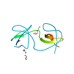
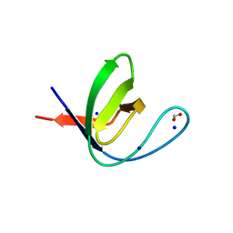
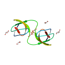
2F2X
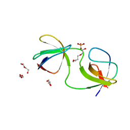
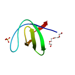
 | | alpha-spectrin SH3 domain R21G mutant | | Descriptor: | SULFATE ION, Spectrin alpha chain, brain | | Authors: | Camara-Artigas, A, Conejero-Lara, F, Casares, S, Lopez-Mayorga, O, Vega, C. | | Deposit date: | 2005-11-18 | | Release date: | 2006-10-31 | | Last modified: | 2023-08-23 | | Method: | X-RAY DIFFRACTION (1.6 Å) | | Cite: | Cooperative propagation of local stability changes from low-stability and high-stability regions in a SH3 domain
Proteins, 67, 2007
|
|
7NET
 | |
7NES
 | |
7NER
 | |
8AH4
 | |
8AH7
 | |
1Z9K
 | |
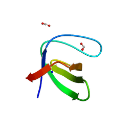
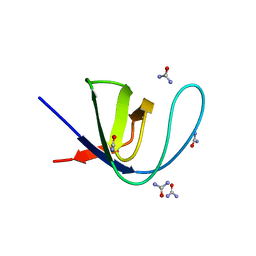
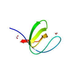
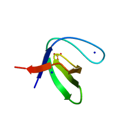
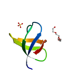
2HDA
 | | Yes SH3 domain | | Descriptor: | Proto-oncogene tyrosine-protein kinase Yes, SULFATE ION | | Authors: | Camara-Artigas, A, Luque, I, Ruiz-Sanz, J, Mateo, P.L, Martin-Garcia, J.M. | | Deposit date: | 2006-06-20 | | Release date: | 2007-04-17 | | Last modified: | 2023-08-30 | | Method: | X-RAY DIFFRACTION (1.9 Å) | | Cite: | Crystallographic structure of the SH3 domain of the human c-Yes tyrosine kinase: Loop flexibility and amyloid aggregation.
Febs Lett., 581, 2007
|
|
3M0U
 | |


