Loading
PDBj
MenuPDBj@FacebookPDBj@TwitterPDBj@YouTubewwPDB FoundationwwPDB
RCSB PDBPDBeBMRBAdv. SearchSearch help
Search by PDB author
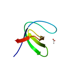
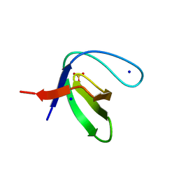
7A33
DownloadVisualize
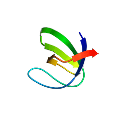
BU of 7a33 by Molmil
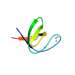
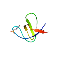
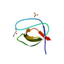
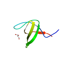
Crystal structure of the c-Src SH3 domain mutant S94A-T98D-V111L-N113S-T114S at pH 3.0
Descriptor: FORMIC ACID, Proto-oncogene tyrosine-protein kinase Src, SODIUM ION
Authors:Camara-Artigas, A, Plaza-Garrido, M, Salinas-Garcia, M.C.
Deposit date:2020-08-18
Release date:2021-08-25
Last modified:2024-01-31
Method:X-RAY DIFFRACTION (0.96 Å)
Cite:Crystal structure of the c-Src SH3 domain mutant S94A-T98D-V111L-N113S-T114S at pH 3.0
To be published
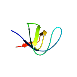
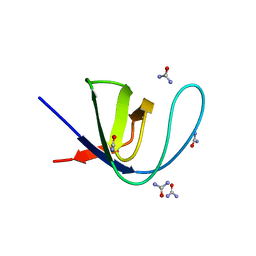
7A32
DownloadVisualize
BU of 7a32 by Molmil
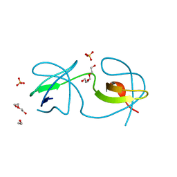
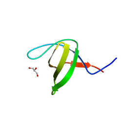
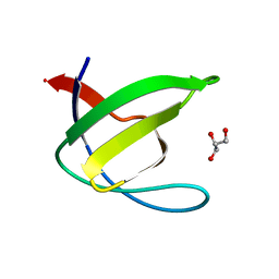
Crystal structure of the c-Src SH3 domain mutant S94A-T98D-V111L-N113S-T114S at pH 7.0
Descriptor: GLYCEROL, Proto-oncogene tyrosine-protein kinase Src, SULFATE ION
Authors:Camara-Artigas, A, Plaza-Garrido, M, Salinas-Garcia, M.C.
Deposit date:2020-08-18
Release date:2021-08-25
Last modified:2024-01-31
Method:X-RAY DIFFRACTION (1.15 Å)
Cite:Crystal structure of the c-Src SH3 domain mutant S94A-T98D-V111L-N113S-T114S at pH 7.0
To be published
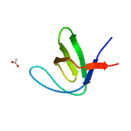
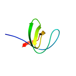
7A2T
DownloadVisualize
BU of 7a2t by Molmil
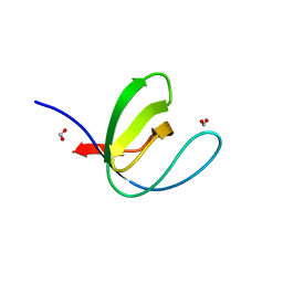
Crystal structure of the Fyn SH3 domain L112V-S114N-S115T-E121L-R123H mutant at pH 4.0
Descriptor: ACETATE ION, Tyrosine-protein kinase Fyn
Authors:Camara-Artigas, A, Plaza-Garrido, M, Salinas-Garcia, M.C.
Deposit date:2020-08-18
Release date:2021-08-25
Last modified:2024-01-31
Method:X-RAY DIFFRACTION (1.22 Å)
Cite:Crystal structure of the Fyn SH3 domain L112V-S114N-S115T-E121L-R123H mutant at pH 4.0
To be published
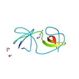
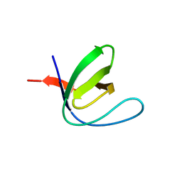
7A35
DownloadVisualize
BU of 7a35 by Molmil
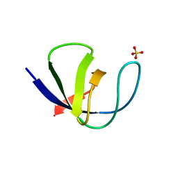
Crystal structure of the c-Src SH3 domain mutant V111L-N113S-T114S at pH 7.0
Descriptor: Proto-oncogene tyrosine-protein kinase Src, SULFATE ION
Authors:Camara-Artigas, A, Plaza-Garrido, M, Salinas-Garcia, M.C.
Deposit date:2020-08-18
Release date:2021-08-25
Last modified:2024-01-31
Method:X-RAY DIFFRACTION (1.314 Å)
Cite:Crystal structure of the c-Src SH3 domain mutant V111L-N113S-T114S at pH 7.0
To be published
7A2U
DownloadVisualize
BU of 7a2u by Molmil
Crystal structure of the Fyn SH3 domain L112V-S114N-S115T at pH 4.5
Descriptor: ACETATE ION, Tyrosine-protein kinase Fyn
Authors:Camara-Artigas, A, Plaza-Garrido, M, Salinas-Garcia, M.C.
Deposit date:2020-08-18
Release date:2021-08-25
Last modified:2024-01-31
Method:X-RAY DIFFRACTION (1.7 Å)
Cite:Crystal structure of the Fyn SH3 domain L112V-S114N-S115T at pH 4.5
To be published
7A34
DownloadVisualize
BU of 7a34 by Molmil
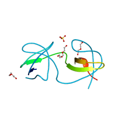
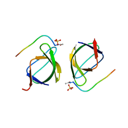
Intertwined dimer of the c-Src SH3 domain mutant V111L-N113S-T114S
Descriptor: DI(HYDROXYETHYL)ETHER, Proto-oncogene tyrosine-protein kinase Src, TRIETHYLENE GLYCOL
Authors:Camara-Artigas, A, Plaza-Garrido, M, Salinas-Garcia, M.C.
Deposit date:2020-08-18
Release date:2021-08-25
Last modified:2024-01-31
Method:X-RAY DIFFRACTION (1.85 Å)
Cite:Intertwined dimer of the c-Src SH3 domain mutant V111L-N113S-T114S
To be published
7A2M
DownloadVisualize
BU of 7a2m by Molmil
Crystal structure of the Fyn SH3 domain A95S-D99T mutant in C21 space group
Descriptor: FORMIC ACID, SODIUM ION, SULFATE ION, ...
Authors:Camara-Artigas, A, Plaza-Garrido, M, Salinas-Garcia, M.C.
Deposit date:2020-08-18
Release date:2021-08-25
Last modified:2024-01-31
Method:X-RAY DIFFRACTION (1.5 Å)
Cite:Crystal structure of the Fyn SH3 domain A95S-D99T mutant in C21 space group
To be published
7A37
DownloadVisualize
BU of 7a37 by Molmil
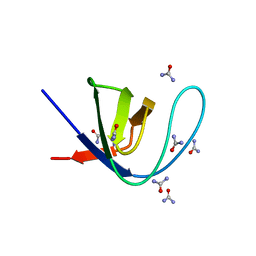
Crystal structure of the c-Src SH3 domain mutant V111L-N113S-T114S in 3 M urea
Descriptor: Proto-oncogene tyrosine-protein kinase Src, UREA
Authors:Camara-Artigas, A, Plaza-Garrido, M, Salinas-Garcia, M.C.
Deposit date:2020-08-18
Release date:2021-08-25
Last modified:2024-01-31
Method:X-RAY DIFFRACTION (1.52 Å)
Cite:Crystal structure of the c-Src SH3 domain mutant V111L-N113S-T114S in 3 M urea
To be published
7A2S
DownloadVisualize
BU of 7a2s by Molmil
Crystal structure of the Fyn SH3 domain L112V-S114N-S115T-E121L-R123H mutant at pH 5.0
Descriptor: Tyrosine-protein kinase Fyn
Authors:Camara-Artigas, A, Plaza-Garrido, M, Salinas-Garcia, M.C.
Deposit date:2020-08-18
Release date:2021-08-25
Last modified:2024-01-31
Method:X-RAY DIFFRACTION (1.02 Å)
Cite:Crystal structure of the Fyn SH3 domain L112V-S114N-S115T-E121L-R123H mutant at pH 5.0
To Be Published
7A2K
DownloadVisualize
BU of 7a2k by Molmil
Crystal structure of the Fyn SH3 domain in space group P1 at pH 7.5
Descriptor: SODIUM ION, Tyrosine-protein kinase Fyn
Authors:Camara-Artigas, A, Plaza-Garrido, M, Salinas-Garcia, M.C.
Deposit date:2020-08-18
Release date:2021-08-25
Last modified:2024-01-31
Method:X-RAY DIFFRACTION (1.5 Å)
Cite:Crystal structure of the Fyn SH3 domain in space group P1 at pH 7.5
To be published
7A3A
DownloadVisualize
BU of 7a3a by Molmil
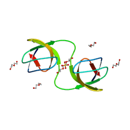
Intertwined dimer of the c-Src SH3 domain mutant V111L-N113S-T114S-Q128E at pH 6.0
Descriptor: 1,2-ETHANEDIOL, DI(HYDROXYETHYL)ETHER, Proto-oncogene tyrosine-protein kinase Src, ...
Authors:Camara-Artigas, A, Plaza-Garrido, M, Salinas-Garcia, M.C.
Deposit date:2020-08-18
Release date:2021-08-25
Last modified:2024-01-31
Method:X-RAY DIFFRACTION (1.8 Å)
Cite:Intertwined dimer of the c-Src SH3 domain mutant V111L-N113S-T114S-Q128E at pH 6.0
To be published
7A36
DownloadVisualize
BU of 7a36 by Molmil
Crystal structure of the c-Src SH3 domain mutant V111L-N113S-T114S in 7 M urea
Descriptor: Proto-oncogene tyrosine-protein kinase Src, SODIUM ION, UREA
Authors:Camara-Artigas, A, Plaza-Garrido, M, Salinas-Garcia, M.C.
Deposit date:2020-08-18
Release date:2021-08-25
Last modified:2024-01-31
Method:X-RAY DIFFRACTION (1.5 Å)
Cite:Crystal structure of the c-Src SH3 domain mutant V111L-N113S-T114S in 7 M urea
To be published
7A2N
DownloadVisualize
BU of 7a2n by Molmil
Crystal structure of the Fyn SH3 domain A95S-D99T mutant in space group P21
Descriptor: FORMIC ACID, SODIUM ION, Tyrosine-protein kinase Fyn
Authors:Camara-Artigas, A, Plaza-Garrido, M, Salinas-Garcia, M.C.
Deposit date:2020-08-18
Release date:2021-08-25
Last modified:2024-01-31
Method:X-RAY DIFFRACTION (1.4 Å)
Cite:Crystal structure of the Fyn SH3 domain A95S-D99T mutant in space group P21
To be published
7A3E
DownloadVisualize
BU of 7a3e by Molmil
Intertwined dimer of the c-Src SH3 domain mutant T126S
Descriptor: DI(HYDROXYETHYL)ETHER, Proto-oncogene tyrosine-protein kinase Src, SULFATE ION, ...
Authors:Camara-Artigas, A, Plaza-Garrido, M, Salinas-Garcia, M.C.
Deposit date:2020-08-18
Release date:2021-08-25
Last modified:2024-01-31
Method:X-RAY DIFFRACTION (1.52 Å)
Cite:Intertwined dimer of the c-Src SH3 domain mutant T126S
To be published
7A2W
DownloadVisualize
BU of 7a2w by Molmil
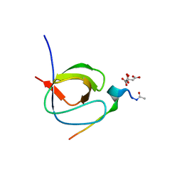
Crystal structure of the Fyn SH3 domain L112V-S114N-S115T-E121L-R123H mutant in complex with VSL12 at pH 3.0
Descriptor: CITRIC ACID, Tyrosine-protein kinase Fyn, VSL12
Authors:Camara-Artigas, A, Plaza-Garrido, M, Salinas-Garcia, M.C.
Deposit date:2020-08-18
Release date:2021-08-25
Last modified:2024-01-31
Method:X-RAY DIFFRACTION (0.99 Å)
Cite:Crystal structure of the Fyn SH3 domain L112V-S114N-S115T-E121L-R123H mutant in complex with VSL12 at pH 3.0
To be published
7A38
DownloadVisualize
BU of 7a38 by Molmil
Crystal structure of the c-Src SH3 domain mutant V111L-N113S-T114S-Q128E at pH 6.0
Descriptor: 2-(N-MORPHOLINO)-ETHANESULFONIC ACID, Proto-oncogene tyrosine-protein kinase Src, SULFATE ION
Authors:Camara-Artigas, A, Plaza-Garrido, M, Salinas-Garcia, M.C.
Deposit date:2020-08-18
Release date:2021-08-25
Last modified:2024-01-31
Method:X-RAY DIFFRACTION (1.62 Å)
Cite:Crystal structure of the c-Src SH3 domain mutant V111L-N113S-T114S-Q128E at pH 6.0
To be published
7NET
DownloadVisualize
BU of 7net by Molmil
Crystal structure of the v-Src SH3 domain W95R-I96T mutant
Descriptor: DI(HYDROXYETHYL)ETHER, SULFATE ION, TRIETHYLENE GLYCOL, ...
Authors:Camara-Artigas, A, Salinas-Garcia, M.C.
Deposit date:2021-02-04
Release date:2021-06-02
Last modified:2024-01-31
Method:X-RAY DIFFRACTION (1.5 Å)
Cite:The impact of oncogenic mutations of the viral Src kinase on the structure and stability of the SH3 domain.
Acta Crystallogr D Struct Biol, 77, 2021
2HDA
DownloadVisualize
BU of 2hda by Molmil
Yes SH3 domain
Descriptor: Proto-oncogene tyrosine-protein kinase Yes, SULFATE ION
Authors:Camara-Artigas, A, Luque, I, Ruiz-Sanz, J, Mateo, P.L, Martin-Garcia, J.M.
Deposit date:2006-06-20
Release date:2007-04-17
Last modified:2023-08-30
Method:X-RAY DIFFRACTION (1.9 Å)
Cite:Crystallographic structure of the SH3 domain of the human c-Yes tyrosine kinase: Loop flexibility and amyloid aggregation.
Febs Lett., 581, 2007
6R2G
DownloadVisualize
BU of 6r2g by Molmil
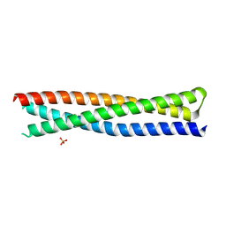
Crystal structure of a single-chain protein mimetic of the gp41 NHR trimer in complex with the synthetic CHR peptide C34
Descriptor: Envelope glycoprotein gp160, PHOSPHATE ION, Single-chain protein mimetics of the N-terminal heptad-repeat region of gp41
Authors:Camara-Artigas, A, Conejero-Lara, F, Jurado, S, Cano-Munoz, M, Morel, B.
Deposit date:2019-03-17
Release date:2019-07-10
Last modified:2024-01-24
Method:X-RAY DIFFRACTION (1.9 Å)
Cite:Structural and Thermodynamic Analysis of HIV-1 Fusion Inhibition Using Small gp41 Mimetic Proteins.
J.Mol.Biol., 431, 2019
3EG2
DownloadVisualize
BU of 3eg2 by Molmil
Crystal structure of the N114Q mutant of ABL-SH3 domain
Descriptor: GLYCEROL, Proto-oncogene tyrosine-protein kinase ABL1
Authors:Camara-Artigas, A.
Deposit date:2008-09-10
Release date:2009-09-15
Last modified:2023-08-30
Method:X-RAY DIFFRACTION (1.8 Å)
Cite:Role of interfacial water molecules in proline-rich ligand recognition by the Src homology 3 domain of Abl.
J.Biol.Chem., 285, 2010
3EG1
DownloadVisualize
BU of 3eg1 by Molmil
Crystal structure of the N114Q mutant of ABL-SH3 domain complexed with a designed high-affinity peptide ligand: implications for SH3-ligand interactions
Descriptor: Proto-oncogene tyrosine-protein kinase ABL1, SULFATE ION, p41 peptide
Authors:Camara-Artigas, A.
Deposit date:2008-09-10
Release date:2009-09-15
Last modified:2023-08-30
Method:X-RAY DIFFRACTION (1.85 Å)
Cite:Role of interfacial water molecules in proline-rich ligand recognition by the Src homology 3 domain of Abl.
J.Biol.Chem., 285, 2010
3EGU
DownloadVisualize
BU of 3egu by Molmil
Crystal structure of the N114A mutant of ABL-SH3 domain
Descriptor: GLYCEROL, Proto-oncogene tyrosine-protein kinase ABL1, SULFATE ION
Authors:Camara-Artigas, A.
Deposit date:2008-09-11
Release date:2009-09-15
Last modified:2024-02-21
Method:X-RAY DIFFRACTION (2.25 Å)
Cite:Role of interfacial water molecules in proline-rich ligand recognition by the Src homology 3 domain of Abl.
J.Biol.Chem., 285, 2010
3EG3
DownloadVisualize
BU of 3eg3 by Molmil
Crystal structure of the N114A mutant of ABL-SH3 domain
Descriptor: GLYCEROL, Proto-oncogene tyrosine-protein kinase ABL1
Authors:Camara-Artigas, A.
Deposit date:2008-09-10
Release date:2009-09-15
Last modified:2023-08-30
Method:X-RAY DIFFRACTION (1.4 Å)
Cite:Role of interfacial water molecules in proline-rich ligand recognition by the Src homology 3 domain of Abl.
J.Biol.Chem., 285, 2010
3EG0
DownloadVisualize
BU of 3eg0 by Molmil
Crystal structure of the N114T mutant of ABL-SH3 domain
Descriptor: GLYCEROL, Proto-oncogene tyrosine-protein kinase ABL1
Authors:Camara-Artigas, A.
Deposit date:2008-09-10
Release date:2009-09-15
Last modified:2023-08-30
Method:X-RAY DIFFRACTION (2.3 Å)
Cite:Role of interfacial water molecules in proline-rich ligand recognition by the Src homology 3 domain of Abl.
J.Biol.Chem., 285, 2010
2F0R
DownloadVisualize
BU of 2f0r by Molmil
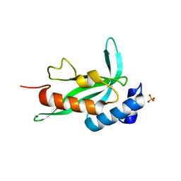
Crystallographic structure of human Tsg101 UEV domain
Descriptor: SULFATE ION, Tumor susceptibility gene 101 protein
Authors:Camara-Artigas, A, Luque, I, Palencia, A, Martinez, J.C, Mateo, P.L.
Deposit date:2005-11-13
Release date:2006-03-28
Last modified:2023-08-23
Method:X-RAY DIFFRACTION (2.26 Å)
Cite:Structure of human TSG101 UEV domain.
Acta Crystallogr.,Sect.D, 62, 2006

220472

数据于2024-05-29公开中

PDB statisticsPDBj update infoContact PDBjnumon