Loading
PDBj
MenuPDBj@FacebookPDBj@X(formerly Twitter)PDBj@BlueSkyPDBj@YouTubewwPDB FoundationwwPDBDonate
RCSB PDBPDBeBMRBAdv. SearchSearch help
Search by PDB author
1VR0
DownloadVisualize
BU of 1vr0 by Molmil
Crystal structure of putative 2-phosphosulfolactate phosphatase (15026306) from Clostridium acetobutylicum at 2.6 A resolution
Descriptor: (2R)-3-SULFOLACTIC ACID, MAGNESIUM ION, Probable 2-phosphosulfolactate phosphatase
Authors:Joint Center for Structural Genomics (JCSG)
Deposit date:2005-01-26
Release date:2005-02-15
Last modified:2024-10-09
Method:X-RAY DIFFRACTION (2.49 Å)
Cite:Crystal structure of 2-phosphosulfolactate phosphatase (ComB) from Clostridium acetobutylicum at 2.6 A resolution reveals a new fold with a novel active site.
Proteins, 65, 2006
1VJL
DownloadVisualize
BU of 1vjl by Molmil
Crystal structure of a duf151 family protein (tm0160) from thermotoga maritima at 1.90 A resolution
Descriptor: CHLORIDE ION, UNKNOWN LIGAND, hypothetical protein TM0160
Authors:Joint Center for Structural Genomics, Joint Center for Structural Genomics (JCSG)
Deposit date:2004-03-10
Release date:2004-03-16
Last modified:2024-10-09
Method:X-RAY DIFFRACTION (1.9 Å)
Cite:On the use of DXMS to produce more crystallizable proteins: structures of the T. maritima proteins TM0160 and TM1171.
Protein Sci., 13, 2004
1VQR
DownloadVisualize
BU of 1vqr by Molmil
Crystal structure of a virulence factor (cj0248) from campylobacter jejuni subsp. jejuni at 2.25 A resolution
Descriptor: hypothetical protein Cj0248
Authors:Joint Center for Structural Genomics (JCSG)
Deposit date:2004-12-17
Release date:2004-12-28
Last modified:2024-10-09
Method:X-RAY DIFFRACTION (2.25 Å)
Cite:Crystal structure of virulence factor CJ0248 from Campylobacter jejuni at 2.25 A resolution reveals a new fold.
Proteins, 62, 2006
1VR8
DownloadVisualize
BU of 1vr8 by Molmil
Crystal structure of GTP binding regulator (TM1622) from Thermotoga Maritima at 1.75 A resolution
Descriptor: AZIDE ION, GLYCEROL, GTP binding regulator
Authors:Joint Center for Structural Genomics (JCSG)
Deposit date:2005-02-16
Release date:2005-03-15
Last modified:2024-11-13
Method:X-RAY DIFFRACTION (1.75 Å)
Cite:Crystal structure of an ORFan protein (TM1622) from Thermotoga maritima at 1.75 A resolution reveals a fold similar to the Ran-binding protein Mog1p.
Proteins, 65, 2006
1VK3
DownloadVisualize
BU of 1vk3 by Molmil
Crystal structure of Phosphoribosylformylglycinamidine synthase II (TM1246) from Thermotoga maritima at 2.15 A resolution
Descriptor: CHLORIDE ION, Phosphoribosylformylglycinamidine synthase II
Authors:Joint Center for Structural Genomics (JCSG)
Deposit date:2004-04-23
Release date:2004-05-11
Last modified:2024-11-20
Method:X-RAY DIFFRACTION (2.15 Å)
Cite:Crystal structure of phosphoribosylformylglycinamidine synthase II (smPurL) from Thermotoga maritima at 2.15 A resolution.
Proteins, 63, 2006
1VPZ
DownloadVisualize
BU of 1vpz by Molmil
Crystal structure of a putative carbon storage regulator protein (csra, pa0905) from pseudomonas aeruginosa at 2.05 A resolution
Descriptor: Carbon storage regulator homolog
Authors:Joint Center for Structural Genomics (JCSG)
Deposit date:2004-11-24
Release date:2004-12-14
Last modified:2024-11-13
Method:X-RAY DIFFRACTION (2.05 Å)
Cite:Crystal structure of the global regulatory protein CsrA from Pseudomonas putida at 2.05 A resolution reveals a new fold.
Proteins, 61, 2005
1VL4
DownloadVisualize
BU of 1vl4 by Molmil
CRYSTAL STRUCTURE OF A PUTATIVE MODULATOR OF A DNA GYRASE (TM0727) FROM THERMOTOGA MARITIMA MSB8 AT 1.95 A RESOLUTION
Descriptor: pmbA-related protein
Authors:Joint Center for Structural Genomics (JCSG)
Deposit date:2004-07-09
Release date:2004-09-21
Last modified:2024-05-22
Method:X-RAY DIFFRACTION (1.95 Å)
Cite:Crystal structure of a putative modulator of DNA gyrase (pmbA) from Thermotoga maritima at 1.95 A resolution reveals a new fold.
Proteins, 61, 2005
2JZF
DownloadVisualize
BU of 2jzf by Molmil
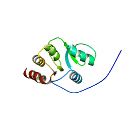
NMR Conformer closest to the mean coordinates of the domain 513-651 of the SARS-CoV nonstructural protein nsp3
Descriptor: Replicase polyprotein 1ab
Authors:Chatterjee, A, Johnson, M.A, Serrano, P, Pedrini, B, Joseph, J, Saikatendu, K, Neuman, B, Stevens, R.C, Wilson, I.A, Buchmeier, M.J, Kuhn, P, Wuthrich, K, Joint Center for Structural Genomics (JCSG)
Deposit date:2008-01-04
Release date:2008-02-05
Last modified:2024-05-29
Method:SOLUTION NMR
Cite:Nuclear magnetic resonance structure shows that the severe acute respiratory syndrome coronavirus-unique domain contains a macrodomain fold.
J.Virol., 83, 2009
1UWD
DownloadVisualize
BU of 1uwd by Molmil
NMR STRUCTURE OF A PROTEIN WITH UNKNOWN FUNCTION FROM THERMOTOGA MARITIMA (TM0487), WHICH BELONGS TO THE DUF59 FAMILY.
Descriptor: HYPOTHETICAL PROTEIN TM0487
Authors:Almeida, M.S, Peti, W, Herrmann, T, Wuthrich, K.
Deposit date:2004-02-03
Release date:2004-12-14
Last modified:2024-05-15
Method:SOLUTION NMR
Cite:NMR Structure of the Conserved Hypothetical Protein Tm0487 from Thermotoga Maritima: Implications for 216 Homologous Duf59 Proteins.
Protein Sci., 14, 2005
2KAF
DownloadVisualize
BU of 2kaf by Molmil
Solution structure of the SARS-unique domain-C from the nonstructural protein 3 (nsp3) of the severe acute respiratory syndrome coronavirus
Descriptor: Non-structural protein 3
Authors:Johnson, M.A, Mohanty, B, Pedrini, B, Serrano, P, Chatterjee, A, Herrmann, T, Joseph, J, Saikatendu, K, Wilson, I.A, Buchmeier, M.J, Kuhn, P, Wuthrich, K, Joint Center for Structural Genomics (JCSG)
Deposit date:2008-11-05
Release date:2008-11-25
Last modified:2024-05-01
Method:SOLUTION NMR
Cite:SARS coronavirus unique domain: three-domain molecular architecture in solution and RNA binding.
J.Mol.Biol., 400, 2010
2KYS
DownloadVisualize
BU of 2kys by Molmil
NMR Structure of the SARS Coronavirus Nonstructural Protein Nsp7 in Solution at pH 6.5
Descriptor: Non-structural protein 7
Authors:Johnson, M.A, Jaudzems, K, Wilson, I.A, Wuthrich, K, Joint Center for Structural Genomics (JCSG)
Deposit date:2010-06-07
Release date:2010-06-16
Last modified:2024-05-15
Method:SOLUTION NMR
Cite:NMR Structure of the SARS-CoV Nonstructural Protein 7 in Solution at pH 6.5.
J.Mol.Biol., 402, 2010
3SXK
DownloadVisualize
BU of 3sxk by Molmil
Zn2+-bound FCD domain of TM0439, a putative transcriptional regulator
Descriptor: BICARBONATE ION, SULFATE ION, Transcriptional regulator, ...
Authors:Czelakowski, G.P, Derewenda, Z.S, Integrated Center for Structure and Function Innovation (ISFI)
Deposit date:2011-07-15
Release date:2011-11-16
Last modified:2023-09-13
Method:X-RAY DIFFRACTION (1.63 Å)
Cite:Zn2+-bound FCD domain of TM0439, a putative transcriptional regulator
To be Published
3SXM
DownloadVisualize
BU of 3sxm by Molmil
Metal-free FCD domain of TM0439 a putative transcriptional regulator
Descriptor: BICARBONATE ION, Transcriptional regulator, GntR family
Authors:Czelakowski, G.P, Derewenda, Z.S, Integrated Center for Structure and Function Innovation (ISFI)
Deposit date:2011-07-15
Release date:2011-11-16
Last modified:2023-09-13
Method:X-RAY DIFFRACTION (1.55 Å)
Cite:Metal-free FCD domain of TM0439 a putative transcriptional regulator
To be Published
3SXY
DownloadVisualize
BU of 3sxy by Molmil
Metal-free full-length structure of Tm0439, a metal-binding FCD family transcriptional regulator
Descriptor: Transcriptional regulator, GntR family
Authors:Czelakowski, G.P, Derewenda, Z.S, Integrated Center for Structure and Function Innovation (ISFI)
Deposit date:2011-07-15
Release date:2011-11-16
Last modified:2023-09-13
Method:X-RAY DIFFRACTION (1.647 Å)
Cite:Metal-free full-length structure of Tm0439, a metal-binding FCD family transcriptional regulator
To be Published
3SXZ
DownloadVisualize
BU of 3sxz by Molmil
Metal-free FCD domain of TM0439 a putative transcriptional regulator
Descriptor: Transcriptional regulator, GntR family
Authors:Czelakowski, G.P, Derewenda, Z.S, Integrated Center for Structure and Function Innovation (ISFI)
Deposit date:2011-07-15
Release date:2011-11-16
Last modified:2023-09-13
Method:X-RAY DIFFRACTION (2.322 Å)
Cite:Metal-free FCD domain of TM0439 a putative transcriptional regulator
To be Published
9C3G
DownloadVisualize
BU of 9c3g by Molmil
human cGAS core domain (K427E/K428E) bound to Cladophorol A
Descriptor: Cyclic GMP-AMP synthase, ZINC ION, cladophorol A
Authors:Kissai, M, Stanfield, R.L, Lairson, L.L.
Deposit date:2024-05-31
Release date:2025-03-05
Method:X-RAY DIFFRACTION (2.75 Å)
Cite:Cladophorol-A is an inhibitor of cyclic GMP-AMP synthase.
Bioorg.Med.Chem.Lett., 115, 2025
2ETS
DownloadVisualize
BU of 2ets by Molmil
CRYSTAL STRUCTURE OF A BACTERIAL DOMAIN OF UNKNOWN FUNCTION FROM DUF1798 FAMILY (MW1337) FROM STAPHYLOCOCCUS AUREUS SUBSP. AUREUS AT 2.25 A RESOLUTION
Descriptor: CHLORIDE ION, PHOSPHATE ION, hypothetical protein
Authors:Joint Center for Structural Genomics (JCSG)
Deposit date:2005-10-27
Release date:2005-11-08
Last modified:2024-10-30
Method:X-RAY DIFFRACTION (2.25 Å)
Cite:Crystal structures of MW1337R and lin2004: representatives of a novel protein family that adopt a four-helical bundle fold.
Proteins, 71, 2008
2FNA
DownloadVisualize
BU of 2fna by Molmil
Crystal structure of an archaeal aaa+ atpase (sso1545) from sulfolobus solfataricus p2 at 2.00 A resolution
Descriptor: 1,2-ETHANEDIOL, ADENOSINE-5'-DIPHOSPHATE, Conserved hypothetical protein, ...
Authors:Joint Center for Structural Genomics (JCSG)
Deposit date:2006-01-10
Release date:2006-02-07
Last modified:2024-10-09
Method:X-RAY DIFFRACTION (2 Å)
Cite:Crystal structure of a novel archaeal AAA+ ATPase SSO1545 from Sulfolobus solfataricus.
Proteins, 74, 2009
2GAZ
DownloadVisualize
BU of 2gaz by Molmil
Mycobacterial lipoglycan presentation by CD1d
Descriptor: (2R)-3-[(HYDROXY{[(2R,3R,5S,6R)-3,4,5-TRIHYDROXY-2,6-BIS(ALPHA-D-MANNOPYRANOSYLOXY)CYCLOHEXYL]OXY}PHOSPHORYL)OXY]PROPAN E-1,2-DIYL DIHEXADECANOATE, 2-acetamido-2-deoxy-beta-D-glucopyranose, T-cell surface glycoprotein CD1d1, ...
Authors:Zajonc, D.M.
Deposit date:2006-03-09
Release date:2006-09-26
Last modified:2024-10-30
Method:X-RAY DIFFRACTION (2.61 Å)
Cite:Structural characterization of mycobacterial phosphatidylinositol mannoside binding to mouse CD1d.
J.Immunol., 177, 2006
2FIK
DownloadVisualize
BU of 2fik by Molmil
Structure of a microbial glycosphingolipid bound to mouse CD1d
Descriptor: (2S,3R)-3-HYDROXY-2-(TETRADECANOYLAMINO)OCTADECYL ALPHA-D-GALACTOPYRANOSIDURONIC ACID, 2-acetamido-2-deoxy-beta-D-glucopyranose, 2-acetamido-2-deoxy-beta-D-glucopyranose-(1-4)-2-acetamido-2-deoxy-beta-D-glucopyranose, ...
Authors:Wu, D, Zajonc, D.M.
Deposit date:2005-12-29
Release date:2006-03-21
Last modified:2024-11-13
Method:X-RAY DIFFRACTION (1.8 Å)
Cite:Design of natural killer T cell activators: structure and function of a microbial glycosphingolipid bound to mouse CD1d.
Proc.Natl.Acad.Sci.Usa, 103, 2006
2G36
DownloadVisualize
BU of 2g36 by Molmil
Crystal structure of Tryptophanyl-tRNA synthetase (EC 6.1.1.2) (Tryptophan-tRNA ligase)(TrpRS) (tm0492) from THERMOTOGA MARITIMA at 2.50 A resolution
Descriptor: IRON/SULFUR CLUSTER, TRYPTOPHAN, Tryptophanyl-tRNA synthetase
Authors:Joint Center for Structural Genomics (JCSG)
Deposit date:2006-02-17
Release date:2006-03-28
Last modified:2024-04-03
Method:X-RAY DIFFRACTION (2.5 Å)
Cite:Structure of a tryptophanyl-tRNA synthetase containing an iron-sulfur cluster.
Acta Crystallogr.,Sect.F, 66, 2010
2FG0
DownloadVisualize
BU of 2fg0 by Molmil
Crystal structure of a putative gamma-d-glutamyl-l-diamino acid endopeptidase (npun_r0659) from nostoc punctiforme pcc 73102 at 1.79 A resolution
Descriptor: COG0791: Cell wall-associated hydrolases (invasion-associated proteins), GLYCEROL
Authors:Joint Center for Structural Genomics (JCSG)
Deposit date:2005-12-20
Release date:2006-01-10
Last modified:2024-11-13
Method:X-RAY DIFFRACTION (1.79 Å)
Cite:Structural Basis of Murein Peptide Specificity of a gamma-D-Glutamyl-L-Diamino Acid Endopeptidase.
Structure, 17, 2009
2FEA
DownloadVisualize
BU of 2fea by Molmil
Crystal structure of MtnX phosphatase from Bacillus Subtilis at 2.00 A resolution
Descriptor: 1,2-ETHANEDIOL, 2-hydroxy-3-keto-5-methylthiopentenyl-1-phosphate phosphatase, MAGNESIUM ION, ...
Authors:Joint Center for Structural Genomics (JCSG)
Deposit date:2005-12-15
Release date:2005-12-27
Last modified:2024-11-06
Method:X-RAY DIFFRACTION (2 Å)
Cite:Crystal structure of MtnX phosphatase from Bacillus subtilis at 2.0 A resolution provides a structural basis for bipartite phosphomonoester hydrolysis of 2-hydroxy-3-keto-5-methylthiopentenyl-1-phosphate.
Proteins, 69, 2007
2F46
DownloadVisualize
BU of 2f46 by Molmil
Crystal structure of a putative phosphatase (nma1982) from neisseria meningitidis z2491 at 1.41 A resolution
Descriptor: CHLORIDE ION, UNKNOWN LIGAND, hypothetical protein
Authors:Joint Center for Structural Genomics (JCSG)
Deposit date:2005-11-22
Release date:2006-02-07
Last modified:2024-10-30
Method:X-RAY DIFFRACTION (1.41 Å)
Cite:Crystal structure of NMA1982 from Neisseria meningitidis at 1.5 A resolution provides a structural scaffold for nonclassical, eukaryotic-like phosphatases.
Proteins, 69, 2007
2EVR
DownloadVisualize
BU of 2evr by Molmil
CRYSTAL STRUCTURE OF A PUTATIVE GAMMA-D-GLUTAMYL-L-DIAMINO ACID ENDOPEPTIDASE (NPUN_R0659) FROM NOSTOC PUNCTIFORME PCC 73102 AT 1.60 A RESOLUTION
Descriptor: 1,2-ETHANEDIOL, CHLORIDE ION, COG0791: Cell wall-associated hydrolases (invasion-associated proteins), ...
Authors:Joint Center for Structural Genomics (JCSG)
Deposit date:2005-10-31
Release date:2005-11-22
Last modified:2024-10-16
Method:X-RAY DIFFRACTION (1.6 Å)
Cite:Structural Basis of Murein Peptide Specificity of a gamma-D-Glutamyl-L-Diamino Acid Endopeptidase.
Structure, 17, 2009

238582

数据于2025-07-09公开中

PDB statisticsPDBj update infoContact PDBjnumon