Loading
PDBj
MenuPDBj@FacebookPDBj@TwitterPDBj@YouTubewwPDB FoundationwwPDB
RCSB PDBPDBeBMRBAdv. SearchSearch help
Search by PDB author
2NAF
DownloadVisualize
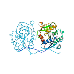
BU of 2naf by Molmil
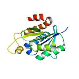
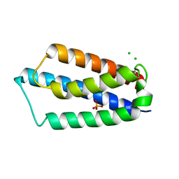
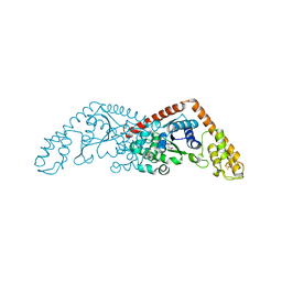
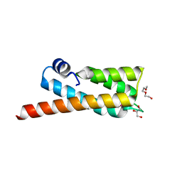
Solution structure of peptidyl-tRNA hydrolase from Mycobacterium smegmatis
Descriptor: Peptidyl-tRNA hydrolase
Authors:Yadav, R, Pathak, P, Fatma, F, Kabra, A, Pulavarti, S, Jain, A, Kumar, A, Shukla, V, Arora, A.
Deposit date:2015-12-23
Release date:2017-01-11
Last modified:2024-05-15
Method:SOLUTION NMR
Cite:Structural characterization of peptidyl-tRNA hydrolase from Mycobacterium smegmatis by NMR spectroscopy.
Biochim.Biophys.Acta, 1864, 2016
2MAU
DownloadVisualize
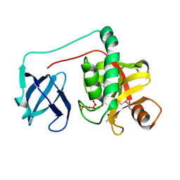
BU of 2mau by Molmil
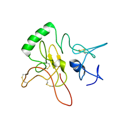
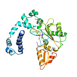
Solution structure of alpha-amylase inhibitor wrightide R1 (wR1) peptide from Wrightia religiosa
Descriptor: Wrightide R1
Authors:Wang, S, Nguyen, Q, Tam, J.
Deposit date:2013-07-17
Release date:2014-07-23
Last modified:2019-12-25
Method:SOLUTION NMR
Cite:Discovery and characterization of pseudocyclic cystine-knot alpha-amylase inhibitors with high resistance to heat and proteolytic degradation.
Febs J., 281, 2014
6A8M
DownloadVisualize
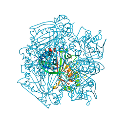
BU of 6a8m by Molmil
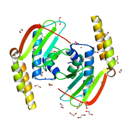
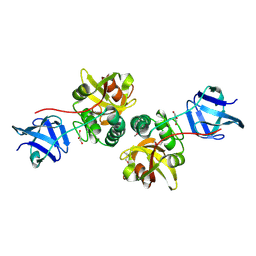
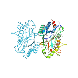
N-terminal domain of FACT complex subunit SPT16 from Eremothecium gossypii (Ashbya gossypii)
Descriptor: FACT complex subunit SPT16
Authors:Gaur, N.K, Are, V.N, Durani, V, Ghosh, B, Kumar, A, Kulkarni, K, Makde, R.D.
Deposit date:2018-07-09
Release date:2018-08-15
Last modified:2023-11-22
Method:X-RAY DIFFRACTION (1.7 Å)
Cite:Evolutionary conservation of protein dynamics: insights from all-atom molecular dynamics simulations of 'peptidase' domain of Spt16.
J.Biomol.Struct.Dyn., 2021
1UNE
DownloadVisualize
BU of 1une by Molmil
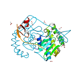
CARBOXYLIC ESTER HYDROLASE, 1.5 ANGSTROM ORTHORHOMBIC FORM OF THE BOVINE RECOMBINANT PLA2
Descriptor: CALCIUM ION, PHOSPHOLIPASE A2
Authors:Sundaralingam, M.
Deposit date:1997-11-05
Release date:1998-05-06
Last modified:2023-08-09
Method:X-RAY DIFFRACTION (1.5 Å)
Cite:High-resolution refinement of orthorhombic bovine pancreatic phospholipase A2.
Acta Crystallogr.,Sect.D, 55, 1999
1KVW
DownloadVisualize
BU of 1kvw by Molmil
CARBOXYLIC ESTER HYDROLASE, SINGLE MUTANT H48Q OF BOVINE PANCREATIC PLA2 ENZYME
Descriptor: CALCIUM ION, PHOSPHOLIPASE A2
Authors:Sundaralingam, M.
Deposit date:1998-04-24
Release date:1998-11-18
Last modified:2023-08-09
Method:X-RAY DIFFRACTION (1.95 Å)
Cite:Structures of the catalytic site mutants D99A and H48Q and the calcium-loop mutant D49E of phospholipase A2.
Acta Crystallogr.,Sect.D, 55, 1999
1KVX
DownloadVisualize
BU of 1kvx by Molmil
CARBOXYLIC ESTER HYDROLASE, SINGLE MUTANT D99A OF BOVINE PANCREATIC PLA2, 1.9 A ORTHORHOMBIC FORM
Descriptor: CALCIUM ION, PHOSPHOLIPASE A2
Authors:Sundaralingam, M.
Deposit date:1998-04-28
Release date:1998-11-18
Last modified:2023-08-09
Method:X-RAY DIFFRACTION (1.9 Å)
Cite:Structures of the catalytic site mutants D99A and H48Q and the calcium-loop mutant D49E of phospholipase A2.
Acta Crystallogr.,Sect.D, 55, 1999
1KVY
DownloadVisualize
BU of 1kvy by Molmil
CARBOXYLIC ESTER HYDROLASE, SINGLE MUTANT D49E COORDINATED TO CALCIUM
Descriptor: CALCIUM ION, PHOSPHOLIPASE A2
Authors:Sundaralingam, M.
Deposit date:1998-04-29
Release date:1998-11-18
Last modified:2023-08-09
Method:X-RAY DIFFRACTION (1.9 Å)
Cite:Structures of the catalytic site mutants D99A and H48Q and the calcium-loop mutant D49E of phospholipase A2.
Acta Crystallogr.,Sect.D, 55, 1999
1MKT
DownloadVisualize
BU of 1mkt by Molmil
CARBOXYLIC ESTER HYDROLASE, 1.72 ANGSTROM TRIGONAL FORM OF THE BOVINE RECOMBINANT PLA2 ENZYME
Descriptor: CALCIUM ION, PHOSPHOLIPASE A2
Authors:Sundaralingam, M.
Deposit date:1997-09-06
Release date:1998-03-11
Last modified:2023-08-09
Method:X-RAY DIFFRACTION (1.72 Å)
Cite:1.72 A resolution refinement of the trigonal form of bovine pancreatic phospholipase A2.
Acta Crystallogr.,Sect.D, 54, 1998
6B4M
DownloadVisualize
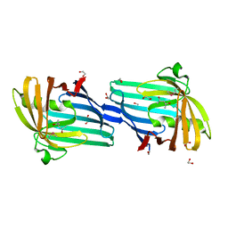
BU of 6b4m by Molmil
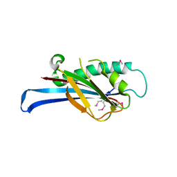
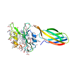
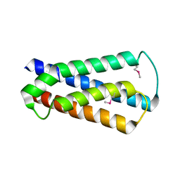
Structural characterization of a novel monotreme-specific protein from the milk of the platypus
Descriptor: 2-acetamido-2-deoxy-beta-D-glucopyranose, GLYCEROL, IODIDE ION, ...
Authors:Peat, T.S, Newman, J, Sharp, J.A, Kumar, A, Nicholas, K.R, Adams, T.E.
Deposit date:2017-09-27
Release date:2018-01-17
Last modified:2023-10-04
Method:X-RAY DIFFRACTION (2.5 Å)
Cite:Structural characterization of a novel monotreme-specific protein with antimicrobial activity from the milk of the platypus.
Acta Crystallogr F Struct Biol Commun, 74, 2018
3SE0
DownloadVisualize
BU of 3se0 by Molmil
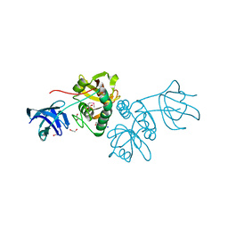
Structural characterization of the subunit A mutant F508W of the A-ATP synthase from Pyrococcus horikoshii
Descriptor: (4S)-2-METHYL-2,4-PENTANEDIOL, 2-AMINO-2-HYDROXYMETHYL-PROPANE-1,3-DIOL, ACETIC ACID, ...
Authors:Tadwal, V.S, Manimekalai, M.S.S, Balakrishna, A.M, Gruber, G.
Deposit date:2011-06-09
Release date:2012-01-25
Last modified:2023-11-01
Method:X-RAY DIFFRACTION (2.62 Å)
Cite:Engineered tryptophan in the adenine-binding pocket of catalytic subunit A of A-ATP synthase demonstrates the importance of aromatic residues in adenine binding, forming a tool for steady-state and time-resolved fluorescence spectroscopy.
Acta Crystallogr.,Sect.F, 67, 2011
227D
DownloadVisualize
BU of 227d by Molmil
A CRYSTALLOGRAPHIC AND SPECTROSCOPIC STUDY OF THE COMPLEX BETWEEN D(CGCGAATTCGCG)2 AND 2,5-BIS(4-GUANYLPHENYL)FURAN, AN ANALOGUE OF BERENIL. STRUCTURAL ORIGINS OF ENHANCED DNA-BINDING AFFINITY
Descriptor: 2,5-BIS(4-GUANYLPHENYL)FURAN, DNA (5'-D(*CP*GP*CP*GP*AP*AP*TP*TP*CP*GP*CP*G)-3')
Authors:Laughton, C.A, Tanious, F, Nunn, C.M, Boykin, D.W, Wilson, W.D, Neidle, S.
Deposit date:1995-08-08
Release date:1995-11-11
Last modified:2024-02-14
Method:X-RAY DIFFRACTION (2.2 Å)
Cite:A crystallographic and spectroscopic study of the complex between d(CGCGAATTCGCG)2 and 2,5-bis(4-guanylphenyl)furan, an analogue of berenil. Structural origins of enhanced DNA-binding affinity.
Biochemistry, 35, 1996
3SDZ
DownloadVisualize
BU of 3sdz by Molmil
Structural characterization of the subunit A mutant F427W of the A-ATP synthase from Pyrococcus horikoshii
Descriptor: (4S)-2-METHYL-2,4-PENTANEDIOL, 2-AMINO-2-HYDROXYMETHYL-PROPANE-1,3-DIOL, ACETIC ACID, ...
Authors:Tadwal, V.S, Manimekalai, M.S.S, Balakrishna, A.M, Gruber, G.
Deposit date:2011-06-09
Release date:2012-01-25
Last modified:2023-11-01
Method:X-RAY DIFFRACTION (2.53 Å)
Cite:Engineered tryptophan in the adenine-binding pocket of catalytic subunit A of A-ATP synthase demonstrates the importance of aromatic residues in adenine binding, forming a tool for steady-state and time-resolved fluorescence spectroscopy.
Acta Crystallogr.,Sect.F, 67, 2011

221051

数据于2024-06-12公开中

PDB statisticsPDBj update infoContact PDBjnumon