Loading
PDBj
MenuPDBj@FacebookPDBj@TwitterPDBj@YouTubewwPDB FoundationwwPDB
RCSB PDBPDBeBMRBAdv. SearchSearch help
Search by PDB author
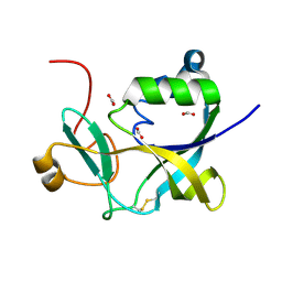
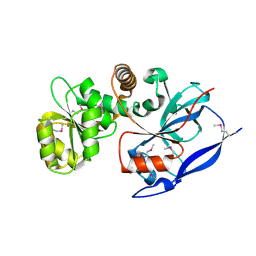
1Z9F
DownloadVisualize
BU of 1z9f by Molmil
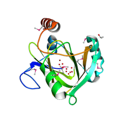
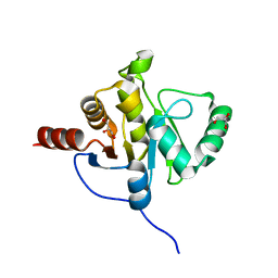
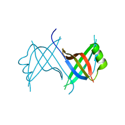
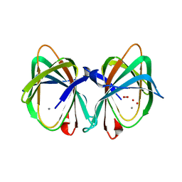
Crystal structure of single stranded DNA-binding protein (TM0604) from Thermotoga maritima at 2.60 A resolution
Descriptor: Single-strand binding protein
Authors:Joint Center for Structural Genomics (JCSG)
Deposit date:2005-04-01
Release date:2005-04-12
Last modified:2023-09-20
Method:X-RAY DIFFRACTION (2.3 Å)
Cite:Crystal structure of a single-stranded DNA-binding protein (TM0604) from Thermotoga maritima at 2.60 A resolution.
Proteins, 63, 2006
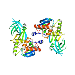
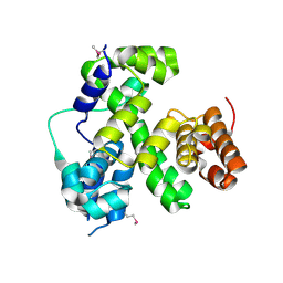
1VJ2
DownloadVisualize
BU of 1vj2 by Molmil
Crystal structure of a novel family of manganese-containing cupin (tm1459) from thermotoga maritima at 1.65 A resolution
Descriptor: MANGANESE (II) ION, UNKNOWN LIGAND, novel manganese-containing cupin TM1459
Authors:Joint Center for Structural Genomics (JCSG)
Deposit date:2003-12-03
Release date:2003-12-09
Last modified:2023-09-20
Method:X-RAY DIFFRACTION (1.65 Å)
Cite:Crystal structure of a novel manganese-containing cupin (TM1459) from Thermotoga maritima at 1.65 A resolution.
Proteins, 56, 2004
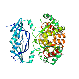
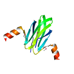
1VKB
DownloadVisualize
BU of 1vkb by Molmil
Crystal structure of an aig2-like protein (a2ld1, ggact, mgc7867) from mus musculus at 1.90 A resolution
Descriptor: FORMIC ACID, hypothetical protein
Authors:Joint Center for Structural Genomics (JCSG)
Deposit date:2004-05-07
Release date:2004-05-18
Last modified:2023-01-25
Method:X-RAY DIFFRACTION (1.9 Å)
Cite:Crystal structure of a conserved hypothetical protein (gi: 13879369) from Mouse at 1.90 A resolution reveals a new fold.
Proteins, 61, 2005
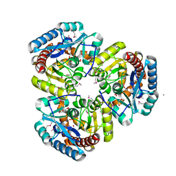
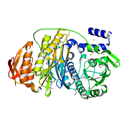
1VKY
DownloadVisualize
BU of 1vky by Molmil
Crystal structure of S-adenosylmethionine tRNA ribosyltransferase (TM0574) from Thermotoga maritima at 2.00 A resolution
Descriptor: S-adenosylmethionine:tRNA ribosyltransferase-isomerase, UNKNOWN LIGAND
Authors:Joint Center for Structural Genomics (JCSG)
Deposit date:2004-06-28
Release date:2004-08-17
Last modified:2024-05-22
Method:X-RAY DIFFRACTION (2 Å)
Cite:Crystal structure of S-adenosylmethionine:tRNA ribosyltransferase-isomerase (QueA) from Thermotoga maritima at 2.0 A resolution reveals a new fold.
Proteins, 59, 2005
1VLR
DownloadVisualize
BU of 1vlr by Molmil
Crystal structure of mRNA decapping enzyme (DcpS) from Mus musculus at 1.83 A resolution
Descriptor: 1,2-ETHANEDIOL, mRNA decapping enzyme
Authors:Joint Center for Structural Genomics (JCSG)
Deposit date:2004-08-10
Release date:2004-08-24
Last modified:2023-09-20
Method:X-RAY DIFFRACTION (1.83 Å)
Cite:Crystal structure of an Apo mRNA decapping enzyme (DcpS) from Mouse at 1.83 A resolution.
Proteins, 60, 2005
1VKM
DownloadVisualize
BU of 1vkm by Molmil
Crystal structure of an indigoidine synthase a (idga)-like protein (tm1464) from thermotoga maritima msb8 at 1.90 A resolution
Descriptor: 1,2-ETHANEDIOL, MANGANESE (II) ION, UNKNOWN LIGAND, ...
Authors:Joint Center for Structural Genomics (JCSG)
Deposit date:2004-06-09
Release date:2004-06-29
Last modified:2023-01-25
Method:X-RAY DIFFRACTION (1.9 Å)
Cite:Crystal structure of an indigoidine synthase A (IndA)-like protein (TM1464) from Thermotoga maritima at 1.90 A resolution reveals a new fold.
Proteins, 59, 2005
1VJ1
DownloadVisualize
BU of 1vj1 by Molmil
Crystal structure of putative NADPH-dependent oxidoreductase from Mus musculus at 2.10 A resolution
Descriptor: putative NADPH-dependent oxidoreductase
Authors:Joint Center for Structural Genomics (JCSG)
Deposit date:2003-12-03
Release date:2003-12-09
Last modified:2023-01-25
Method:X-RAY DIFFRACTION (2.1 Å)
Cite:Crystal structure of a putative NADPH-dependent oxidoreductase (GI: 18204011) from mouse at 2.10 A resolution
Proteins, 56, 2004
1VQR
DownloadVisualize
BU of 1vqr by Molmil
Crystal structure of a virulence factor (cj0248) from campylobacter jejuni subsp. jejuni at 2.25 A resolution
Descriptor: hypothetical protein Cj0248
Authors:Joint Center for Structural Genomics (JCSG)
Deposit date:2004-12-17
Release date:2004-12-28
Last modified:2023-01-25
Method:X-RAY DIFFRACTION (2.25 Å)
Cite:Crystal structure of virulence factor CJ0248 from Campylobacter jejuni at 2.25 A resolution reveals a new fold.
Proteins, 62, 2006
1VPZ
DownloadVisualize
BU of 1vpz by Molmil
Crystal structure of a putative carbon storage regulator protein (csra, pa0905) from pseudomonas aeruginosa at 2.05 A resolution
Descriptor: Carbon storage regulator homolog
Authors:Joint Center for Structural Genomics (JCSG)
Deposit date:2004-11-24
Release date:2004-12-14
Last modified:2023-01-25
Method:X-RAY DIFFRACTION (2.05 Å)
Cite:Crystal structure of the global regulatory protein CsrA from Pseudomonas putida at 2.05 A resolution reveals a new fold.
Proteins, 61, 2005
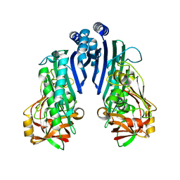
1VK3
DownloadVisualize
BU of 1vk3 by Molmil
Crystal structure of Phosphoribosylformylglycinamidine synthase II (TM1246) from Thermotoga maritima at 2.15 A resolution
Descriptor: CHLORIDE ION, Phosphoribosylformylglycinamidine synthase II
Authors:Joint Center for Structural Genomics (JCSG)
Deposit date:2004-04-23
Release date:2004-05-11
Last modified:2023-01-25
Method:X-RAY DIFFRACTION (2.15 Å)
Cite:Crystal structure of phosphoribosylformylglycinamidine synthase II (smPurL) from Thermotoga maritima at 2.15 A resolution.
Proteins, 63, 2006
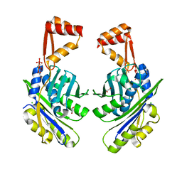
1VL4
DownloadVisualize
BU of 1vl4 by Molmil
CRYSTAL STRUCTURE OF A PUTATIVE MODULATOR OF A DNA GYRASE (TM0727) FROM THERMOTOGA MARITIMA MSB8 AT 1.95 A RESOLUTION
Descriptor: pmbA-related protein
Authors:Joint Center for Structural Genomics (JCSG)
Deposit date:2004-07-09
Release date:2004-09-21
Last modified:2024-05-22
Method:X-RAY DIFFRACTION (1.95 Å)
Cite:Crystal structure of a putative modulator of DNA gyrase (pmbA) from Thermotoga maritima at 1.95 A resolution reveals a new fold.
Proteins, 61, 2005
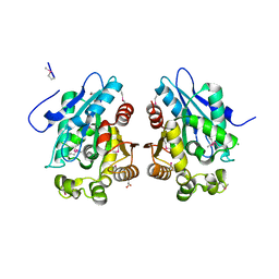
1VKH
DownloadVisualize
BU of 1vkh by Molmil
CRYSTAL STRUCTURE OF A PUTATIVE SERINE HYDROLASE (YDR428C) FROM SACCHAROMYCES CEREVISIAE AT 1.85 A RESOLUTION
Descriptor: CHLORIDE ION, GLYCEROL, putative serine hydrolase
Authors:Joint Center for Structural Genomics (JCSG)
Deposit date:2004-05-20
Release date:2004-06-08
Last modified:2023-01-25
Method:X-RAY DIFFRACTION (1.85 Å)
Cite:Crystal structure of an alpha/beta serine hydrolase (YDR428C) from Saccharomyces cerevisiae at 1.85 A resolution
Proteins, 58, 2005
1VQ0
DownloadVisualize
BU of 1vq0 by Molmil
Crystal structure of 33 kDa chaperonin (Heat shock protein 33 homolog) (HSP33) (TM1394) from Thermotoga maritima at 2.20 A resolution
Descriptor: 1,2-ETHANEDIOL, 33 kDa chaperonin, CHLORIDE ION, ...
Authors:Joint Center for Structural Genomics (JCSG)
Deposit date:2004-11-30
Release date:2004-12-14
Last modified:2023-09-20
Method:X-RAY DIFFRACTION (2.2 Å)
Cite:Crystal structure of Hsp33 chaperone (TM1394) from Thermotoga maritima at 2.20 A resolution.
Proteins, 61, 2005
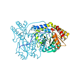
1VJO
DownloadVisualize
BU of 1vjo by Molmil
Crystal structure of Alanine--glyoxylate aminotransferase (ALR1004) from Nostoc sp. at 1.70 A resolution
Descriptor: PYRIDOXAL-5'-PHOSPHATE, alanine--glyoxylate aminotransferase
Authors:Joint Center for Structural Genomics (JCSG)
Deposit date:2004-03-16
Release date:2004-03-23
Last modified:2023-01-25
Method:X-RAY DIFFRACTION (1.7 Å)
Cite:Crystal structure of an alanine-glyoxylate aminotransferase from Anabaena sp. at 1.70 A resolution reveals a noncovalently linked PLP cofactor
Proteins, 58, 2005
1VR3
DownloadVisualize
BU of 1vr3 by Molmil
Crystal structure of Acireductone dioxygenase (13543033) from Mus musculus at 2.06 A resolution
Descriptor: Acireductone dioxygenase, ISOPROPYL ALCOHOL, NICKEL (II) ION, ...
Authors:Joint Center for Structural Genomics (JCSG)
Deposit date:2005-01-28
Release date:2005-02-15
Last modified:2023-01-25
Method:X-RAY DIFFRACTION (2.06 Å)
Cite:Crystal structure of acireductone dioxygenase (ARD) from Mus musculus at 2.06 angstrom resolution.
Proteins, 64, 2006
2ACF
DownloadVisualize
BU of 2acf by Molmil
NMR STRUCTURE OF SARS-COV NON-STRUCTURAL PROTEIN NSP3A (SARS1) FROM SARS CORONAVIRUS
Descriptor: GLYCEROL, Replicase polyprotein 1ab
Authors:Saikatendu, K.S, Joseph, J.S, Subramanian, V, Neuman, B.W, Buchmeier, M.J, Stevens, R.C, Kuhn, P, Joint Center for Structural Genomics (JCSG)
Deposit date:2005-07-18
Release date:2006-02-14
Last modified:2024-02-14
Method:X-RAY DIFFRACTION (1.4 Å)
Cite:Structural basis of severe acute respiratory syndrome coronavirus ADP-ribose-1''-phosphate dephosphorylation by a conserved domain of nsP3.
Structure, 13, 2005
<12


221051

数据于2024-06-12公开中

PDB statisticsPDBj update infoContact PDBjnumon