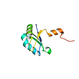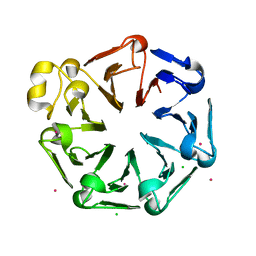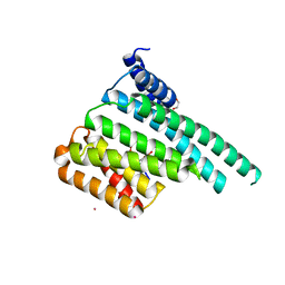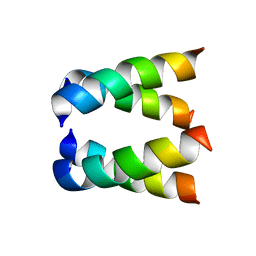
Loading
PDBj
MenuPDBj@FacebookPDBj@TwitterPDBj@YouTubewwPDB FoundationwwPDB
RCSB PDBPDBeBMRBAdv. SearchSearch help
Search by PDB author
2H95
DownloadVisualize
BU of 2h95 by Molmil
Structure of the Amantadine-Blocked Influenza A M2 Proton Channel Trans-membrane Domain by Solid-state NMR spectroscopy
Descriptor: Matrix protein 2
Authors:Hu, J, Asbury, T, Cross, T.A.
Deposit date:2006-06-08
Release date:2007-04-24
Last modified:2024-05-29
Method:SOLID-STATE NMR
Cite:Backbone structure of the amantadine-blocked trans-membrane domain m2 proton channel from influenza a virus.
Biophys.J., 92, 2007
3S0X
DownloadVisualize
BU of 3s0x by Molmil
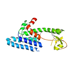
The crystal structure of GxGD membrane protease FlaK
Descriptor: Peptidase A24B, FlaK domain protein
Authors:Hu, J, Xue, Y, Ha, Y.
Deposit date:2011-05-13
Release date:2011-07-20
Last modified:2024-10-16
Method:X-RAY DIFFRACTION (3.6 Å)
Cite:The crystal structure of GXGD membrane protease FlaK.
Nature, 475, 2011
6DWB
DownloadVisualize
BU of 6dwb by Molmil
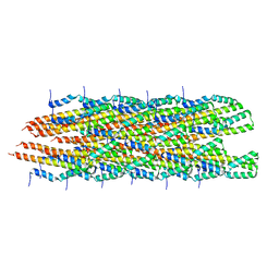
Structure of the Salmonella SPI-1 type III secretion injectisome needle filament
Descriptor: Protein PrgI
Authors:Hu, J, Hong, C, Worrall, L.J, Vuckovic, M, Yu, Z, Strynadka, N.C.J.
Deposit date:2018-06-26
Release date:2018-10-03
Last modified:2024-03-13
Method:ELECTRON MICROSCOPY (3.3 Å)
Cite:Cryo-EM analysis of the T3S injectisome reveals the structure of the needle and open secretin.
Nat Commun, 9, 2018
6DV3
DownloadVisualize
BU of 6dv3 by Molmil
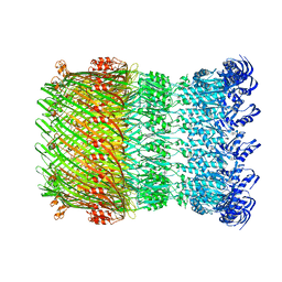
Structure of the Salmonella SPI-1 type III secretion injectisome secretin InvG in the open gate state
Descriptor: Protein InvG
Authors:Hu, J, Worrall, L.J, Vuckovic, M, Atkinson, C.E, Strynadka, N.C.J.
Deposit date:2018-06-22
Release date:2018-10-03
Last modified:2024-03-13
Method:ELECTRON MICROSCOPY (4.1 Å)
Cite:Cryo-EM analysis of the T3S injectisome reveals the structure of the needle and open secretin.
Nat Commun, 9, 2018
6DV6
DownloadVisualize
BU of 6dv6 by Molmil
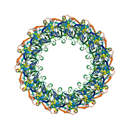
Structure of the Salmonella SPI-1 type III secretion injectisome secretin InvG (residues 176-end) in the open gate state
Descriptor: Protein InvG
Authors:Hu, J, Worrall, L.J, Vuckovic, M, Atkinson, C.E, Strynadka, N.C.J.
Deposit date:2018-06-22
Release date:2018-10-03
Last modified:2024-03-13
Method:ELECTRON MICROSCOPY (3.9 Å)
Cite:Cryo-EM analysis of the T3S injectisome reveals the structure of the needle and open secretin.
Nat Commun, 9, 2018
6DUZ
DownloadVisualize
BU of 6duz by Molmil
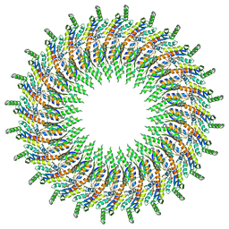
Structure of the periplasmic domains of PrgH and PrgK from the assembled Salmonella type III secretion injectisome needle complex
Descriptor: Lipoprotein PrgK, Protein PrgH
Authors:Hu, J, Worrall, L.J, Vuckovic, M, Atkinson, C.E, Strynadka, N.C.J.
Deposit date:2018-06-22
Release date:2018-10-03
Last modified:2024-03-13
Method:ELECTRON MICROSCOPY (3.6 Å)
Cite:Cryo-EM analysis of the T3S injectisome reveals the structure of the needle and open secretin.
Nat Commun, 9, 2018
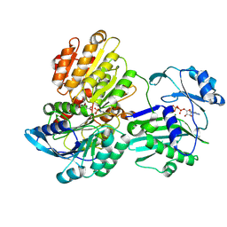
5TE1
DownloadVisualize
BU of 5te1 by Molmil
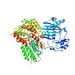
C20S, C293G Mutant N-terminal Human ATP Citrate Lyase Bound to 4R-Hydroxycitrate
Descriptor: 3-C-carboxy-2-deoxy-L-threo-pentaric acid, ATP-citrate synthase, PHOSPHATE ION, ...
Authors:Hu, J, Fraser, M.E.
Deposit date:2016-09-20
Release date:2017-08-09
Last modified:2024-03-06
Method:X-RAY DIFFRACTION (2.25 Å)
Cite:Binding of hydroxycitrate to human ATP-citrate lyase.
Acta Crystallogr D Struct Biol, 73, 2017
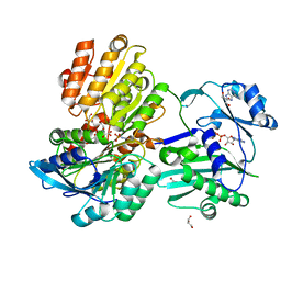
5TDE
DownloadVisualize
BU of 5tde by Molmil
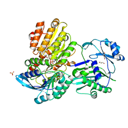
TEV Cleaved Human ATP Citrate Lyase Bound to Citrate
Descriptor: ATP-citrate synthase, CITRATE ANION, DIHYDROGENPHOSPHATE ION, ...
Authors:Hu, J, Fraser, M.E.
Deposit date:2016-09-19
Release date:2017-08-09
Last modified:2024-03-06
Method:X-RAY DIFFRACTION (1.7 Å)
Cite:Binding of hydroxycitrate to human ATP-citrate lyase.
Acta Crystallogr D Struct Biol, 73, 2017
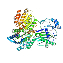
5TDM
DownloadVisualize
BU of 5tdm by Molmil
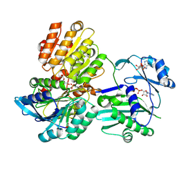
TEV Cleaved Human ATP Citrate Lyase Bound to 4R-Hydroxycitrate and ADP
Descriptor: 3-C-carboxy-2-deoxy-L-threo-pentaric acid, ADENOSINE, ADENOSINE-5'-DIPHOSPHATE, ...
Authors:Hu, J, Fraser, M.E.
Deposit date:2016-09-19
Release date:2017-08-09
Last modified:2017-08-16
Method:X-RAY DIFFRACTION (2.1 Å)
Cite:Binding of hydroxycitrate to human ATP-citrate lyase.
Acta Crystallogr D Struct Biol, 73, 2017
5TDZ
DownloadVisualize
BU of 5tdz by Molmil
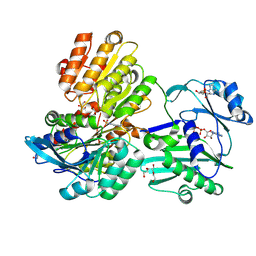
TEV Cleaved Human ATP Citrate Lyase Bound to Tartrate and ADP
Descriptor: 2-AMINO-2-HYDROXYMETHYL-PROPANE-1,3-DIOL, ADENOSINE, ADENOSINE-5'-DIPHOSPHATE, ...
Authors:Hu, J, Fraser, M.E.
Deposit date:2016-09-20
Release date:2017-08-09
Last modified:2017-08-16
Method:X-RAY DIFFRACTION (2 Å)
Cite:Binding of hydroxycitrate to human ATP-citrate lyase.
Acta Crystallogr D Struct Biol, 73, 2017
5TES
DownloadVisualize
BU of 5tes by Molmil
TEV Cleaved Human ATP Citrate Lyase Bound to Citrate and ADP
Descriptor: ADENOSINE-5'-DIPHOSPHATE, ATP-citrate synthase, CITRATE ANION, ...
Authors:Hu, J, Fraser, M.E.
Deposit date:2016-09-22
Release date:2017-08-09
Last modified:2017-08-16
Method:X-RAY DIFFRACTION (2.4 Å)
Cite:Binding of hydroxycitrate to human ATP-citrate lyase.
Acta Crystallogr D Struct Biol, 73, 2017
5TDF
DownloadVisualize
BU of 5tdf by Molmil
TEV Cleaved Human ATP Citrate Lyase Bound to 4S hydroxycitrate
Descriptor: 3-C-carboxy-2-deoxy-D-erythro-pentaric acid, ADENINE, ADENOSINE-5'-DIPHOSPHATE, ...
Authors:Hu, J, Fraser, M.E.
Deposit date:2016-09-19
Release date:2017-08-09
Last modified:2017-08-16
Method:X-RAY DIFFRACTION (1.8 Å)
Cite:Binding of hydroxycitrate to human ATP-citrate lyase.
Acta Crystallogr D Struct Biol, 73, 2017
5TET
DownloadVisualize
BU of 5tet by Molmil
TEV Cleaved Human ATP Citrate Lyase Bound to 4S Hydroxycitrate
Descriptor: 3-C-carboxy-2-deoxy-D-erythro-pentaric acid, ATP-citrate synthase, GLYCEROL, ...
Authors:Hu, J, Fraser, M.E.
Deposit date:2016-09-22
Release date:2017-08-09
Last modified:2024-03-06
Method:X-RAY DIFFRACTION (2.2 Å)
Cite:Binding of hydroxycitrate to human ATP-citrate lyase.
Acta Crystallogr D Struct Biol, 73, 2017
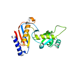
5YDU
DownloadVisualize
BU of 5ydu by Molmil
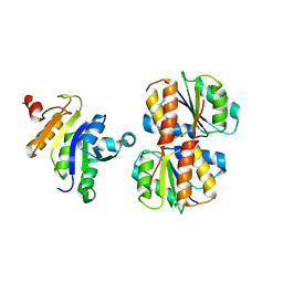
Crystal structure of Utp30
Descriptor: PHOSPHATE ION, Ribosome biogenesis protein UTP30
Authors:Hu, J, Zhu, X, Ye, K.
Deposit date:2017-09-14
Release date:2017-11-01
Last modified:2024-03-27
Method:X-RAY DIFFRACTION (2.646 Å)
Cite:Structure and RNA recognition of ribosome assembly factor Utp30.
RNA, 23, 2017
4MVT
DownloadVisualize
BU of 4mvt by Molmil
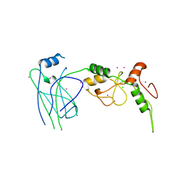
Crystal structure of SUMO E3 Ligase PIAS3
Descriptor: CHLORIDE ION, E3 SUMO-protein ligase PIAS3, UNKNOWN ATOM OR ION, ...
Authors:Dong, A, Hu, J, Li, Y, Tempel, W, Bountra, C, Arrowsmith, C.H, Edwards, A.M, Tong, Y, Structural Genomics Consortium (SGC)
Deposit date:2013-09-24
Release date:2013-10-16
Last modified:2023-09-20
Method:X-RAY DIFFRACTION (2.3 Å)
Cite:Crystal structure of SUMO E3 Ligase PIAS3
to be published
7X4B
DownloadVisualize
BU of 7x4b by Molmil
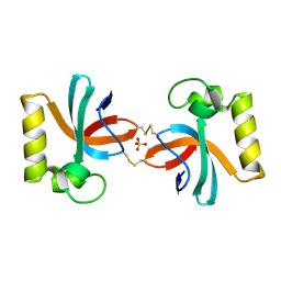
Crystal Structure of An Anti-CRISPR Protein
Descriptor: Anti-CRISPR protein (AcrIIC1), SULFATE ION
Authors:Hu, J, Zhang, S, Gao, J.Y, Liu, X, Liu, J.
Deposit date:2022-03-02
Release date:2022-10-26
Last modified:2024-10-23
Method:X-RAY DIFFRACTION (1.61 Å)
Cite:A redox switch regulates the assembly and anti-CRISPR activity of AcrIIC1.
Nat Commun, 13, 2022
1VSQ
DownloadVisualize
BU of 1vsq by Molmil
Solution NMR structure of the productive complex between IIAMannose and IIBMannose of the mannose transporter of the E. coli phosphotransferase system
Descriptor: Mannose-specific phosphotransferase enzyme IIA component, Mannose-specific phosphotransferase enzyme IIB component
Authors:Clore, G.M, Hu, J, Hu, K.
Deposit date:2008-01-10
Release date:2008-02-19
Last modified:2023-12-27
Method:SOLUTION NMR
Cite:Solution NMR Structures of Productive and Non-productive Complexes between the A and B Domains of the Cytoplasmic Subunit of the Mannose Transporter of the Escherichia coli Phosphotransferase System.
J.Biol.Chem., 283, 2008
2JZO
DownloadVisualize
BU of 2jzo by Molmil
Solution NMR structure of the non-productive complex between IIAMannose and IIBMannose of the mannose transporter of the E. coli phosphotransferase system
Descriptor: PTS system mannose-specific EIIAB component
Authors:Clore, G, Hu, J, Hu, K.
Deposit date:2008-01-10
Release date:2008-02-19
Last modified:2024-05-29
Method:SOLUTION NMR
Cite:Solution NMR Structures of Productive and Non-productive Complexes between the A and B Domains of the Cytoplasmic Subunit of the Mannose Transporter of the Escherichia coli Phosphotransferase System.
J.Biol.Chem., 283, 2008
2JZN
DownloadVisualize
BU of 2jzn by Molmil
Solution NMR structure of the productive complex between IIAMannose and IIBMannose of the mannose transporter of the E. coli phosphotransferase system
Descriptor: Mannose-specific phosphotransferase enzyme IIA component, Mannose-specific phosphotransferase enzyme IIB component
Authors:Clore, G.M, Hu, J, Hu, K.
Deposit date:2008-01-10
Release date:2008-02-19
Last modified:2024-05-29
Method:SOLUTION NMR
Cite:Solution NMR Structures of Productive and Non-productive Complexes between the A and B Domains of the Cytoplasmic Subunit of the Mannose Transporter of the Escherichia coli Phosphotransferase System.
J.Biol.Chem., 283, 2008
4QT6
DownloadVisualize
BU of 4qt6 by Molmil
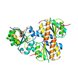
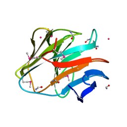
Crystal structure of the SPRY domain of human HERC1
Descriptor: FORMAMIDE, Probable E3 ubiquitin-protein ligase HERC1, UNKNOWN ATOM OR ION
Authors:Dong, A, Hu, J, Guan, X, Wernimont, A, Li, Y, Bountra, C, Arrowsmith, C.H, Edwards, A.M, Tong, Y, Structural Genomics Consortium (SGC)
Deposit date:2014-07-07
Release date:2015-01-07
Last modified:2017-11-22
Method:X-RAY DIFFRACTION (1.64 Å)
Cite:Crystal structure of the SPRY domain of human HERC1
To be Published
4TTB
DownloadVisualize
BU of 4ttb by Molmil
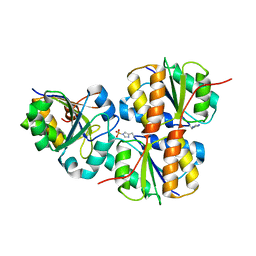
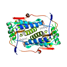
Crystal structure of homo sapiens IODOTYROSINE DEIODINASE (IYD) bound to FMN
Descriptor: FLAVIN MONONUCLEOTIDE, Iodotyrosine dehalogenase 1
Authors:Chuenchor, W, Hu, J, Rokita, S.
Deposit date:2014-06-20
Release date:2014-11-26
Last modified:2023-09-27
Method:X-RAY DIFFRACTION (2.447 Å)
Cite:A Switch between One- and Two-electron Chemistry of the Human Flavoprotein Iodotyrosine Deiodinase Is Controlled by Substrate.
J.Biol.Chem., 290, 2015
4TTC
DownloadVisualize
BU of 4ttc by Molmil
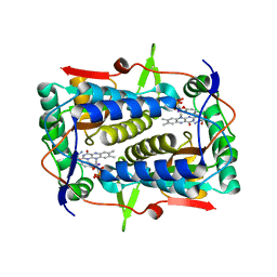
Crystal structure of homo sapiens IODOTYROSINE DEIODINASE bound to FMN and mono-iodotyrosine (MIT)
Descriptor: 3-IODO-TYROSINE, FLAVIN MONONUCLEOTIDE, Iodotyrosine dehalogenase 1
Authors:Chuenchor, W, Hu, J, Rokita, S.
Deposit date:2014-06-20
Release date:2014-11-26
Last modified:2024-04-03
Method:X-RAY DIFFRACTION (2.65 Å)
Cite:A Switch between One- and Two-electron Chemistry of the Human Flavoprotein Iodotyrosine Deiodinase Is Controlled by Substrate.
J.Biol.Chem., 290, 2015
2K3K
DownloadVisualize
BU of 2k3k by Molmil
Solution structure of Drosophila melanogaster SNF RBD1
Descriptor: U1 small nuclear ribonucleoprotein A
Authors:Liu, C, Cui, G, Hu, J, Jin, C, Xia, B.
Deposit date:2008-05-12
Release date:2008-06-03
Last modified:2024-05-29
Method:SOLUTION NMR
Cite:Structural and Functional Studies of RNA Splicing Factor Sans-Fille by Multidimentional NMR Spectroscopy
To be Published
4O2W
DownloadVisualize
BU of 4o2w by Molmil
Crystal structure of the third RCC1-like domain of HERC1
Descriptor: CHLORIDE ION, E3 ubiquitin-protein ligase HERC1, MAGNESIUM ION, ...
Authors:Dong, A, Hu, J, Li, Y, Walker, J.R, Bountra, C, Arrowsmith, C.H, Edwards, A.M, Tong, Y, Structural Genomics Consortium (SGC)
Deposit date:2013-12-17
Release date:2014-01-15
Last modified:2023-09-20
Method:X-RAY DIFFRACTION (2 Å)
Cite:Crystal structure of the third RCC1-like domain of HERC1
To be Published
5ULO
DownloadVisualize
BU of 5ulo by Molmil
Crystal Structure of 14-3-3 zeta in Complex with a Serine 124-phosphorylated TBC1D7 peptide
Descriptor: 1,2-ETHANEDIOL, 14-3-3 protein zeta/delta, TBC1 domain family member 7 peptide, ...
Authors:DONG, A, HU, J, MADIGAN, J, WALKER, J.R, Bountra, C, Arrowsmith, C.H, Edwards, A.M, TONG, Y, Structural Genomics Consortium (SGC)
Deposit date:2017-01-25
Release date:2018-01-31
Last modified:2024-10-09
Method:X-RAY DIFFRACTION (2.14 Å)
Cite:Crystal Structure of 14-3-3 zeta in Complex with a Serine 124-phosphorylated TBC1D7 peptide
to be published

226707

数据于2024-10-30公开中

PDB statisticsPDBj update infoContact PDBjnumon