1RCT
 
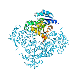
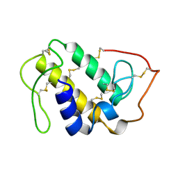
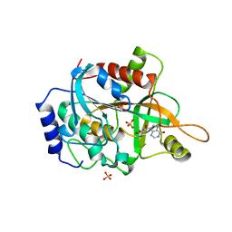 | | Crystal structure of Human purine nucleoside phosphorylase complexed with INOSINE | | Descriptor: | INOSINE, Purine nucleoside phosphorylase, SULFATE ION | | Authors: | Canduri, F, dos Santos, D.M, Silva, R.G, Mendes, M.A, Palma, M.S, de Azevedo Jr, W.F, Basso, L.A, Santos, D.S. | | Deposit date: | 2003-11-04 | | Release date: | 2004-01-20 | | Last modified: | 2023-10-25 | | Method: | X-RAY DIFFRACTION (2.8 Å) | | Cite: | Structures of human purine nucleoside phosphorylase complexed with inosine and ddI
Biochem.Biophys.Res.Commun., 313, 2004
|
|
2P34
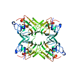
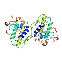
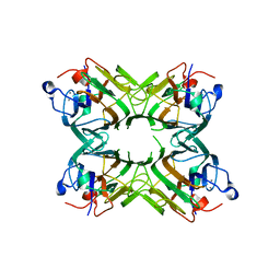 | | Crystal structure of a lectin from Canavalia maritima seeds (CML) in complex with man1-4man-OMe | | Descriptor: | CALCIUM ION, Lectin, MANGANESE (II) ION, ... | | Authors: | Moreno, F.B.M.B, Bezerra, G.A, Oliveira, T.M, Souza, E.P, Rocha, B.A.M, Benevides, R.G, Delatorre, P, Cavada, B.S, de Azevedo Jr, W. | | Deposit date: | 2007-03-08 | | Release date: | 2008-01-22 | | Last modified: | 2024-02-21 | | Method: | X-RAY DIFFRACTION (2.1 Å) | | Cite: | Structural analysis of Canavalia maritima and Canavalia gladiata lectins complexed with different dimannosides: new insights into the understanding of the structure-biological activity relationship in legume lectins.
J.Struct.Biol., 160, 2007
|
|
2IDZ
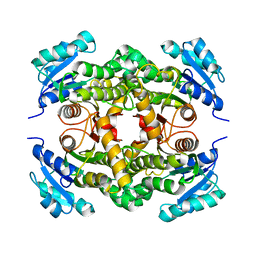
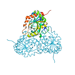
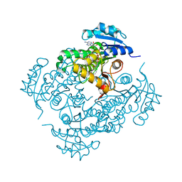 | | Crystal structure of wild type Enoyl-ACP(CoA) reductase from Mycobacterium tuberculosis in complex with NADH-INH | | Descriptor: | Enoyl-[acyl-carrier-protein] reductase [NADH], ISONICOTINIC-ACETYL-NICOTINAMIDE-ADENINE DINUCLEOTIDE | | Authors: | Dias, M.V.B, Prado, M.P.X, Vasconcelos, I.B, Valmir, F, Basso, L.A, Santos, D.S, Azevedo Jr, W.F. | | Deposit date: | 2006-09-15 | | Release date: | 2007-07-24 | | Last modified: | 2023-08-30 | | Method: | X-RAY DIFFRACTION (2 Å) | | Cite: | Crystallographic studies on the binding of isonicotinyl-NAD adduct to wild-type and isoniazid resistant 2-trans-enoyl-ACP (CoA) reductase from Mycobacterium tuberculosis.
J.Struct.Biol., 159, 2007
|
|
2IE0
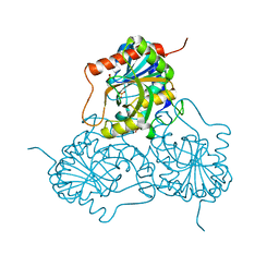
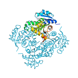 | | Crystal Structure of Isoniazid-resistant I21V Enoyl-ACP(COA) Reductase Mutant Enzyme From MYCOBACTERIUM TUBERCULOSIS in Complex with NADH-INH | | Descriptor: | Enoyl-[acyl-carrier-protein] reductase [NADH], ISONICOTINIC-ACETYL-NICOTINAMIDE-ADENINE DINUCLEOTIDE | | Authors: | Dias, M.V.B, Prado, A.M.X, Vasconceles, I.B, Fadel, F, Basso, L.A, Santos, D.S, Azevedo Jr, W.F. | | Deposit date: | 2006-09-15 | | Release date: | 2007-07-24 | | Last modified: | 2023-08-30 | | Method: | X-RAY DIFFRACTION (2.2 Å) | | Cite: | Crystallographic studies on the binding of isonicotinyl-NAD adduct to wild-type and isoniazid resistant 2-trans-enoyl-ACP (CoA) reductase from Mycobacterium tuberculosis.
J.Struct.Biol., 159, 2007
|
|
2IEB
 | | Crystal Structure of Isoniazid-resistant S94A ENOYL-ACP(COA) Reductase Mutant Enzyme from MYCOBACTERIUM TUBERCULOSIS in Complex with NADH-INH | | Descriptor: | Enoyl-[acyl-carrier-protein] reductase [NADH], ISONICOTINIC-ACETYL-NICOTINAMIDE-ADENINE DINUCLEOTIDE | | Authors: | Dias, M.V.B, Prado, A.M.X, Vasconcelos, I.B, Fadel, V, Basso, L.A, Santos, D.S, Azevedo Jr, W.F. | | Deposit date: | 2006-09-18 | | Release date: | 2007-07-24 | | Last modified: | 2023-08-30 | | Method: | X-RAY DIFFRACTION (2.2 Å) | | Cite: | Crystallographic studies on the binding of isonicotinyl-NAD adduct to wild-type and isoniazid resistant 2-trans-enoyl-ACP (CoA) reductase from Mycobacterium tuberculosis.
J.Struct.Biol., 159, 2007
|
|
2IED
 | | CRYSTAL STRUCTURE of ISONIAZID-RESISTANT S94A ENOYL-ACP(COA) REDUCTASE MUTANT ENZYME FROM MYCOBACTERIUM TUBERCULOSIS UNCOMPLEXED | | Descriptor: | Enoyl-[acyl-carrier-protein] reductase [NADH] | | Authors: | Dias, M.V.B, Prado, A.M.X, Vasconcelos, I.B, Fadel, V, Basso, L.A, Santos, D.S, Azevedo Jr, W.F. | | Deposit date: | 2006-09-18 | | Release date: | 2007-07-24 | | Last modified: | 2023-08-30 | | Method: | X-RAY DIFFRACTION (2.14 Å) | | Cite: | Crystallographic studies on the binding of isonicotinyl-NAD adduct to wild-type and isoniazid resistant 2-trans-enoyl-ACP (CoA) reductase from Mycobacterium tuberculosis.
J.Struct.Biol., 159, 2007
|
|
2P2K
 | | Crystal structure of a lectin from Canavalia gladiata seeds (CGL) in complex with man1-4man-OMe | | Descriptor: | CALCIUM ION, Canavalia gladiata lectin, MANGANESE (II) ION, ... | | Authors: | Moreno, F.B.M.B, Bezerra, G.A, Oliveira, T.M, Souza, E.P, Rocha, B.A.M, Benevides, G.B, Delatorre, P, Cavada, B.S, de Azevedo Jr, W.F. | | Deposit date: | 2007-03-07 | | Release date: | 2007-10-23 | | Last modified: | 2024-02-21 | | Method: | X-RAY DIFFRACTION (1.98 Å) | | Cite: | Structural analysis of Canavalia maritima and Canavalia gladiata lectins complexed with different dimannosides: New insights into the understanding of the structure-biological activity relationship in legume lectins.
J.Struct.Biol., 160, 2007
|
|
2OW4
 | | Crystal structure of a lectin from Canavalia maritima seeds (ConM) in complex with man1-2man-OMe | | Descriptor: | CALCIUM ION, Canavalia maritima lectin, MANGANESE (II) ION, ... | | Authors: | Moreno, F.B.M.B, Bezerra, G.A, de Oliveira, T.M, de Souza, E.M, da Rocha, B.A.M, Benevides, R.G, Delatorre, P, Cavada, B.S, de Azevedo Jr, W.F. | | Deposit date: | 2007-02-15 | | Release date: | 2007-10-23 | | Last modified: | 2024-02-21 | | Method: | X-RAY DIFFRACTION (1.6 Å) | | Cite: | Structural analysis of Canavalia maritima and Canavalia gladiata lectins complexed with different dimannosides: New insights into the understanding of the structure-biological activity relationship in legume lectins.
J.Struct.Biol., 160, 2007
|
|
2OVU
 | | Crystal structure of a lectin from Canavalia gladiata (CGL) in complex with man1-2man-OMe | | Descriptor: | CALCIUM ION, Canavalia gladiata lectin, MANGANESE (II) ION, ... | | Authors: | Moreno, F.B.M.B, Bezerra, G.A, de Oliveira, T.M, de Souza, E.P, da Rocha, B.A.M, Benevides, R.G, Delatorre, P, Cavada, B.S, de Azevedo Jr, W.F. | | Deposit date: | 2007-02-15 | | Release date: | 2007-10-23 | | Last modified: | 2024-02-21 | | Method: | X-RAY DIFFRACTION (1.5 Å) | | Cite: | Structural analysis of Canavalia maritima and Canavalia gladiata lectins complexed with different dimannosides: New insights into the understanding of the structure-biological activity relationship in legume lectins.
J.Struct.Biol., 160, 2007
|
|
1WE2
 | | Crystal structure of shikimate kinase from mycobacterium tuberculosis in complex with MGADP and shikimic acid | | Descriptor: | 3-DEHYDROSHIKIMATE, ADENOSINE-5'-DIPHOSPHATE, CHLORIDE ION, ... | | Authors: | Pereira, J.H, de Oliveira, J.S, Canduri, F, Dias, M.V, Palma, M.S, Basso, L.A, Santos, D.S, de Azevedo Jr, W.F. | | Deposit date: | 2004-05-22 | | Release date: | 2005-05-17 | | Last modified: | 2023-10-25 | | Method: | X-RAY DIFFRACTION (2.3 Å) | | Cite: | Structure of shikimate kinase from Mycobacterium tuberculosis reveals the binding of shikimic acid.
Acta Crystallogr.,Sect.D, 60, 2004
|
|
3IJF
 | |
3IOM
 | |
1FHJ
 | |
1RFG
 | | Crystal Structure of Human Purine Nucleoside Phosphorylase Complexed with Guanosine | | Descriptor: | GUANOSINE, Purine nucleoside phosphorylase, SULFATE ION | | Authors: | Canduri, F, Silva, R.G, Dos Santos, D.M, Palma, M.S, Basso, L.A, Santos, D.S, de Azevedo, W.F. | | Deposit date: | 2003-11-09 | | Release date: | 2004-12-14 | | Last modified: | 2023-08-23 | | Method: | X-RAY DIFFRACTION (2.9 Å) | | Cite: | Structure of human PNP complexed with ligands.
Acta Crystallogr.,Sect.D, 61, 2005
|
|
1M73
 | | CRYSTAL STRUCTURE OF HUMAN PNP AT 2.3A RESOLUTION | | Descriptor: | PURINE NUCLEOSIDE PHOSPHORYLASE, SULFATE ION | | Authors: | De Azevedo Jr, W.F, Marangoni Dos Santos, D, Canduri, F, Santos, G.C, Olivieri, J.R, Silva, R.G, Basso, L.A, Palma, M.S, Santos, D.S. | | Deposit date: | 2002-07-18 | | Release date: | 2003-09-16 | | Last modified: | 2023-10-25 | | Method: | X-RAY DIFFRACTION (2.3 Å) | | Cite: | Crystal structure of human purine nucleoside phosphorylase at 2.3A resolution.
Biochem.Biophys.Res.Commun., 308, 2003
|
|
1XXS
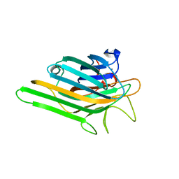
 | | Structural insights for fatty acid binding in a Lys49 phospholipase A2: crystal structure of myotoxin II from Bothrops moojeni complexed with stearic acid | | Descriptor: | Phospholipase A2 homolog 2, STEARIC ACID, SULFATE ION | | Authors: | Watanabe, L, Soares, A.M, Ward, R.J, Fontes, M.R, Arni, R.K. | | Deposit date: | 2004-11-08 | | Release date: | 2005-03-29 | | Last modified: | 2024-04-03 | | Method: | X-RAY DIFFRACTION (1.8 Å) | | Cite: | Structural insights for fatty acid binding in a Lys49-phospholipase A(2): crystal structure of myotoxin II from Bothrops moojeni complexed with stearic acid
Biochimie, 87, 2005
|
|
1GOD
 | | MONOMERIC LYS-49 PHOSPHOLIPASE A2 HOMOLOGUE ISOLATED FROM THE VENOM OF CERROPHIDION (BOTHROPS) GODMANI | | Descriptor: | PROTEIN (PHOSPHOLIPASE A2) | | Authors: | Arni, R.K, Fontes, M.R.M, Barberato, C, Gutierrez, J.M, Diaz-Oreiro, C, Ward, R.J. | | Deposit date: | 1999-04-16 | | Release date: | 1999-04-23 | | Last modified: | 2023-08-02 | | Method: | X-RAY DIFFRACTION (2.8 Å) | | Cite: | Crystal structure of myotoxin II, a monomeric Lys49-phospholipase A2 homologue isolated from the venom of Cerrophidion (Bothrops) godmani.
Arch.Biochem.Biophys., 366, 1999
|
|


