Loading
PDBj
MenuPDBj@FacebookPDBj@TwitterPDBj@YouTubewwPDB FoundationwwPDB
RCSB PDBPDBeBMRBAdv. SearchSearch help
Search by PDB author
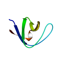
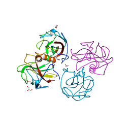
2O2W
DownloadVisualize
BU of 2o2w by Molmil
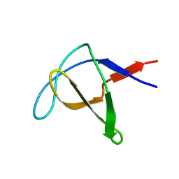
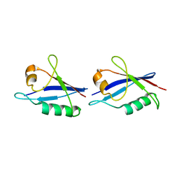
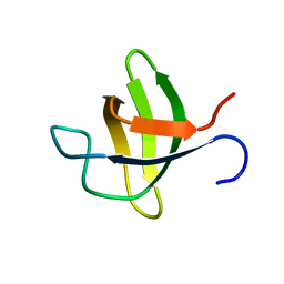
Extending powder diffraction to proteins: structure solution of the second SH3 domain from ponsin
Descriptor: Ponsin
Authors:Pinotsis, N, Margiolaki, I.
Deposit date:2006-11-30
Release date:2007-10-23
Last modified:2023-12-27
Method:POWDER DIFFRACTION
Cite:Second SH3 domain of ponsin solved from powder diffraction
J.Am.Chem.Soc., 129, 2007
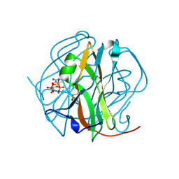
4BEG
DownloadVisualize
BU of 4beg by Molmil
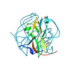
Structure of Rv2140c, a phosphatidyl-ethanolamine binding protein from Mycobacterium tuberculosis in complex with sulphate
Descriptor: GLYCEROL, PHOSPHATIDYLETHANOLAMINE BINDING PROTEIN, SULFATE ION
Authors:Holton, S.J, Williams, M.
Deposit date:2013-03-09
Release date:2013-08-14
Last modified:2023-12-20
Method:X-RAY DIFFRACTION (1.42 Å)
Cite:Structural and Biochemical Characterization of Rv2140C, a Phosphatidylethanolamine-Binding Protein from Mycobacterium Tuberculosis.
FEBS Lett., 587, 2013
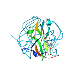
2BZR
DownloadVisualize
BU of 2bzr by Molmil
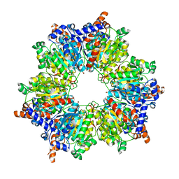
Crystal structure of accD5 (Rv3280), an acyl-CoA carboxylase beta- subunit from Mycobacterium tuberculosis
Descriptor: PROPIONYL-COA CARBOXYLASE BETA CHAIN 5
Authors:Holton, S.J.
Deposit date:2005-08-22
Release date:2007-01-02
Last modified:2024-05-08
Method:X-RAY DIFFRACTION (2.2 Å)
Cite:Structural Diversity in the Six-Fold Redundant Set of Acyl-Coa Carboxyltransferases in Mycobacterium Tuberculosis.
FEBS Lett., 580, 2006
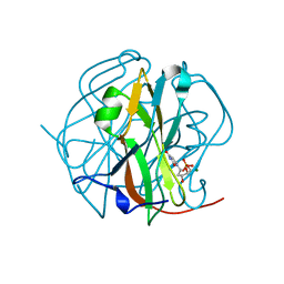
1AWO
DownloadVisualize
BU of 1awo by Molmil
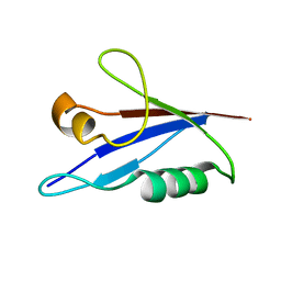
THE SOLUTION NMR STRUCTURE OF ABL SH3 AND ITS RELATIONSHIP TO SH2 IN THE SH(32) CONSTRUCT, 20 STRUCTURES
Descriptor: ABL TYROSINE KINASE
Authors:Cowburn, D.
Deposit date:1997-10-03
Release date:1998-01-28
Last modified:2024-05-22
Method:SOLUTION NMR
Cite:The solution structure of Abl SH3, and its relationship to SH2 in the SH(32) construct.
Structure, 3, 1995
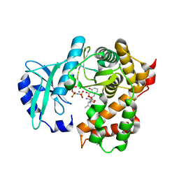
6SUI
DownloadVisualize
BU of 6sui by Molmil
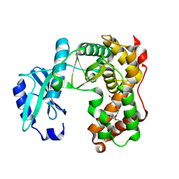
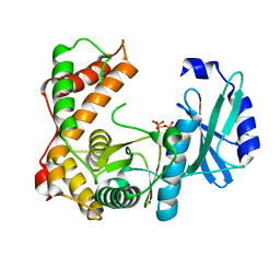
AMICOUMACIN KINASE AMIN
Descriptor: PENTAETHYLENE GLYCOL, Phosphotransferase enzyme family protein
Authors:Bourenkov, G.P, Mokrushina, Y.A, Terekhov, S.S, Smirnov, I.V, Gabibov, A.G, Altman, S.
Deposit date:2019-09-14
Release date:2020-07-22
Last modified:2024-05-15
Method:X-RAY DIFFRACTION (1.6 Å)
Cite:A kinase bioscavenger provides antibiotic resistance by extremely tight substrate binding.
Sci Adv, 6, 2020
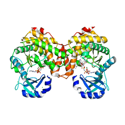
6SUN
DownloadVisualize
BU of 6sun by Molmil
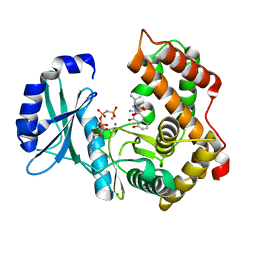
Amicoumacin kinase hAmiN in complex with AMP-PNP, Ca2+ and Ami
Descriptor: APH domain-containing protein, amicoumacin kinase, Amicoumacin A, ...
Authors:Bourenkov, G.P, Mokrushina, Y.A, Terekhov, S.S, Smirnov, I.V, Gabibov, A.G, Altman, S.
Deposit date:2019-09-16
Release date:2020-07-22
Last modified:2024-05-15
Method:X-RAY DIFFRACTION (1.35 Å)
Cite:A kinase bioscavenger provides antibiotic resistance by extremely tight substrate binding.
Sci Adv, 6, 2020
6TGS
DownloadVisualize
BU of 6tgs by Molmil
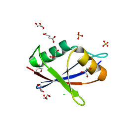
AtNBR1-PB1 domain
Descriptor: CHLORIDE ION, DI(HYDROXYETHYL)ETHER, GLYCEROL, ...
Authors:Jakobi, A.J, Sachse, C.
Deposit date:2019-11-17
Release date:2020-02-12
Last modified:2024-05-01
Method:X-RAY DIFFRACTION (1.53 Å)
Cite:Structural basis of p62/SQSTM1 helical filaments and their role in cellular cargo uptake.
Nat Commun, 11, 2020
6SV5
DownloadVisualize
BU of 6sv5 by Molmil
Amicoumacin kinase AmiN in complex with ATP
Descriptor: ADENOSINE-5'-TRIPHOSPHATE, Phosphotransferase enzyme family protein, amicoumacin kinase
Authors:Bourenkov, G.P, Mokrushina, Y.A, Terekhov, S.S, Smirnov, I.V, Gabibov, A.G, Altman, S.
Deposit date:2019-09-17
Release date:2020-07-22
Last modified:2024-05-15
Method:X-RAY DIFFRACTION (2 Å)
Cite:A kinase bioscavenger provides antibiotic resistance by extremely tight substrate binding.
Sci Adv, 6, 2020
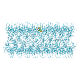
6TGN
DownloadVisualize
BU of 6tgn by Molmil
Cryo-EM structure of AtNBR1-PB1 filament (L-type)
Descriptor: Protein NBR1 homolog
Authors:Jakobi, A.J, Sachse, C.
Deposit date:2019-11-17
Release date:2020-02-12
Last modified:2024-05-22
Method:ELECTRON MICROSCOPY (3.9 Å)
Cite:Structural basis of p62/SQSTM1 helical filaments and their role in cellular cargo uptake.
Nat Commun, 11, 2020
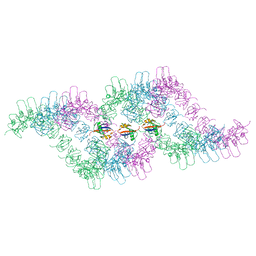
6TGP
DownloadVisualize
BU of 6tgp by Molmil
Cryo-EM structure of AtNBR1-PB1 filament (S-type)
Descriptor: Protein NBR1 homolog
Authors:Jakobi, A.J, Sachse, C.
Deposit date:2019-11-17
Release date:2020-02-19
Last modified:2024-05-22
Method:ELECTRON MICROSCOPY (4.4 Å)
Cite:Structural basis of p62/SQSTM1 helical filaments and their role in cellular cargo uptake.
Nat Commun, 11, 2020
6SUM
DownloadVisualize
BU of 6sum by Molmil
Amicoumacin kinase hAmiN in complex with AMP-PNP, MG2+ and Ami
Descriptor: ACETATE ION, AMICOUMACIN KINASE, Amicoumacin A, ...
Authors:Bourenkov, G.P, Mokrushina, Y.A, Terekhov, S.S, Smirnov, I.V, Gabibov, A.G, Altman, S.
Deposit date:2019-09-16
Release date:2020-07-22
Last modified:2024-05-15
Method:X-RAY DIFFRACTION (1.35 Å)
Cite:A kinase bioscavenger provides antibiotic resistance by extremely tight substrate binding.
Sci Adv, 6, 2020
6SUL
DownloadVisualize
BU of 6sul by Molmil
Amicoumacin kinase AmiN in complex with AMP-PNP, Mg2+ and Ami
Descriptor: Amicoumacin A, CHLORIDE ION, MAGNESIUM ION, ...
Authors:Bourenkov, G.P, Mokrushina, Y.A, Terekhov, S.S, Smirnov, I.V, Gabibov, A.G, Altman, S.
Deposit date:2019-09-15
Release date:2020-07-22
Last modified:2024-05-15
Method:X-RAY DIFFRACTION (1.35 Å)
Cite:A kinase bioscavenger provides antibiotic resistance by extremely tight substrate binding.
Sci Adv, 6, 2020
6TGY
DownloadVisualize
BU of 6tgy by Molmil
Cryo-EM structure of p62-PB1 filament (L-type)
Descriptor: Sequestosome-1
Authors:Jakobi, A.J, Huber, S.T, Mortensen, S.A, Sachse, C.
Deposit date:2019-11-18
Release date:2020-02-12
Last modified:2024-05-22
Method:ELECTRON MICROSCOPY (3.5 Å)
Cite:Structural basis of p62/SQSTM1 helical filaments and their role in cellular cargo uptake.
Nat Commun, 11, 2020
6TH3
DownloadVisualize
BU of 6th3 by Molmil
Cryo-EM structure of p62-PB1 filament (S-type)
Descriptor: Sequestosome-1
Authors:Jakobi, A.J, Huber, S.T, Mortensen, S.A, Sachse, C.
Deposit date:2019-11-18
Release date:2020-02-12
Last modified:2024-05-22
Method:ELECTRON MICROSCOPY (4 Å)
Cite:Structural basis of p62/SQSTM1 helical filaments and their role in cellular cargo uptake.
Nat Commun, 11, 2020
1AEY
DownloadVisualize
BU of 1aey by Molmil
ALPHA-SPECTRIN SRC HOMOLOGY 3 DOMAIN, SOLUTION NMR, 15 STRUCTURES
Descriptor: ALPHA-SPECTRIN
Authors:Blanco, F.J, Ortiz, A.R, Serrano, L.
Deposit date:1997-03-02
Release date:1997-05-15
Last modified:2024-05-22
Method:SOLUTION NMR
Cite:1H and 15N NMR assignment and solution structure of the SH3 domain of spectrin: comparison of unrefined and refined structure sets with the crystal structure.
J.Biomol.NMR, 9, 1997
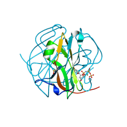
3TPS
DownloadVisualize
BU of 3tps by Molmil
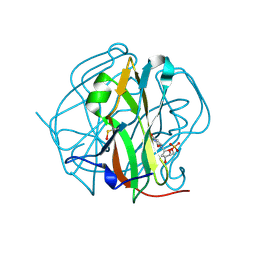
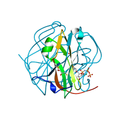
Crystal structure of M-PMV dUTPASE complexed with dUPNPP substrate
Descriptor: 2'-DEOXYURIDINE 5'-ALPHA,BETA-IMIDO-TRIPHOSPHATE, 2-AMINO-2-HYDROXYMETHYL-PROPANE-1,3-DIOL, DEOXYURIDINE 5'-TRIPHOSPHATE NUCLEOTIDO HYDROLASE, ...
Authors:Barabas, O, Nemeth, V, Vertessy, B.G.
Deposit date:2011-09-08
Release date:2011-10-12
Last modified:2023-12-06
Method:X-RAY DIFFRACTION (1.85 Å)
Cite:Structural Snapshots of Enzyme-Catalysed Phosphate Ester Hydrolysis Directly Visualize In-line Attack and Inversion
To be Published
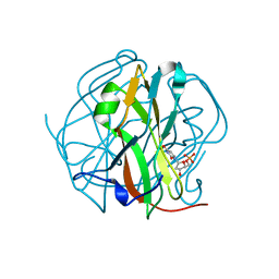
3TPY
DownloadVisualize
BU of 3tpy by Molmil
Crystal structure of M-PMV dUTPase with a mixed population of substrate (dUPNPP) and post-inversion product (dUMP) in the active sites
Descriptor: 2'-DEOXYURIDINE 5'-ALPHA,BETA-IMIDO-TRIPHOSPHATE, 2'-DEOXYURIDINE 5'-MONOPHOSPHATE, 2-AMINO-2-HYDROXYMETHYL-PROPANE-1,3-DIOL, ...
Authors:Barabas, O, Nemeth, V, Vertessy, B.G.
Deposit date:2011-09-08
Release date:2011-10-12
Last modified:2023-09-13
Method:X-RAY DIFFRACTION (1.75 Å)
Cite:Structural Snapshots of Enzyme-Catalysed Phosphate Ester Hydrolysis Directly Visualize In-line Attack and Inversion
to be published
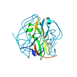
3TQ3
DownloadVisualize
BU of 3tq3 by Molmil
Crystal structure of M-PMV dUTPase with a mixed population of substrate (dUPNPP) and post-inversion product (dUMP) in the active sites
Descriptor: 2'-DEOXYURIDINE 5'-ALPHA,BETA-IMIDO-TRIPHOSPHATE, 2'-DEOXYURIDINE 5'-MONOPHOSPHATE, DEOXYURIDINE 5'-TRIPHOSPHATE NUCLEOTIDO HYDROLASE, ...
Authors:Barabas, O, Nemeth, V, Vertessy, B.G.
Deposit date:2011-09-09
Release date:2011-10-12
Last modified:2023-09-13
Method:X-RAY DIFFRACTION (1.85 Å)
Cite:Structural Snapshots of Enzyme-Catalysed Phosphate Ester Hydrolysis Directly Visualize In-line Attack and Inversion
to be published
3TRN
DownloadVisualize
BU of 3trn by Molmil
Crystal structure of M-PMV dUTPASE post-inversion product (dUMP) complex
Descriptor: 2'-DEOXYURIDINE 5'-MONOPHOSPHATE, DEOXYURIDINE 5'-TRIPHOSPHATE NUCLEOTIDO HYDROLASE
Authors:Nemeth, V, Barabas, O, Vertessy, G.B.
Deposit date:2011-09-09
Release date:2011-10-12
Last modified:2024-10-16
Method:X-RAY DIFFRACTION (1.83 Å)
Cite:Structural Snapshots of Enzyme-Catalysed Phosphate Ester Hydrolysis Directly Visualize in-Line Attack and Inversion
To be Published
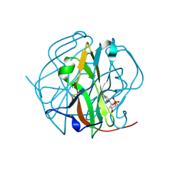
3TP1
DownloadVisualize
BU of 3tp1 by Molmil
Crystal Structure of the precatalytic M-PMV dUTPase - substrate (dUPNPP) complex
Descriptor: 2'-DEOXYURIDINE 5'-ALPHA,BETA-IMIDO-TRIPHOSPHATE, DEOXYURIDINE 5'-TRIPHOSPHATE NUCLEOTIDO HYDROLASE, MAGNESIUM ION
Authors:Barabas, O, Nemeth, V, Vertessy, B.G.
Deposit date:2011-09-07
Release date:2011-09-21
Last modified:2024-10-16
Method:X-RAY DIFFRACTION (1.6 Å)
Cite:Structural Snapshots of Enzyme-Catalysed Phosphate Ester Hydrolysis Directly Visualize In-line Attack and Inversion
To be Published
3TTA
DownloadVisualize
BU of 3tta by Molmil
Crystal structure of M-PMV DUTPASE relaxed end-product (dUMP) complex
Descriptor: 2'-DEOXYURIDINE 5'-MONOPHOSPHATE, DEOXYURIDINE 5'-TRIPHOSPHATE NUCLEOTIDO HYDROLASE
Authors:Nemeth, V, Barabas, O, Vertessy, G.B.
Deposit date:2011-09-14
Release date:2011-10-12
Last modified:2023-09-13
Method:X-RAY DIFFRACTION (2 Å)
Cite:Structural Snapshots of Enzyme-Catalysed Phosphate Ester Hydrolysis Directly Visualize in-Line Attack and Inversion
To be Published
3TSL
DownloadVisualize
BU of 3tsl by Molmil
Crystal structure of M-PMV DUTPASE relaxed end-product (dUMP) complex
Descriptor: 2'-DEOXYURIDINE 5'-MONOPHOSPHATE, DEOXYURIDINE 5'-TRIPHOSPHATE NUCLEOTIDO HYDROLASE
Authors:Nemeth, V, Barabas, O, Vertessy, G.B.
Deposit date:2011-09-13
Release date:2011-10-12
Last modified:2023-09-13
Method:X-RAY DIFFRACTION (2.2 Å)
Cite:Structural Snapshots of Enzyme-Catalysed Phosphate Ester Hydrolysis Directly Visualize in-Line Attack and Inversion
To be Published
3TQ5
DownloadVisualize
BU of 3tq5 by Molmil
Crystal structure of M-PMV dUTPASE post-inversion product (dUMP) COMPLEX
Descriptor: 2'-DEOXYURIDINE 5'-MONOPHOSPHATE, 2-AMINO-2-HYDROXYMETHYL-PROPANE-1,3-DIOL, DEOXYURIDINE 5'-TRIPHOSPHATE NUCLEOTIDO HYDROLASE
Authors:Barabas, O, Nemeth, V, Vertessy, B.G.
Deposit date:2011-09-09
Release date:2011-10-12
Last modified:2023-09-13
Method:X-RAY DIFFRACTION (1.4 Å)
Cite:Structural Snapshots of Enzyme-Catalysed Phosphate Ester Hydrolysis Directly Visualize In-line Attack and Inversion
to be published
3TS6
DownloadVisualize
BU of 3ts6 by Molmil
Crystal structure of M-PMV DUTPASE relaxed end-product (dUMP) complex
Descriptor: 2'-DEOXYURIDINE 5'-MONOPHOSPHATE, DEOXYURIDINE 5'-TRIPHOSPHATE NUCLEOTIDO HYDROLASE
Authors:Nemeth, V, Barabas, O, Vertessy, G.B.
Deposit date:2011-09-12
Release date:2011-10-12
Last modified:2023-12-06
Method:X-RAY DIFFRACTION (1.84 Å)
Cite:Structural Snapshots of Enzyme-Catalysed Phosphate Ester Hydrolysis Directly Visualize in-Line Attack and Inversion
To be Published
3TPN
DownloadVisualize
BU of 3tpn by Molmil
Crystal structure of M-PMV dUTPASE complexed with dUPNPP, substrate
Descriptor: 2'-DEOXYURIDINE 5'-ALPHA,BETA-IMIDO-TRIPHOSPHATE, 2,3-DIHYDROXY-1,4-DITHIOBUTANE, 2-AMINO-2-HYDROXYMETHYL-PROPANE-1,3-DIOL, ...
Authors:Nemeth, V, Barabas, O, Vertessy, G.B.
Deposit date:2011-09-08
Release date:2011-10-12
Last modified:2024-10-16
Method:X-RAY DIFFRACTION (1.65 Å)
Cite:Structural Snapshots of Enzyme-Catalysed Phosphate Ester Hydrolysis Directly Visualize In-line Attack and Inversion
To be Published

226707

数据于2024-10-30公开中

PDB statisticsPDBj update infoContact PDBjnumon