Loading
PDBj
MenuPDBj@FacebookPDBj@TwitterPDBj@YouTubewwPDB FoundationwwPDB
RCSB PDBPDBeBMRBAdv. SearchSearch help
Search by PDB author
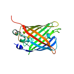
1ACX
DownloadVisualize
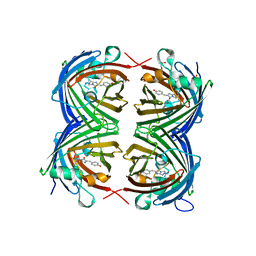
BU of 1acx by Molmil
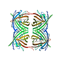
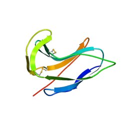
ACTINOXANTHIN STRUCTURE AT THE ATOMIC LEVEL (RUSSIAN)
Descriptor: ACTINOXANTHIN
Authors:Pletnev, V.Z, Kuzin, A.P.
Deposit date:1982-12-17
Release date:1983-03-09
Last modified:2017-11-29
Method:X-RAY DIFFRACTION (2 Å)
Cite:Actinoxanthin Structure at the Atomic Level (Russian)
Bioorg.Khim., 8, 1982
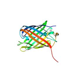
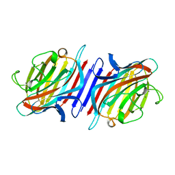
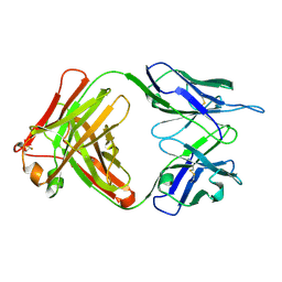
1S5I
DownloadVisualize
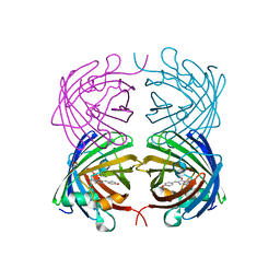
BU of 1s5i by Molmil
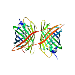
Fab (LNKB-2) of monoclonal antibody to Human Interleukin-2, crystal structure
Descriptor: Fab-fragment of monoclonal antibody
Authors:Pletnev, V.Z, Goryacheva, E.A, Tsygannik, I.N, Nesmeyanov, V.A, Pletnev, S.V, Pangborn, W, Duax, W.
Deposit date:2004-01-21
Release date:2004-05-25
Last modified:2024-10-30
Method:X-RAY DIFFRACTION (2.7 Å)
Cite:[A new crystal form of the Fab fragment of a monoclonal antibody to human interleukin-2: the three-dimensional structure at 2.7 A resolution].
Bioorg. Khim., 30
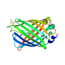
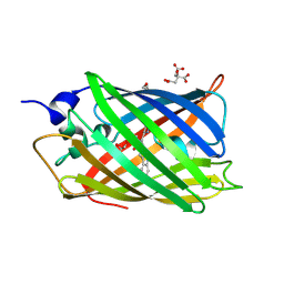
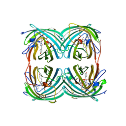
7BHG
DownloadVisualize
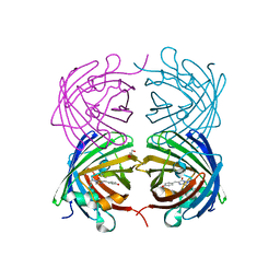
BU of 7bhg by Molmil
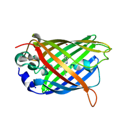
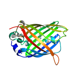
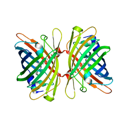
Gene-engineered variant of FusionRed with Trp based chromophore - 2.1A
Descriptor: Fluorescent protein FP480
Authors:Pletnev, V.Z, Pletnev, S.V, Pletneva, N.V.
Deposit date:2021-01-11
Release date:2022-03-02
Last modified:2024-07-10
Method:X-RAY DIFFRACTION (2.09 Å)
Cite:Gene-engineered variant of FusionRed with Trp based chromophore - 2.1A
To Be Published
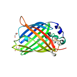
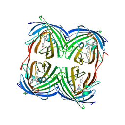
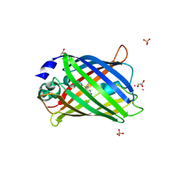
4RTC
DownloadVisualize
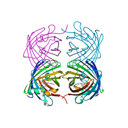
BU of 4rtc by Molmil
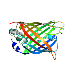
Crystal structure of the green fluorescent variant, nowGFP, of the cyan Cerulean at pH 9.0
Descriptor: GLYCEROL, nowGFP
Authors:Pletnev, V.Z, Pletneva, N.V, Pletnev, S.V.
Deposit date:2014-11-14
Release date:2015-09-02
Last modified:2023-12-06
Method:X-RAY DIFFRACTION (1.35 Å)
Cite:Structure of the green fluorescent protein NowGFP with an anionic tryptophan-based chromophore.
Acta Crystallogr.,Sect.D, 71, 2015
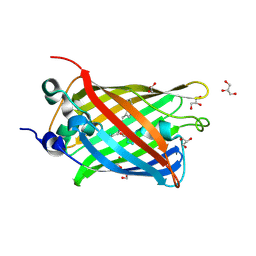
4RYW
DownloadVisualize
BU of 4ryw by Molmil
Crystal structure of the photoconverted green fluorescent protein NowGFP_conv (the variant of cyan Cerulean) at pH 7.0
Descriptor: GLYCEROL, NowGFP_conv
Authors:Pletnev, V.Z, Pletneva, N.V, Pletnev, S.V.
Deposit date:2014-12-17
Release date:2015-09-02
Last modified:2024-11-06
Method:X-RAY DIFFRACTION (2.5 Å)
Cite:Structure of the green fluorescent protein NowGFP with an anionic tryptophan-based chromophore.
Acta Crystallogr.,Sect.D, 71, 2015
4RYS
DownloadVisualize
BU of 4rys by Molmil
Crystal structure of the green fluorescent rotein NowGFP (the variant of cyan Cerulean) at pH 4.8
Descriptor: GLYCEROL, NowGFP
Authors:Pletnev, V.Z, Pletneva, N.V, Pletnev, S.V.
Deposit date:2014-12-17
Release date:2015-09-02
Last modified:2023-12-06
Method:X-RAY DIFFRACTION (1.18 Å)
Cite:Structure of the green fluorescent protein NowGFP with an anionic tryptophan-based chromophore.
Acta Crystallogr.,Sect.D, 71, 2015
4HVF
DownloadVisualize
BU of 4hvf by Molmil
Crystal structure of green fluorescent protein lanGFP(Branchiostoma Lanceolatum)
Descriptor: GLYCEROL, Green fluorescent protein blFP-Y6
Authors:Pletnev, V.Z, Pletneva, N.V, Pletnev, S.V.
Deposit date:2012-11-06
Release date:2013-09-18
Last modified:2024-10-30
Method:X-RAY DIFFRACTION (1.7 Å)
Cite:Structure of the red fluorescent protein from a lancelet (Branchiostoma lanceolatum): a novel GYG chromophore covalently bound to a nearby tyrosine.
Acta Crystallogr.,Sect.D, 69, 2013
4JEO
DownloadVisualize
BU of 4jeo by Molmil
Crystal structure of red fluorescent protein lanRFPdam exposed to prolonged X-ray irradiation
Descriptor: GLYCEROL, Red fluorescent protein blFP-R5
Authors:Pletnev, V.Z, Pletneva, N.V, Pletnev, S.V.
Deposit date:2013-02-27
Release date:2013-09-18
Last modified:2024-11-06
Method:X-RAY DIFFRACTION (2.35 Å)
Cite:Structure of the red fluorescent protein from a lancelet (Branchiostoma lanceolatum): a novel GYG chromophore covalently bound to a nearby tyrosine.
Acta Crystallogr.,Sect.D, 69, 2013
4JGE
DownloadVisualize
BU of 4jge by Molmil
Crystal structure of red fluorescent gene-engineered variant with improved folding - lanRFP_delS83 (Branchiostoma lanceolatum)
Descriptor: GLYCEROL, Red fluorescent protein blFP-R5
Authors:Pletnev, V.Z, Pletneva, N.V, Pletnev, S.V.
Deposit date:2013-03-01
Release date:2013-09-18
Last modified:2023-12-06
Method:X-RAY DIFFRACTION (1.94 Å)
Cite:Structure of the red fluorescent protein from a lancelet (Branchiostoma lanceolatum): a novel GYG chromophore covalently bound to a nearby tyrosine.
Acta Crystallogr.,Sect.D, 69, 2013
4JF9
DownloadVisualize
BU of 4jf9 by Molmil
Crystal structure of the wild type red fluorescent protein lanRFP (Branchiostoma Lanceolatum)
Descriptor: Red fluorescent protein blFP-R5
Authors:Pletnev, V.Z, Pletneva, N.V, Pletnev, S.V.
Deposit date:2013-02-28
Release date:2013-09-18
Last modified:2024-10-16
Method:X-RAY DIFFRACTION (2.33 Å)
Cite:Structure of the red fluorescent protein from a lancelet (Branchiostoma lanceolatum): a novel GYG chromophore covalently bound to a nearby tyrosine.
Acta Crystallogr.,Sect.D, 69, 2013
5DRF
DownloadVisualize
BU of 5drf by Molmil
Green/cyan WasCFP-pH5.5 at pH 5.5
Descriptor: GLYCEROL, SODIUM ION, WasCFP-pH5.5 at pH 5.5
Authors:Pletnev, V.Z, Pletneva, N.V, Pletnev, S.V.
Deposit date:2015-09-15
Release date:2016-07-27
Last modified:2024-10-23
Method:X-RAY DIFFRACTION (1.14 Å)
Cite:Crystal structure of pH and T dependent green fluorescent protein WasCFP with Trp based chromophore
Russ.J.Bioorganic Chem., 42 (6), 2016
5DQM
DownloadVisualize
BU of 5dqm by Molmil
Green/cyan WasCFP at pH 2.0
Descriptor: WasCFP_pH2
Authors:Pletnev, V.Z, Pletneva, N.V, Pletnev, S.V.
Deposit date:2015-09-14
Release date:2016-07-27
Last modified:2024-10-09
Method:X-RAY DIFFRACTION (1.3 Å)
Cite:Crystal structure of pH and T dependent green fluorescent protein WasCFP with Trp based chromophore
Russ.J.Bioorganic Chem., 42 (6), 2016
5DRG
DownloadVisualize
BU of 5drg by Molmil
Green/cyan WasCFP at pH 10.0
Descriptor: GLYCEROL, Green/cyan WasCFP_pH10 at pH 10.0, SODIUM ION
Authors:Pletnev, V.Z, Pletneva, N.V, Pletnev, S.V.
Deposit date:2015-09-15
Release date:2016-07-27
Last modified:2024-11-06
Method:X-RAY DIFFRACTION (1.14 Å)
Cite:Crystal structure of pH and T dependent green fluorescent protein WasCFP with Trp based chromophore
Russ.J.Bioorganic Chem., 42 (6), 2016
1EUF
DownloadVisualize
BU of 1euf by Molmil
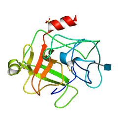
BOVINE DUODENASE(NEW SERINE PROTEASE), CRYSTAL STRUCTURE
Descriptor: 2-acetamido-2-deoxy-beta-D-glucopyranose, DUODENASE, PHOSPHATE ION
Authors:Pletnev, V.Z, Zamolodchikova, T.S, Pangborn, W.A, Duax, W.L.
Deposit date:2000-04-14
Release date:2001-04-14
Last modified:2024-10-30
Method:X-RAY DIFFRACTION (2.4 Å)
Cite:Crystal structure of bovine duodenase, a serine protease, with dual trypsin and chymotrypsin-like specificities.
Proteins, 41, 2000
6ZQO
DownloadVisualize
BU of 6zqo by Molmil
EYFP mutant - F165G
Descriptor: G protein/GFP fusion protein, SULFATE ION
Authors:Pletnev, V.Z, Pletnev, S.V, Pletneva, N.V.
Deposit date:2020-07-10
Release date:2021-06-16
Last modified:2024-10-16
Method:X-RAY DIFFRACTION (2.2 Å)
Cite:Amino acid residue at the 165th position tunes EYFP chromophore maturation. A structure-based design.
Comput Struct Biotechnol J, 19, 2021
2BQP
DownloadVisualize
BU of 2bqp by Molmil
THE STRUCTURE OF THE PEA LECTIN-D-GLUCOPYRANOSE COMPLEX
Descriptor: CALCIUM ION, MANGANESE (II) ION, PROTEIN (PEA LECTIN), ...
Authors:Ruzeinikov, S.N, Mikhailova Yu, I, Tsygannik, I.N, Pangborn, W, Duax, W, Pletnev, V.Z.
Deposit date:1998-12-08
Release date:1998-12-16
Last modified:2023-08-23
Method:X-RAY DIFFRACTION (1.9 Å)
Cite:The Structure of Pea Lectin-D-Glucopyranose Complex at a 1.9 A Resolution
RUSS.J.BIOORGANIC CHEM., 23, 1997
4ZBL
DownloadVisualize
BU of 4zbl by Molmil
Phototoxic fluorescent protein mKillerOrange
Descriptor: CITRIC ACID, GLYCEROL, KillerOrange
Authors:Pletnev, V.Z, Pletneva, N.V, Pletnev, S.V.
Deposit date:2015-04-14
Release date:2015-12-23
Last modified:2024-11-06
Method:X-RAY DIFFRACTION (1.57 Å)
Cite:Crystal Structure of Phototoxic Orange Fluorescent Proteins with a Tryptophan-Based Chromophore.
Plos One, 10, 2015
5EXB
DownloadVisualize
BU of 5exb by Molmil
Wild type green fluorescent protein DendFP (Dendronephthya sp.)
Descriptor: GLYCEROL, Green fluorescent protein
Authors:Pletnev, V.Z, Pletneva, N.V, Pletnev, S.V.
Deposit date:2015-11-23
Release date:2016-08-03
Last modified:2024-11-06
Method:X-RAY DIFFRACTION (1.81 Å)
Cite:Crystal structure of the fluorescent protein from Dendronephthya sp. in both green and photoconverted red forms.
Acta Crystallogr D Struct Biol, 72, 2016
5EXC
DownloadVisualize
BU of 5exc by Molmil
Photoconverted red fluorescent protein DendRFP
Descriptor: GLYCEROL, Green fluorescent protein, MAGNESIUM ION
Authors:Pletnev, V.Z, Pletneva, N.V, Pletnev, S.V.
Deposit date:2015-11-23
Release date:2016-08-03
Last modified:2023-11-15
Method:X-RAY DIFFRACTION (2.14 Å)
Cite:Crystal structure of the fluorescent protein from Dendronephthya sp. in both green and photoconverted red forms.
Acta Crystallogr D Struct Biol, 72, 2016
4N3D
DownloadVisualize
BU of 4n3d by Molmil
Crystal structure of the dimeric variant EGFP-K162Q in P61 space group
Descriptor: GLYCEROL, Green fluorescent protein, PHOSPHATE ION, ...
Authors:Pletneva, N.V, Pletnev, V.Z, Pletnev, S.V.
Deposit date:2013-10-07
Release date:2014-08-27
Method:X-RAY DIFFRACTION (1.34 Å)
Cite:Three dimensional structure of the dimeric gene-engineered variant of green fluorescent protein egfp-K162Q in P61 crystal space group
Rus.J.Bioorg.Chem., 40, 2014
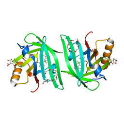
6UBO
DownloadVisualize
BU of 6ubo by Molmil
Fluorogen Activating Protein Dib1
Descriptor: 12-(diethylamino)-2,2-bis(fluoranyl)-4,5-dimethyl-5-aza-3-azonia-2-boranuidatricyclo[7.4.0.0^{3,7}]trideca-1(13),3,7,9,11-pentaen-6-one, CITRIC ACID, Outer membrane lipoprotein Blc, ...
Authors:Muslinkina, L, Pletneva, N, Pletnev, V.Z, Pletnev, S.
Deposit date:2019-09-12
Release date:2020-09-16
Last modified:2023-10-11
Method:X-RAY DIFFRACTION (1.58 Å)
Cite:Structure-Based Rational Design of Two Enhanced Bacterial Lipocalin Blc Tags for Protein-PAINT Super-resolution Microscopy.
Acs Chem.Biol., 15, 2020
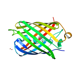
6U1A
DownloadVisualize
BU of 6u1a by Molmil
Crystal Structure of Fluorescent Protein FusionRed
Descriptor: CALCIUM ION, NICKEL (II) ION, Red fluorescent protein
Authors:Pletnev, S, Muslinkina, L, Pletneva, N, Pletnev, V.Z.
Deposit date:2019-08-15
Release date:2020-04-22
Last modified:2023-11-15
Method:X-RAY DIFFRACTION (1.09 Å)
Cite:Two independent routes of post-translational chemistry in fluorescent protein FusionRed.
Int.J.Biol.Macromol., 155, 2020
2OGR
DownloadVisualize
BU of 2ogr by Molmil
Crystal Structure of Yellow Fluorescent Protein from Zoanthus sp. at 1.8 A Resolution
Descriptor: FLUORESCENT PROTEIN FP538
Authors:Pletneva, N.V, Pletnev, S.V, Tikhonova, T.V, Pletnev, V.Z.
Deposit date:2007-01-08
Release date:2007-09-25
Last modified:2023-12-27
Method:X-RAY DIFFRACTION (1.8 Å)
Cite:Three-dimensional structure of yellow fluorescent protein zYFP538 from Zoanthus sp. at the resolution 1.8 angstrom
Bioorg.Khim., 33
4ZFS
DownloadVisualize
BU of 4zfs by Molmil
Phototoxic Fluorescent Protein KillerOrange
Descriptor: KillerOrange
Authors:Pletneva, N.V, Pletnev, V.Z, Pletnev, S.
Deposit date:2015-04-21
Release date:2015-12-23
Last modified:2024-11-06
Method:X-RAY DIFFRACTION (2.01 Å)
Cite:Crystal Structure of Phototoxic Orange Fluorescent Proteins with a Tryptophan-Based Chromophore.
Plos One, 10, 2015
4HE4
DownloadVisualize
BU of 4he4 by Molmil
Crystal structure of the yellow fluorescent protein phiYFP (Phialidium sp.)
Descriptor: Yellow fluorescent protein
Authors:Pletneva, N, Pletnev, S, Pletnev, V.Z.
Deposit date:2012-10-03
Release date:2013-05-29
Last modified:2024-11-06
Method:X-RAY DIFFRACTION (2.05 Å)
Cite:Yellow fluorescent protein phiYFPv (Phialidium): structure and structure-based mutagenesis.
Acta Crystallogr.,Sect.D, 69, 2013


123>

227111

건을2024-11-06부터공개중

PDB statisticsPDBj update infoContact PDBjnumon