Loading
PDBj
MenuPDBj@FacebookPDBj@X(formerly Twitter)PDBj@BlueSkyPDBj@YouTubewwPDB FoundationwwPDB
RCSB PDBPDBeBMRBAdv. SearchSearch help
Search by PDB author
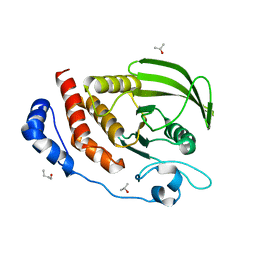
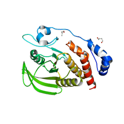
3S3E
DownloadVisualize
BU of 3s3e by Molmil
Crystal structure of the catalytic domain of PTP10D from Drosophila melanogaster
Descriptor: 1,4-BUTANEDIOL, 1-BUTANOL, ISOPROPYL ALCOHOL, ...
Authors:Madan, L.L, Gopal, B.
Deposit date:2011-05-18
Release date:2011-11-02
Last modified:2023-11-01
Method:X-RAY DIFFRACTION (2.4 Å)
Cite:Conformational basis for substrate recruitment in protein tyrosine phosphatase 10D
Biochemistry, 50, 2011
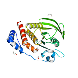
3S3H
DownloadVisualize
BU of 3s3h by Molmil
Crystal structure of the catalytic domain of PTP10D from Drosophila melanogaster with a phosphopeptide substrate GP4
Descriptor: 1,4-BUTANEDIOL, 1-BUTANOL, Tyrosine-protein phosphatase 10D, ...
Authors:Madan, L.L, Gopal, B.
Deposit date:2011-05-18
Release date:2011-11-02
Last modified:2024-10-16
Method:X-RAY DIFFRACTION (2.8 Å)
Cite:Conformational basis for substrate recruitment in protein tyrosine phosphatase 10D
Biochemistry, 50, 2011
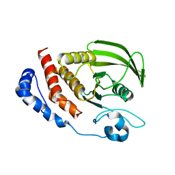
3S3F
DownloadVisualize
BU of 3s3f by Molmil
Crystal Structure of the catalytic domain of PTP10D from Drosophila melanogaster with a small molecule inhibitor Vanadate
Descriptor: 1,4-BUTANEDIOL, 1-BUTANOL, ISOPROPYL ALCOHOL, ...
Authors:Madan, L.L, Gopal, B.
Deposit date:2011-05-18
Release date:2011-11-02
Last modified:2023-11-01
Method:X-RAY DIFFRACTION (2.7 Å)
Cite:Conformational basis for substrate recruitment in protein tyrosine phosphatase 10D
Biochemistry, 50, 2011
3S3K
DownloadVisualize
BU of 3s3k by Molmil
Crystal structure of the catalytic domain of PTP10D from Drosophila melanogaster with a small molecular inhibitor para-NitroCatechol Sulphate
Descriptor: N,4-DIHYDROXY-N-OXO-3-(SULFOOXY)BENZENAMINIUM, Tyrosine-protein phosphatase 10D
Authors:Madan, L.L, Gopal, B.
Deposit date:2011-05-18
Release date:2011-11-02
Last modified:2023-11-01
Method:X-RAY DIFFRACTION (3.2 Å)
Cite:Conformational basis for substrate recruitment in protein tyrosine phosphatase 10D
Biochemistry, 50, 2011
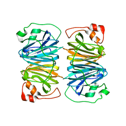
1Y3T
DownloadVisualize
BU of 1y3t by Molmil
Crystal structure of YxaG, a dioxygenase from Bacillus subtilis
Descriptor: FE (III) ION, Hypothetical protein yxaG
Authors:Gopal, B, Madan, L.L, Betz, S.F, Kossiakoff, A.A.
Deposit date:2004-11-26
Release date:2005-01-18
Last modified:2023-08-23
Method:X-RAY DIFFRACTION (2.4 Å)
Cite:The Crystal Structure of a Quercetin 2,3-Dioxygenase from Bacillus subtilis Suggests Modulation of Enzyme Activity by a Change in the Metal Ion at the Active Site(s)
Biochemistry, 44, 2005

227344

건을2024-11-13부터공개중

PDB statisticsPDBj update infoContact PDBjnumon