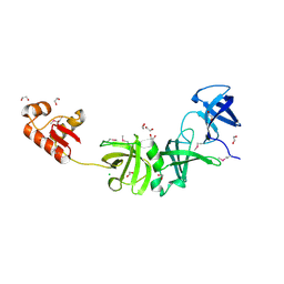
Loading
PDBj
MenuPDBj@FacebookPDBj@TwitterPDBj@YouTubewwPDB FoundationwwPDB
RCSB PDBPDBeBMRBAdv. SearchSearch help
Search by PDB author
1S96
DownloadVisualize
BU of 1s96 by Molmil
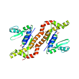
The 2.0 A X-ray structure of Guanylate Kinase from E.coli
Descriptor: Guanylate kinase, PHOSPHATE ION, UNKNOWN ATOM OR ION
Authors:Kreusch, A, Spraggon, G, Klock, H.E, McMullan, D, Vincent, J, Rodrigues, K, Lesley, S.A.
Deposit date:2004-02-03
Release date:2004-02-10
Last modified:2011-07-13
Method:X-RAY DIFFRACTION (2 Å)
Cite:The Structure of Guanylate Kinase from Escherichia coli at 2.0 A resolution
To be Published
1R73
DownloadVisualize
BU of 1r73 by Molmil
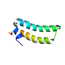
Solution Structure of TM1492, the L29 ribosomal protein from Thermotoga maritima
Descriptor: 50S ribosomal protein L29
Authors:Peti, W, Etezady-Esfarjani, T, Herrmann, T, Klock, H.E, Lesley, S.A, Wuethrich, K, Joint Center for Structural Genomics (JCSG)
Deposit date:2003-10-17
Release date:2004-08-10
Last modified:2024-05-22
Method:SOLUTION NMR
Cite:NMR for structural proteomics of Thermotoga maritima: Screening and structure determination
J.STRUCT.FUNCT.GENOM., 5, 2004
1RDU
DownloadVisualize
BU of 1rdu by Molmil
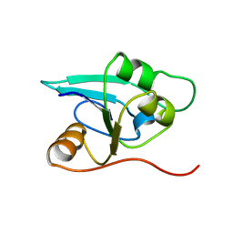
NMR STRUCTURE OF A PUTATIVE NIFB PROTEIN FROM THERMOTOGA (TM1290), WHICH BELONGS TO THE DUF35 FAMILY
Descriptor: conserved hypothetical protein
Authors:Etezady-Esfarjani, T, Herrmann, T, Peti, W, Klock, H.E, Lesley, S.A, Wuthrich, K, Joint Center for Structural Genomics (JCSG)
Deposit date:2003-11-06
Release date:2004-07-06
Last modified:2024-05-22
Method:SOLUTION NMR
Cite:NMR Structure Determination of the Hypothetical Protein TM1290 from Thermotoga Maritima using Automated NOESY Analysis.
J.Biomol.NMR, 29, 2004
1SJ5
DownloadVisualize
BU of 1sj5 by Molmil
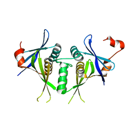
Crystal structure of a duf151 family protein (tm0160) from thermotoga maritima at 2.8 A resolution
Descriptor: conserved hypothetical protein TM0160
Authors:Spraggon, G, Panatazatos, D, Klock, H.E, Wilson, I.A, Woods Jr, V.L, Lesley, S.A, Joint Center for Structural Genomics (JCSG)
Deposit date:2004-03-02
Release date:2005-03-01
Last modified:2023-08-23
Method:X-RAY DIFFRACTION (2.8 Å)
Cite:On the use of DXMS to produce more crystallizable proteins: structures of the T. maritima proteins TM0160 and TM1171.
Protein Sci., 13, 2004
1VKB
DownloadVisualize
BU of 1vkb by Molmil
Crystal structure of an aig2-like protein (a2ld1, ggact, mgc7867) from mus musculus at 1.90 A resolution
Descriptor: FORMIC ACID, hypothetical protein
Authors:Joint Center for Structural Genomics (JCSG)
Deposit date:2004-05-07
Release date:2004-05-18
Last modified:2023-01-25
Method:X-RAY DIFFRACTION (1.9 Å)
Cite:Crystal structure of a conserved hypothetical protein (gi: 13879369) from Mouse at 1.90 A resolution reveals a new fold.
Proteins, 61, 2005
3HBZ
DownloadVisualize
BU of 3hbz by Molmil
Crystal structure of a putative glycoside hydrolase (bt_2081) from bacteroides thetaiotaomicron vpi-5482 at 2.05 A resolution
Descriptor: ACETATE ION, CACODYLATE ION, CALCIUM ION, ...
Authors:Joint Center for Structural Genomics (JCSG)
Deposit date:2009-05-05
Release date:2009-05-26
Last modified:2023-02-01
Method:X-RAY DIFFRACTION (2.05 Å)
Cite:Structure of Bacteroides thetaiotaomicron BT2081 at 2.05 A resolution: the first structural representative of a new protein family that may play a role in carbohydrate metabolism.
Acta Crystallogr.,Sect.F, 66, 2010
3GO5
DownloadVisualize
BU of 3go5 by Molmil
Crystal structure of a multidomain protein with nucleic acid binding domains (sp_0946) from streptococcus pneumoniae tigr4 at 1.40 A resolution
Descriptor: 1,2-ETHANEDIOL, CHLORIDE ION, Multidomain protein with S1 RNA-binding domains
Authors:Joint Center for Structural Genomics (JCSG)
Deposit date:2009-03-18
Release date:2009-04-07
Last modified:2023-02-01
Method:X-RAY DIFFRACTION (1.4 Å)
Cite:Structure of a virulence regulatory factor CvfB reveals a novel winged helix RNA binding module.
Structure, 18, 2010
2FNO
DownloadVisualize
BU of 2fno by Molmil
Crystal structure of a glutathione s-transferase (atu5508) from agrobacterium tumefaciens str. c58 at 2.00 A resolution
Descriptor: AGR_pAT_752p, THIOCYANATE ION
Authors:Joint Center for Structural Genomics (JCSG)
Deposit date:2006-01-11
Release date:2006-02-14
Last modified:2023-01-25
Method:X-RAY DIFFRACTION (2 Å)
Cite:Comparative structural analysis of a novel glutathioneS-transferase (ATU5508) from Agrobacterium tumefaciens at 2.0 A resolution.
Proteins, 65, 2006
3PXP
DownloadVisualize
BU of 3pxp by Molmil
Crystal structure of a PAS and DNA binding domain containing protein (Caur_2278) from CHLOROFLEXUS AURANTIACUS J-10-FL at 2.30 A resolution
Descriptor: 1,2-ETHANEDIOL, CHLORIDE ION, Helix-turn-helix domain protein, ...
Authors:Joint Center for Structural Genomics (JCSG)
Deposit date:2010-12-10
Release date:2011-01-19
Last modified:2023-02-01
Method:X-RAY DIFFRACTION (2.3 Å)
Cite:Structure of an MmyB-Like Regulator from C. aurantiacus, Member of a New Transcription Factor Family Linked to Antibiotic Metabolism in Actinomycetes.
Plos One, 7, 2012
3U21
DownloadVisualize
BU of 3u21 by Molmil
Crystal structure of a Fragment of Nuclear factor related to kappa-B-binding protein (residues 370-495) (NFRKB) from Homo sapiens at 2.18 A resolution
Descriptor: Nuclear factor related to kappa-B-binding protein, SODIUM ION
Authors:Joint Center for Structural Genomics (JCSG), Partnership for Stem Cell Biology, Partnership for Stem Cell Biology (STEMCELL)
Deposit date:2011-09-30
Release date:2011-11-02
Last modified:2023-02-01
Method:X-RAY DIFFRACTION (2.18 Å)
Cite:Structure of a Novel Winged-Helix Like Domain from Human NFRKB Protein.
Plos One, 7, 2012
1VK3
DownloadVisualize
BU of 1vk3 by Molmil
Crystal structure of Phosphoribosylformylglycinamidine synthase II (TM1246) from Thermotoga maritima at 2.15 A resolution
Descriptor: CHLORIDE ION, Phosphoribosylformylglycinamidine synthase II
Authors:Joint Center for Structural Genomics (JCSG)
Deposit date:2004-04-23
Release date:2004-05-11
Last modified:2023-01-25
Method:X-RAY DIFFRACTION (2.15 Å)
Cite:Crystal structure of phosphoribosylformylglycinamidine synthase II (smPurL) from Thermotoga maritima at 2.15 A resolution.
Proteins, 63, 2006
1VL4
DownloadVisualize
BU of 1vl4 by Molmil
CRYSTAL STRUCTURE OF A PUTATIVE MODULATOR OF A DNA GYRASE (TM0727) FROM THERMOTOGA MARITIMA MSB8 AT 1.95 A RESOLUTION
Descriptor: pmbA-related protein
Authors:Joint Center for Structural Genomics (JCSG)
Deposit date:2004-07-09
Release date:2004-09-21
Last modified:2024-05-22
Method:X-RAY DIFFRACTION (1.95 Å)
Cite:Crystal structure of a putative modulator of DNA gyrase (pmbA) from Thermotoga maritima at 1.95 A resolution reveals a new fold.
Proteins, 61, 2005
1VJO
DownloadVisualize
BU of 1vjo by Molmil
Crystal structure of Alanine--glyoxylate aminotransferase (ALR1004) from Nostoc sp. at 1.70 A resolution
Descriptor: PYRIDOXAL-5'-PHOSPHATE, alanine--glyoxylate aminotransferase
Authors:Joint Center for Structural Genomics (JCSG)
Deposit date:2004-03-16
Release date:2004-03-23
Last modified:2023-01-25
Method:X-RAY DIFFRACTION (1.7 Å)
Cite:Crystal structure of an alanine-glyoxylate aminotransferase from Anabaena sp. at 1.70 A resolution reveals a noncovalently linked PLP cofactor
Proteins, 58, 2005
1VQ0
DownloadVisualize
BU of 1vq0 by Molmil
Crystal structure of 33 kDa chaperonin (Heat shock protein 33 homolog) (HSP33) (TM1394) from Thermotoga maritima at 2.20 A resolution
Descriptor: 1,2-ETHANEDIOL, 33 kDa chaperonin, CHLORIDE ION, ...
Authors:Joint Center for Structural Genomics (JCSG)
Deposit date:2004-11-30
Release date:2004-12-14
Last modified:2023-09-20
Method:X-RAY DIFFRACTION (2.2 Å)
Cite:Crystal structure of Hsp33 chaperone (TM1394) from Thermotoga maritima at 2.20 A resolution.
Proteins, 61, 2005
1VR3
DownloadVisualize
BU of 1vr3 by Molmil
Crystal structure of Acireductone dioxygenase (13543033) from Mus musculus at 2.06 A resolution
Descriptor: Acireductone dioxygenase, ISOPROPYL ALCOHOL, NICKEL (II) ION, ...
Authors:Joint Center for Structural Genomics (JCSG)
Deposit date:2005-01-28
Release date:2005-02-15
Last modified:2023-01-25
Method:X-RAY DIFFRACTION (2.06 Å)
Cite:Crystal structure of acireductone dioxygenase (ARD) from Mus musculus at 2.06 angstrom resolution.
Proteins, 64, 2006
1VRM
DownloadVisualize
BU of 1vrm by Molmil
Crystal structure of the apbe protein (tm1553) from thermotoga maritima msb8 at 1.58 A resolution
Descriptor: (4R)-2-METHYLPENTANE-2,4-DIOL, (4S)-2-METHYL-2,4-PENTANEDIOL, UNKNOWN LIGAND, ...
Authors:Joint Center for Structural Genomics (JCSG)
Deposit date:2005-03-10
Release date:2005-03-22
Last modified:2023-01-25
Method:X-RAY DIFFRACTION (1.58 Å)
Cite:Crystal structure of the ApbE protein (TM1553) from Thermotoga maritima at 1.58 A resolution.
Proteins, 64, 2006
2RE3
DownloadVisualize
BU of 2re3 by Molmil
CRYSTAL STRUCTURE OF a DUF1285 family protein (SPO_0140) FROM SILICIBACTER POMEROYI DSS-3 AT 2.50 A RESOLUTION
Descriptor: GLYCEROL, Uncharacterized protein
Authors:Joint Center for Structural Genomics (JCSG)
Deposit date:2007-09-25
Release date:2007-10-09
Last modified:2023-01-25
Method:X-RAY DIFFRACTION (2.5 Å)
Cite:Structures of the first representatives of Pfam family PF06938 (DUF1285) reveal a new fold with repeated structural motifs and possible involvement in signal transduction.
Acta Crystallogr.,Sect.F, 66, 2010
2RA9
DownloadVisualize
BU of 2ra9 by Molmil
Crystal structure of a duf1285 family protein (sbal_2486) from shewanella baltica os155 at 1.40 A resolution
Descriptor: 1,2-ETHANEDIOL, SODIUM ION, Uncharacterized protein DUF1285
Authors:Joint Center for Structural Genomics (JCSG)
Deposit date:2007-09-14
Release date:2007-10-16
Last modified:2023-01-25
Method:X-RAY DIFFRACTION (1.4 Å)
Cite:Structures of the first representatives of Pfam family PF06938 (DUF1285) reveal a new fold with repeated structural motifs and possible involvement in signal transduction.
Acta Crystallogr.,Sect.F, 66, 2010
2QTP
DownloadVisualize
BU of 2qtp by Molmil
Crystal structure of a duf1185 family protein (spo0826) from silicibacter pomeroyi dss-3 at 2.10 A resolution
Descriptor: Uncharacterized protein
Authors:Joint Center for Structural Genomics (JCSG)
Deposit date:2007-08-02
Release date:2007-08-21
Last modified:2023-01-25
Method:X-RAY DIFFRACTION (2.1 Å)
Cite:Structures of the first representatives of Pfam family PF06684 (DUF1185) reveal a novel variant of the Bacillus chorismate mutase fold and suggest a role in amino-acid metabolism.
Acta Crystallogr.,Sect.F, 66, 2010
2OOC
DownloadVisualize
BU of 2ooc by Molmil
Crystal structure of Histidine Phosphotransferase ShpA (NP_419930.1) from Caulobacter crescentus at 1.52 A resolution
Descriptor: GLYCEROL, Histidine phosphotransferase, TETRAETHYLENE GLYCOL
Authors:Joint Center for Structural Genomics (JCSG)
Deposit date:2007-01-25
Release date:2007-02-06
Last modified:2023-01-25
Method:X-RAY DIFFRACTION (1.52 Å)
Cite:Crystal structure of histidine phosphotransfer protein ShpA, an essential regulator of stalk biogenesis in Caulobacter crescentus.
J.Mol.Biol., 390, 2009
3BYQ
DownloadVisualize
BU of 3byq by Molmil
Crystal structure of a duf1185 family protein (bb2672) from bordetella bronchiseptica rb50 at 1.70 A resolution
Descriptor: 1,2-ETHANEDIOL, CHLORIDE ION, TETRAETHYLENE GLYCOL, ...
Authors:Joint Center for Structural Genomics (JCSG)
Deposit date:2008-01-16
Release date:2008-01-29
Last modified:2023-02-01
Method:X-RAY DIFFRACTION (1.7 Å)
Cite:Structures of the first representatives of Pfam family PF06684 (DUF1185) reveal a novel variant of the Bacillus chorismate mutase fold and suggest a role in amino-acid metabolism.
Acta Crystallogr.,Sect.F, 66, 2010
3B77
DownloadVisualize
BU of 3b77 by Molmil
Crystal structure of a ph domain containing bacterial protein (exig_2160) from exiguobacterium sibiricum 255-15 at 2.42 A resolution
Descriptor: Uncharacterized protein
Authors:Joint Center for Structural Genomics (JCSG)
Deposit date:2007-10-30
Release date:2007-11-13
Last modified:2023-01-25
Method:X-RAY DIFFRACTION (2.42 Å)
Cite:Bacterial pleckstrin homology domains: a prokaryotic origin for the PH domain.
J.Mol.Biol., 396, 2010
2PV7
DownloadVisualize
BU of 2pv7 by Molmil
Crystal structure of chorismate mutase / prephenate dehydrogenase (tyrA) (1574749) from Haemophilus influenzae RD at 2.00 A resolution
Descriptor: NICOTINAMIDE-ADENINE-DINUCLEOTIDE, T-protein [Includes: Chorismate mutase (EC 5.4.99.5) (CM) and Prephenate dehydrogenase (EC 1.3.1.12) (PDH)], TYROSINE
Authors:Joint Center for Structural Genomics (JCSG)
Deposit date:2007-05-09
Release date:2007-05-22
Last modified:2023-01-25
Method:X-RAY DIFFRACTION (2 Å)
Cite:The structure of Haemophilus influenzae prephenate dehydrogenase suggests unique features of bifunctional TyrA enzymes.
Acta Crystallogr.,Sect.F, 66, 2010
2Q3L
DownloadVisualize
BU of 2q3l by Molmil
CRYSTAL STRUCTURE OF AN UNCHARACTERIZED PROTEIN FROM DUF3478 FAMILY WITH A SPOIIAA-LIKE FOLD (SHEW_3102) FROM SHEWANELLA LOIHICA PV-4 AT 2.25 A RESOLUTION
Descriptor: (4S)-2-METHYL-2,4-PENTANEDIOL, CHLORIDE ION, SODIUM ION, ...
Authors:Joint Center for Structural Genomics (JCSG)
Deposit date:2007-05-30
Release date:2007-06-12
Last modified:2023-01-25
Method:X-RAY DIFFRACTION (2.25 Å)
Cite:Open and closed conformations of two SpoIIAA-like proteins (YP_749275.1 and YP_001095227.1) provide insights into membrane association and ligand binding.
Acta Crystallogr.,Sect.F, 66, 2010
3BY7
DownloadVisualize
BU of 3by7 by Molmil
CRYSTAL STRUCTURE OF A PROTEIN STRUCTURALLY SIMILAR TO SM/LSM-LIKE RNA-BINDING PROTEINS (JCVI_PEP_1096686650277) FROM UNCULTURED MARINE ORGANISM AT 2.60 A RESOLUTION
Descriptor: uncharacterized protein
Authors:Joint Center for Structural Genomics (JCSG)
Deposit date:2008-01-15
Release date:2008-01-29
Last modified:2023-02-01
Method:X-RAY DIFFRACTION (2.6 Å)
Cite:Crystal structure of a novel Sm-like protein of putative cyanophage origin at 2.60 A resolution.
Proteins, 75, 2009

222415

건을2024-07-10부터공개중

PDB statisticsPDBj update infoContact PDBjnumon