Loading
PDBj
MenuPDBj@FacebookPDBj@TwitterPDBj@YouTubewwPDB FoundationwwPDB
RCSB PDBPDBeBMRBAdv. SearchSearch help
Search by PDB author
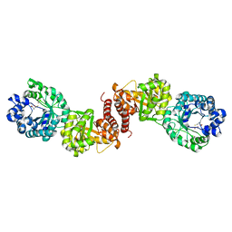
5WPR
DownloadVisualize
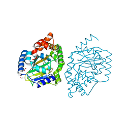
BU of 5wpr by Molmil
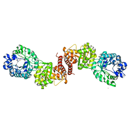
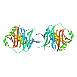
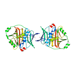
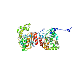
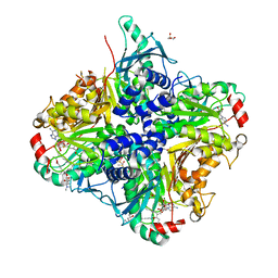
Crystal structure HpiC1 in C2 space group
Descriptor: 12-epi-hapalindole C/U synthase, 2-AMINO-2-HYDROXYMETHYL-PROPANE-1,3-DIOL, CALCIUM ION, ...
Authors:Newmister, S.A, Li, S, Garcia-Borras, M, Sanders, J.N, Yang, S, Lowell, A.N, Yu, F, Smith, J.L, Williams, R.M, Houk, K.N, Sherman, D.H.
Deposit date:2017-08-07
Release date:2018-03-07
Last modified:2023-10-04
Method:X-RAY DIFFRACTION (1.49 Å)
Cite:Structural basis of the Cope rearrangement and cyclization in hapalindole biogenesis.
Nat. Chem. Biol., 14, 2018
5WM3
DownloadVisualize
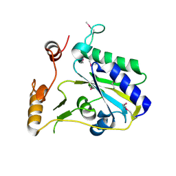
BU of 5wm3 by Molmil
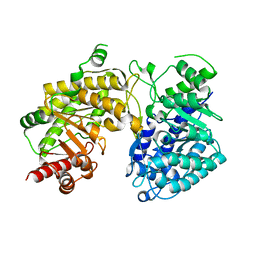
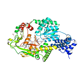
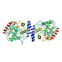
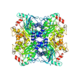
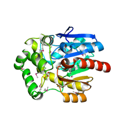
Crystal Structure of CahJ in Complex with Salicyl Adenylate
Descriptor: 9-(5-O-{(S)-hydroxy[(2-hydroxybenzene-1-carbonyl)oxy]phosphoryl}-alpha-L-lyxofuranosyl)-9H-purin-6-amine, ACETATE ION, GLYCEROL, ...
Authors:Sikkema, A.P, Smith, J.L.
Deposit date:2017-07-28
Release date:2018-05-23
Last modified:2023-10-04
Method:X-RAY DIFFRACTION (1.679 Å)
Cite:A Defined and Flexible Pocket Explains Aryl Substrate Promiscuity of the Cahuitamycin Starter Unit-Activating Enzyme CahJ.
Chembiochem, 19, 2018
5WPP
DownloadVisualize
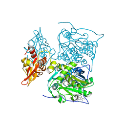
BU of 5wpp by Molmil
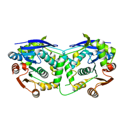
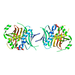
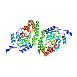
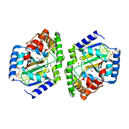
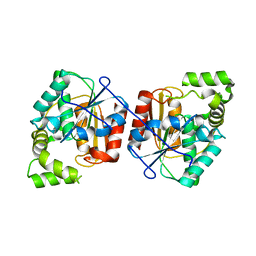
Crystal structure HpiC1 W73M/K132M
Descriptor: 12-epi-hapalindole C/U synthase, CALCIUM ION, TETRAETHYLENE GLYCOL, ...
Authors:Newmister, S.A, Li, S, Garcia-Borras, M, Sanders, J.N, Yang, S, Lowell, A.N, Yu, F, Smith, J.L, Williams, R.M, Houk, K.N, Sherman, D.H.
Deposit date:2017-08-07
Release date:2018-03-07
Last modified:2024-03-13
Method:X-RAY DIFFRACTION (1.7 Å)
Cite:Structural basis of the Cope rearrangement and cyclization in hapalindole biogenesis.
Nat. Chem. Biol., 14, 2018
5W7R
DownloadVisualize
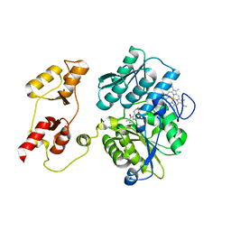
BU of 5w7r by Molmil
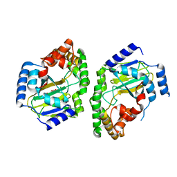
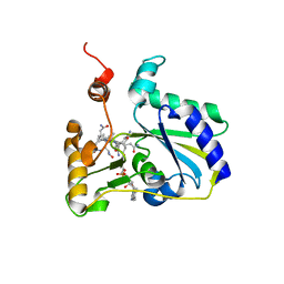
Crystal structure of OxaC in complex with SAH and oxaline
Descriptor: (3E,7aR,12aS)-3-[(1H-imidazol-4-yl)methylidene]-6,12-dimethoxy-7a-(2-methylbut-3-en-2-yl)-7a,12-dihydro-1H,5H-imidazo[1 ',2':1,2]pyrido[2,3-b]indole-2,5(3H)-dione, OxaC, S-ADENOSYL-L-HOMOCYSTEINE
Authors:Newmister, S.A, Romminger, S, Schmidt, J.J, Williams, R.M, Smith, J.L, Berlinck, R.G.S, Sherman, D.H.
Deposit date:2017-06-20
Release date:2018-07-04
Last modified:2023-10-04
Method:X-RAY DIFFRACTION (2.497 Å)
Cite:Unveiling sequential late-stage methyltransferase reactions in the meleagrin/oxaline biosynthetic pathway.
Org. Biomol. Chem., 16, 2018
5WGY
DownloadVisualize
BU of 5wgy by Molmil
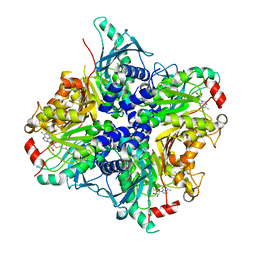
Crystal Structure of MalA' C112S/C128S, malbrancheamide B complex
Descriptor: (5aS,12aS,13aS)-9-chloro-12,12-dimethyl-2,3,11,12,12a,13-hexahydro-1H,5H,6H-5a,13a-(epiminomethano)indolizino[7,6-b]carbazol-14-one, CADMIUM ION, CHLORIDE ION, ...
Authors:Fraley, A.E, Smith, J.L.
Deposit date:2017-07-14
Release date:2017-08-16
Last modified:2023-10-04
Method:X-RAY DIFFRACTION (2 Å)
Cite:Function and Structure of MalA/MalA', Iterative Halogenases for Late-Stage C-H Functionalization of Indole Alkaloids.
J. Am. Chem. Soc., 139, 2017
5WM6
DownloadVisualize
BU of 5wm6 by Molmil
Crystal Structure of CahJ in Complex with Benzoyl Adenylate
Descriptor: 5'-O-[(R)-(benzoyloxy)(hydroxy)phosphoryl]adenosine, ACETATE ION, MAGNESIUM ION, ...
Authors:Sikkema, A.P, Smith, J.L.
Deposit date:2017-07-28
Release date:2018-05-23
Last modified:2023-10-04
Method:X-RAY DIFFRACTION (2 Å)
Cite:A Defined and Flexible Pocket Explains Aryl Substrate Promiscuity of the Cahuitamycin Starter Unit-Activating Enzyme CahJ.
Chembiochem, 19, 2018
5WPU
DownloadVisualize
BU of 5wpu by Molmil
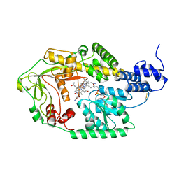
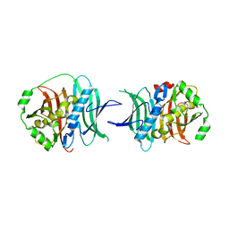
Crystal structure HpiC1 Y101S
Descriptor: 12-epi-hapalindole C/U synthase, 2-AMINO-2-HYDROXYMETHYL-PROPANE-1,3-DIOL, CALCIUM ION
Authors:Newmister, S.A, Li, S, Garcia-Borras, M, Sanders, J.N, Yang, S, Lowell, A.N, Yu, F, Smith, J.L, Williams, R.M, Houk, K.N, Sherman, D.H.
Deposit date:2017-08-07
Release date:2018-03-07
Last modified:2023-10-04
Method:X-RAY DIFFRACTION (1.39 Å)
Cite:Structural basis of the Cope rearrangement and cyclization in hapalindole biogenesis.
Nat. Chem. Biol., 14, 2018
5WGW
DownloadVisualize
BU of 5wgw by Molmil
Crystal Structure of Wild-type MalA', malbrancheamide B complex
Descriptor: (5aS,12aS,13aS)-9-chloro-12,12-dimethyl-2,3,11,12,12a,13-hexahydro-1H,5H,6H-5a,13a-(epiminomethano)indolizino[7,6-b]carbazol-14-one, CADMIUM ION, CHLORIDE ION, ...
Authors:Fraley, A.E, Smith, J.L.
Deposit date:2017-07-14
Release date:2017-08-16
Last modified:2019-11-27
Method:X-RAY DIFFRACTION (2.092 Å)
Cite:Function and Structure of MalA/MalA', Iterative Halogenases for Late-Stage C-H Functionalization of Indole Alkaloids.
J. Am. Chem. Soc., 139, 2017
5W7P
DownloadVisualize
BU of 5w7p by Molmil
Crystal structure of OxaC
Descriptor: OxaC, S-ADENOSYLMETHIONINE
Authors:Newmister, S.A, Romminger, S, Schmidt, J.J, Williams, R.M, Smith, J.L, Berlinck, R.G.S, Sherman, D.H.
Deposit date:2017-06-20
Release date:2018-06-27
Last modified:2024-03-13
Method:X-RAY DIFFRACTION (2.4 Å)
Cite:Unveiling sequential late-stage methyltransferase reactions in the meleagrin/oxaline biosynthetic pathway.
Org. Biomol. Chem., 16, 2018
5WGT
DownloadVisualize
BU of 5wgt by Molmil
Crystal Structure of MalA' H253A, premalbrancheamide complex
Descriptor: (5aS,12aS,13aS)-12,12-dimethyl-2,3,11,12,12a,13-hexahydro-1H,5H,6H-5a,13a-(epiminomethano)indolizino[7,6-b]carbazol-14-one, CADMIUM ION, CHLORIDE ION, ...
Authors:Fraley, A.E, Smith, J.L.
Deposit date:2017-07-14
Release date:2017-08-16
Last modified:2023-10-04
Method:X-RAY DIFFRACTION (2.093 Å)
Cite:Function and Structure of MalA/MalA', Iterative Halogenases for Late-Stage C-H Functionalization of Indole Alkaloids.
J. Am. Chem. Soc., 139, 2017
5WM4
DownloadVisualize
BU of 5wm4 by Molmil
Crystal Structure of CahJ in Complex with 6-Methylsalicyl Adenylate
Descriptor: 9-(5-O-{(S)-hydroxy[(2-hydroxy-6-methylbenzene-1-carbonyl)oxy]phosphoryl}-alpha-L-lyxofuranosyl)-9H-purin-6-amine, ACETATE ION, GLYCEROL, ...
Authors:Sikkema, A.P, Smith, J.L.
Deposit date:2017-07-28
Release date:2018-05-23
Last modified:2023-10-04
Method:X-RAY DIFFRACTION (1.781 Å)
Cite:A Defined and Flexible Pocket Explains Aryl Substrate Promiscuity of the Cahuitamycin Starter Unit-Activating Enzyme CahJ.
Chembiochem, 19, 2018
5W7K
DownloadVisualize
BU of 5w7k by Molmil
Crystal structure of OxaG
Descriptor: CHLORIDE ION, OxaG, S-ADENOSYL-L-HOMOCYSTEINE
Authors:Newmister, S.A, Romminger, S, Schmidt, J.J, Williams, R.M, Smith, J.L, Berlinck, R.G.S, Sherman, D.H.
Deposit date:2017-06-20
Release date:2018-06-27
Last modified:2024-03-13
Method:X-RAY DIFFRACTION (1.994 Å)
Cite:Unveiling sequential late-stage methyltransferase reactions in the meleagrin/oxaline biosynthetic pathway.
Org. Biomol. Chem., 16, 2018
5WM7
DownloadVisualize
BU of 5wm7 by Molmil
Crystal Structure of CahJ in Complex with AMP
Descriptor: ACETATE ION, ADENOSINE MONOPHOSPHATE, GLYCEROL, ...
Authors:Sikkema, A.P, Smith, J.L.
Deposit date:2017-07-28
Release date:2018-05-23
Last modified:2023-10-04
Method:X-RAY DIFFRACTION (1.777 Å)
Cite:A Defined and Flexible Pocket Explains Aryl Substrate Promiscuity of the Cahuitamycin Starter Unit-Activating Enzyme CahJ.
Chembiochem, 19, 2018
5WPS
DownloadVisualize
BU of 5wps by Molmil
Crystal structure HpiC1 Y101F
Descriptor: 1,2-ETHANEDIOL, 12-epi-hapalindole C/U synthase, 2-AMINO-2-HYDROXYMETHYL-PROPANE-1,3-DIOL, ...
Authors:Newmister, S.A, Li, S, Garcia-Borras, M, Sanders, J.N, Yang, S, Lowell, A.N, Yu, F, Smith, J.L, Williams, R.M, Houk, K.N, Sherman, D.H.
Deposit date:2017-08-07
Release date:2018-04-04
Last modified:2023-10-04
Method:X-RAY DIFFRACTION (1.389 Å)
Cite:Structural basis of the Cope rearrangement and cyclization in hapalindole biogenesis.
Nat. Chem. Biol., 14, 2018
5THY
DownloadVisualize
BU of 5thy by Molmil
Crystal structure of SeMet-Substituted CurJ carbon methyltransferase
Descriptor: CurJ, OXIDIZED GLUTATHIONE DISULFIDE, S-ADENOSYL-L-HOMOCYSTEINE
Authors:Skiba, M.A, Smith, J.L.
Deposit date:2016-09-30
Release date:2016-10-19
Last modified:2019-12-04
Method:X-RAY DIFFRACTION (2.087 Å)
Cite:Domain Organization and Active Site Architecture of a Polyketide Synthase C-methyltransferase.
ACS Chem. Biol., 11, 2016
7LO1
DownloadVisualize
BU of 7lo1 by Molmil
FAD-dependent monooxygenase AfoD from A. nidulans
Descriptor: FAD-dependent monooxygenase afoD, FLAVIN-ADENINE DINUCLEOTIDE, SULFATE ION
Authors:Rodriguez Benitez, A, Smith, J.L, Narayan, A.R.H.
Deposit date:2021-02-08
Release date:2022-04-27
Last modified:2023-10-25
Method:X-RAY DIFFRACTION (2.09 Å)
Cite:Deciphering the evolution of flavin-dependent monooxygenase stereoselectivity using ancestral sequence reconstruction.
Proc.Natl.Acad.Sci.USA, 120, 2023
7K93
DownloadVisualize
BU of 7k93 by Molmil
DENV2 NS1 in complex with neutralizing 2B7 single chain Fab variable region (scFv)
Descriptor: 2-acetamido-2-deoxy-beta-D-glucopyranose, 2B7 single chain fab variable region, Non-structural protein 1
Authors:Akey, D.L, Smith, J.L.
Deposit date:2020-09-28
Release date:2020-12-23
Last modified:2023-10-18
Method:X-RAY DIFFRACTION (2.89 Å)
Cite:Structural basis for antibody inhibition of flavivirus NS1-triggered endothelial dysfunction.
Science, 371, 2021
2Q2X
DownloadVisualize
BU of 2q2x by Molmil
Crystal Structure of the ECH2 decarboxylase domain of CurF from Lyngbya majuscula
Descriptor: CurF, GLYCEROL
Authors:Geders, T.W, Mowers, J.C, Smith, J.L.
Deposit date:2007-05-29
Release date:2007-10-09
Last modified:2017-10-18
Method:X-RAY DIFFRACTION (2 Å)
Cite:Crystal structure of the ECH2 catalytic domain of CurF from Lyngbya majuscula. Insights into a decarboxylase involved in polyketide chain beta-branching.
J.Biol.Chem., 282, 2007
2REF
DownloadVisualize
BU of 2ref by Molmil
Crystal structure of the loading GNATL domain of CurA from Lyngbya majuscula soaked with malonyl-CoA
Descriptor: ACETYL COENZYME *A, CurA
Authors:Geders, T.W, Smith, J.L.
Deposit date:2007-09-26
Release date:2007-11-20
Last modified:2024-04-03
Method:X-RAY DIFFRACTION (2.75 Å)
Cite:GNAT-like strategy for polyketide chain initiation.
Science, 318, 2007
1AO0
DownloadVisualize
BU of 1ao0 by Molmil
GLUTAMINE PHOSPHORIBOSYLPYROPHOSPHATE (PRPP) AMIDOTRANSFERASE FROM B. SUBTILIS COMPLEXED WITH ADP AND GMP
Descriptor: ADENOSINE-5'-DIPHOSPHATE, GLUTAMINE PHOSPHORIBOSYLPYROPHOSPHATE AMIDOTRANSFERASE, GUANOSINE-5'-MONOPHOSPHATE, ...
Authors:Tomchick, D.R, Smith, J.L.
Deposit date:1997-07-15
Release date:1997-11-12
Last modified:2024-05-22
Method:X-RAY DIFFRACTION (2.8 Å)
Cite:Mechanism of the synergistic end-product regulation of Bacillus subtilis glutamine phosphoribosylpyrophosphate amidotransferase by nucleotides.
Biochemistry, 36, 1997
2REE
DownloadVisualize
BU of 2ree by Molmil
Crystal structure of the loading GNATL domain of CurA from Lyngbya majuscula
Descriptor: CHLORIDE ION, CurA, GLYCEROL, ...
Authors:Geders, T.W, Smith, J.L.
Deposit date:2007-09-26
Release date:2007-11-20
Last modified:2024-02-21
Method:X-RAY DIFFRACTION (1.95 Å)
Cite:GNAT-like strategy for polyketide chain initiation.
Science, 318, 2007
1A3C
DownloadVisualize
BU of 1a3c by Molmil
PYRR, THE BACILLUS SUBTILIS PYRIMIDINE BIOSYNTHETIC OPERON REPRESSOR, DIMERIC FORM
Descriptor: PYRIMIDINE OPERON REGULATORY PROTEIN PYRR, SAMARIUM (III) ION, SULFATE ION
Authors:Tomchick, D.R, Turner, R.J, Switzer, R.W, Smith, J.L.
Deposit date:1998-01-20
Release date:1998-08-05
Last modified:2024-02-07
Method:X-RAY DIFFRACTION (1.6 Å)
Cite:Adaptation of an enzyme to regulatory function: structure of Bacillus subtilis PyrR, a pyr RNA-binding attenuation protein and uracil phosphoribosyltransferase.
Structure, 6, 1998
1A4X
DownloadVisualize
BU of 1a4x by Molmil
PYRR, THE BACILLUS SUBTILIS PYRIMIDINE BIOSYNTHETIC OPERON REPRESSOR, HEXAMERIC FORM
Descriptor: PYRIMIDINE OPERON REGULATORY PROTEIN PYRR, SULFATE ION
Authors:Tomchick, D.R, Turner, R.J, Switzer, R.W, Smith, J.L.
Deposit date:1998-02-08
Release date:1998-08-05
Last modified:2024-05-22
Method:X-RAY DIFFRACTION (2.3 Å)
Cite:Adaptation of an enzyme to regulatory function: structure of Bacillus subtilis PyrR, a pyr RNA-binding attenuation protein and uracil phosphoribosyltransferase.
Structure, 6, 1998
1ECJ
DownloadVisualize
BU of 1ecj by Molmil
ESCHERICHIA COLI GLUTAMINE PHOSPHORIBOSYLPYROPHOSPHATE (PRPP) AMIDOTRANSFERASE COMPLEXED WITH 2 AMP PER TETRAMER
Descriptor: ADENOSINE MONOPHOSPHATE, GLUTAMINE PHOSPHORIBOSYLPYROPHOSPHATE AMIDOTRANSFERASE
Authors:Muchmore, C.R, Krahn, J.M, Smith, J.L.
Deposit date:1997-07-16
Release date:1998-04-15
Last modified:2024-05-22
Method:X-RAY DIFFRACTION (2.5 Å)
Cite:Crystal structure of glutamine phosphoribosylpyrophosphate amidotransferase from Escherichia coli.
Protein Sci., 7, 1998
1RHY
DownloadVisualize
BU of 1rhy by Molmil
Crystal structure of Imidazole Glycerol Phosphate Dehydratase
Descriptor: ACETIC ACID, ETHYL MERCURY ION, GLYCEROL, ...
Authors:Sinha, S.C, Chaudhuri, B.N, Burgner, J.W, Yakovleva, G, Davisson, V.J, Smith, J.L.
Deposit date:2003-11-14
Release date:2004-05-04
Last modified:2024-02-14
Method:X-RAY DIFFRACTION (2.3 Å)
Cite:Crystal structure of imidazole glycerol-phosphate dehydratase: duplication of an unusual fold
J.Biol.Chem., 279, 2004

221371

건을2024-06-19부터공개중

PDB statisticsPDBj update infoContact PDBjnumon