Loading
PDBj
MenuPDBj@FacebookPDBj@TwitterPDBj@YouTubewwPDB FoundationwwPDB
RCSB PDBPDBeBMRBAdv. SearchSearch help
Search by PDB author
7TPC
DownloadVisualize
BU of 7tpc by Molmil
Delta (B.1.617.2) SARS-CoV-2 variant spike protein (S-GSAS-Delta) in the 1-RBD-up conformation; Subclassification D15 state
Descriptor: 2-acetamido-2-deoxy-beta-D-glucopyranose, 2-acetamido-2-deoxy-beta-D-glucopyranose-(1-4)-2-acetamido-2-deoxy-beta-D-glucopyranose, Spike glycoprotein
Authors:Gobeil, S, Acharya, P.
Deposit date:2022-01-25
Release date:2022-02-09
Last modified:2023-04-12
Method:ELECTRON MICROSCOPY (3.91 Å)
Cite:Structural diversity of the SARS-CoV-2 Omicron spike.
Mol.Cell, 82, 2022
7TOV
DownloadVisualize
BU of 7tov by Molmil
Delta (B.1.617.2) SARS-CoV-2 variant spike protein (S-GSAS-Delta) in the 1-RBD-up conformation; consensus state D2
Descriptor: 2-acetamido-2-deoxy-beta-D-glucopyranose, 2-acetamido-2-deoxy-beta-D-glucopyranose-(1-4)-2-acetamido-2-deoxy-beta-D-glucopyranose, Spike glycoprotein
Authors:Gobeil, S, Acharya, P.
Deposit date:2022-01-24
Release date:2022-02-09
Last modified:2023-04-12
Method:ELECTRON MICROSCOPY (3.16 Å)
Cite:Structural diversity of the SARS-CoV-2 Omicron spike.
Mol.Cell, 82, 2022
7TP8
DownloadVisualize
BU of 7tp8 by Molmil
Delta (B.1.617.2) SARS-CoV-2 variant spike protein (S-GSAS-Delta) in the 1-RBD-up conformation; Subclassification D12 state
Descriptor: 2-acetamido-2-deoxy-beta-D-glucopyranose, 2-acetamido-2-deoxy-beta-D-glucopyranose-(1-4)-2-acetamido-2-deoxy-beta-D-glucopyranose, Spike glycoprotein
Authors:Gobeil, S, Acharya, P.
Deposit date:2022-01-25
Release date:2022-02-09
Last modified:2023-04-12
Method:ELECTRON MICROSCOPY (3.4 Å)
Cite:Structural diversity of the SARS-CoV-2 Omicron spike.
Mol.Cell, 82, 2022
7TP0
DownloadVisualize
BU of 7tp0 by Molmil
Delta (B.1.617.2) SARS-CoV-2 variant spike protein (S-GSAS-Delta) in the 3-RBD-down conformation; Subclassification D8 state
Descriptor: 2-acetamido-2-deoxy-beta-D-glucopyranose, 2-acetamido-2-deoxy-beta-D-glucopyranose-(1-4)-2-acetamido-2-deoxy-beta-D-glucopyranose, Spike glycoprotein
Authors:Gobeil, S, Acharya, P.
Deposit date:2022-01-24
Release date:2022-02-09
Last modified:2023-04-12
Method:ELECTRON MICROSCOPY (3.57 Å)
Cite:Structural diversity of the SARS-CoV-2 Omicron spike.
Mol.Cell, 82, 2022
7TOY
DownloadVisualize
BU of 7toy by Molmil
Delta (B.1.617.2) SARS-CoV-2 variant spike protein (S-GSAS-Delta) in the 3-RBD-down conformation; Subclassification D6 state
Descriptor: 2-acetamido-2-deoxy-beta-D-glucopyranose, 2-acetamido-2-deoxy-beta-D-glucopyranose-(1-4)-2-acetamido-2-deoxy-beta-D-glucopyranose, Spike glycoprotein
Authors:Gobeil, S, Acharya, P.
Deposit date:2022-01-24
Release date:2022-02-09
Last modified:2023-04-12
Method:ELECTRON MICROSCOPY (3.53 Å)
Cite:Structural diversity of the SARS-CoV-2 Omicron spike.
Mol.Cell, 82, 2022
7TPF
DownloadVisualize
BU of 7tpf by Molmil
Delta (B.1.617.2) SARS-CoV-2 variant spike protein (S-GSAS-Delta) in the 1-RBD-up conformation; Subclassification D17 state
Descriptor: 2-acetamido-2-deoxy-beta-D-glucopyranose, 2-acetamido-2-deoxy-beta-D-glucopyranose-(1-4)-2-acetamido-2-deoxy-beta-D-glucopyranose, Spike glycoprotein
Authors:Gobeil, S, Acharya, P.
Deposit date:2022-01-25
Release date:2022-02-09
Last modified:2023-04-12
Method:ELECTRON MICROSCOPY (3.4 Å)
Cite:Structural diversity of the SARS-CoV-2 Omicron spike.
Mol.Cell, 82, 2022
7TPE
DownloadVisualize
BU of 7tpe by Molmil
Delta (B.1.617.2) SARS-CoV-2 variant spike protein (S-GSAS-Delta) in the 1-RBD-up conformation; Subclassification D16 state
Descriptor: 2-acetamido-2-deoxy-beta-D-glucopyranose, 2-acetamido-2-deoxy-beta-D-glucopyranose-(1-4)-2-acetamido-2-deoxy-beta-D-glucopyranose, Spike glycoprotein
Authors:Gobeil, S, Acharya, P.
Deposit date:2022-01-25
Release date:2022-02-09
Last modified:2023-04-12
Method:ELECTRON MICROSCOPY (4 Å)
Cite:Structural diversity of the SARS-CoV-2 Omicron spike.
Mol.Cell, 82, 2022
7TOU
DownloadVisualize
BU of 7tou by Molmil
Delta (B.1.617.2) SARS-CoV-2 variant spike protein (S-GSAS-Delta) in the 3-RBD-down conformation; consensus state D1
Descriptor: 2-acetamido-2-deoxy-beta-D-glucopyranose, 2-acetamido-2-deoxy-beta-D-glucopyranose-(1-4)-2-acetamido-2-deoxy-beta-D-glucopyranose, Spike glycoprotein
Authors:Gobeil, S, Acharya, P.
Deposit date:2022-01-24
Release date:2022-02-09
Last modified:2023-04-12
Method:ELECTRON MICROSCOPY (3.24 Å)
Cite:Structural diversity of the SARS-CoV-2 Omicron spike.
Mol.Cell, 82, 2022
7TP1
DownloadVisualize
BU of 7tp1 by Molmil
Delta (B.1.617.2) SARS-CoV-2 variant spike protein (S-GSAS-Delta) in the 3-RBD-down conformation; Subclassification D9 state
Descriptor: 2-acetamido-2-deoxy-beta-D-glucopyranose, 2-acetamido-2-deoxy-beta-D-glucopyranose-(1-4)-2-acetamido-2-deoxy-beta-D-glucopyranose, Spike glycoprotein
Authors:Gobeil, S, Acharya, P.
Deposit date:2022-01-24
Release date:2022-02-09
Last modified:2023-04-12
Method:ELECTRON MICROSCOPY (3.81 Å)
Cite:Structural diversity of the SARS-CoV-2 Omicron spike.
Mol.Cell, 82, 2022
7TPL
DownloadVisualize
BU of 7tpl by Molmil
Delta (B.1.617.2) SARS-CoV-2 variant spike protein (S-GSAS-Delta) in the M1 conformation, D4
Descriptor: 2-acetamido-2-deoxy-beta-D-glucopyranose, 2-acetamido-2-deoxy-beta-D-glucopyranose-(1-4)-2-acetamido-2-deoxy-beta-D-glucopyranose, Spike glycoprotein
Authors:Gobeil, S, Acharya, P.
Deposit date:2022-01-25
Release date:2022-02-09
Last modified:2023-04-12
Method:ELECTRON MICROSCOPY (3.87 Å)
Cite:Structural diversity of the SARS-CoV-2 Omicron spike.
Mol.Cell, 82, 2022
7TP2
DownloadVisualize
BU of 7tp2 by Molmil
Delta (B.1.617.2) SARS-CoV-2 variant spike protein (S-GSAS-Delta) in the 3-RBD-down conformation; Subclassification D10 state
Descriptor: 2-acetamido-2-deoxy-beta-D-glucopyranose, 2-acetamido-2-deoxy-beta-D-glucopyranose-(1-4)-2-acetamido-2-deoxy-beta-D-glucopyranose, Spike glycoprotein
Authors:Gobeil, S, Acharya, P.
Deposit date:2022-01-24
Release date:2022-02-09
Last modified:2023-04-12
Method:ELECTRON MICROSCOPY (3.72 Å)
Cite:Structural diversity of the SARS-CoV-2 Omicron spike.
Mol.Cell, 82, 2022
7TOW
DownloadVisualize
BU of 7tow by Molmil
Antibody DH1058 Fab fragment bound to SARS-CoV-2 fusion peptide
Descriptor: CALCIUM ION, DH1058 Fab Light chain, DH1058 Fab heavy chain, ...
Authors:Gobeil, S, Acharya, P.
Deposit date:2022-01-24
Release date:2022-02-16
Last modified:2023-10-25
Method:X-RAY DIFFRACTION (2.15 Å)
Cite:Structural diversity of the SARS-CoV-2 Omicron spike.
Mol.Cell, 82, 2022
7UB5
DownloadVisualize
BU of 7ub5 by Molmil
SARS-CoV-2 Omicron-BA.2 3-RBD down Spike Protein Trimer without the P986-P987 stabilizing mutations (S-GSAS-Omicron-BA.2)
Descriptor: 2-acetamido-2-deoxy-beta-D-glucopyranose, 2-acetamido-2-deoxy-beta-D-glucopyranose-(1-4)-2-acetamido-2-deoxy-beta-D-glucopyranose, Spike glycoprotein
Authors:Stalls, V, Acharya, P.
Deposit date:2022-03-14
Release date:2022-04-20
Last modified:2022-07-13
Method:ELECTRON MICROSCOPY (3.35 Å)
Cite:Cryo-EM structures of SARS-CoV-2 Omicron BA.2 spike.
Cell Rep, 39, 2022
7UB6
DownloadVisualize
BU of 7ub6 by Molmil
SARS-CoV-2 Omicron-BA.2 3-RBD down Spike Protein Trimer without the P986-P987 stabilizing mutations (S-GSAS-Omicron-BA.2)
Descriptor: 2-acetamido-2-deoxy-beta-D-glucopyranose, 2-acetamido-2-deoxy-beta-D-glucopyranose-(1-4)-2-acetamido-2-deoxy-beta-D-glucopyranose, Spike glycoprotein
Authors:Stalls, V, Acharya, P.
Deposit date:2022-03-14
Release date:2022-04-20
Last modified:2022-07-13
Method:ELECTRON MICROSCOPY (3.52 Å)
Cite:Cryo-EM structures of SARS-CoV-2 Omicron BA.2 spike.
Cell Rep, 39, 2022
7UB0
DownloadVisualize
BU of 7ub0 by Molmil
SARS-CoV-2 Omicron-BA.2 3-RBD down Spike Protein Trimer without the P986-P987 stabilizing mutations (S-GSAS-Omicron-BA.2)
Descriptor: 2-acetamido-2-deoxy-beta-D-glucopyranose, 2-acetamido-2-deoxy-beta-D-glucopyranose-(1-4)-2-acetamido-2-deoxy-beta-D-glucopyranose, Spike glycoprotein
Authors:Stalls, V, Acharya, P.
Deposit date:2022-03-14
Release date:2022-04-20
Last modified:2022-07-13
Method:ELECTRON MICROSCOPY (3.31 Å)
Cite:Cryo-EM structures of SARS-CoV-2 Omicron BA.2 spike.
Cell Rep, 39, 2022
1T8O
DownloadVisualize
BU of 1t8o by Molmil
CRYSTAL STRUCTURE OF THE P1 TRP BPTI MUTANT- BOVINE CHYMOTRYPSIN COMPLEX
Descriptor: Chymotrypsin A, Pancreatic trypsin inhibitor, SULFATE ION
Authors:Czapinska, H, Helland, R, Otlewski, J, Smalas, A.O.
Deposit date:2004-05-13
Release date:2005-03-08
Last modified:2023-08-23
Method:X-RAY DIFFRACTION (1.7 Å)
Cite:Crystal structures of five bovine chymotrypsin complexes with P1 BPTI variants.
J.Mol.Biol., 344, 2004
1T8L
DownloadVisualize
BU of 1t8l by Molmil
CRYSTAL STRUCTURE OF THE P1 MET BPTI MUTANT- BOVINE CHYMOTRYPSIN COMPLEX
Descriptor: Chymotrypsin A, Pancreatic trypsin inhibitor, SULFATE ION
Authors:Czapinska, H, Helland, R, Otlewski, J, Smalas, A.O.
Deposit date:2004-05-13
Release date:2005-03-08
Last modified:2023-08-23
Method:X-RAY DIFFRACTION (1.75 Å)
Cite:Crystal structures of five bovine chymotrypsin complexes with P1 BPTI variants.
J.Mol.Biol., 344, 2004
1T8N
DownloadVisualize
BU of 1t8n by Molmil
CRYSTAL STRUCTURE OF THE P1 THR BPTI MUTANT- BOVINE CHYMOTRYPSIN COMPLEX
Descriptor: Chymotrypsin A, Pancreatic trypsin inhibitor, SULFATE ION
Authors:Czapinska, H, Helland, R, Otlewski, J, Smalas, A.O.
Deposit date:2004-05-13
Release date:2005-03-08
Last modified:2023-08-23
Method:X-RAY DIFFRACTION (1.75 Å)
Cite:Crystal structures of five bovine chymotrypsin complexes with P1 BPTI variants.
J.Mol.Biol., 344, 2004
1T7C
DownloadVisualize
BU of 1t7c by Molmil
CRYSTAL STRUCTURE OF THE P1 GLU BPTI MUTANT- BOVINE CHYMOTRYPSIN COMPLEX
Descriptor: Chymotrypsin A, Pancreatic trypsin inhibitor, SULFATE ION
Authors:Czapinska, H, Helland, R, Otlewski, J, Smalas, A.O.
Deposit date:2004-05-09
Release date:2005-03-08
Last modified:2023-08-23
Method:X-RAY DIFFRACTION (1.85 Å)
Cite:Crystal structures of five bovine chymotrypsin complexes with P1 BPTI variants.
J.Mol.Biol., 344, 2004
1T8M
DownloadVisualize
BU of 1t8m by Molmil
CRYSTAL STRUCTURE OF THE P1 HIS BPTI MUTANT- BOVINE CHYMOTRYPSIN COMPLEX
Descriptor: Chymotrypsin A, Pancreatic trypsin inhibitor, SULFATE ION
Authors:Czapinska, H, Helland, R, Otlewski, J, Smalas, A.O.
Deposit date:2004-05-13
Release date:2005-03-08
Last modified:2023-08-23
Method:X-RAY DIFFRACTION (1.8 Å)
Cite:Crystal structures of five bovine chymotrypsin complexes with P1 BPTI variants.
J.Mol.Biol., 344, 2004
1E12
DownloadVisualize
BU of 1e12 by Molmil
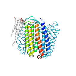
Halorhodopsin, a light-driven chloride pump
Descriptor: (2R)-2,3-dihydroxypropyl (9Z)-octadec-9-enoate, CHLORIDE ION, HALORHODOPSIN, ...
Authors:Essen, L.-O, Kolbe, M, Oesterhelt, D.
Deposit date:2000-04-14
Release date:2000-06-02
Last modified:2023-12-06
Method:X-RAY DIFFRACTION (1.8 Å)
Cite:Structure of Light-Driven Chloride Pump Halorhodopsin at 1.8 A Resolution
Science, 288, 2000
1DLC
DownloadVisualize
BU of 1dlc by Molmil
CRYSTAL STRUCTURE OF INSECTICIDAL DELTA-ENDOTOXIN FROM BACILLUS THURINGIENSIS AT 2.5 ANGSTROMS RESOLUTION
Descriptor: DELTA-ENDOTOXIN CRYIIIA
Authors:Li, J.
Deposit date:1994-06-22
Release date:1994-09-30
Last modified:2024-02-07
Method:X-RAY DIFFRACTION (2.5 Å)
Cite:Crystal structure of insecticidal delta-endotoxin from Bacillus thuringiensis at 2.5 A resolution.
Nature, 353, 1991
7TCN
DownloadVisualize
BU of 7tcn by Molmil
Cryo-EM structure of CH235.12 in complex with HIV-1 Env trimer CH505TF.N279K.SOSIP.664 with high-mannose glycans
Descriptor: 2-acetamido-2-deoxy-beta-D-glucopyranose, 2-acetamido-2-deoxy-beta-D-glucopyranose-(1-4)-2-acetamido-2-deoxy-beta-D-glucopyranose, CH235.12 Fab Heavy Chain, ...
Authors:Manne, K, Henderson, R, Acharya, P.
Deposit date:2021-12-27
Release date:2023-08-02
Method:ELECTRON MICROSCOPY (4.1 Å)
Cite:Cryo-EM structure of CH235.12 in complex with HIV-1 Env trimer CH505TF.N279K.G458Y.SOSIP.664
To Be Published
7TCO
DownloadVisualize
BU of 7tco by Molmil
Cryo-EM structure of CH235.12 in complex with HIV-1 Env trimer CH505TF.N279K.G458Y.SOSIP.664 with high-mannose glycans
Descriptor: 2-acetamido-2-deoxy-beta-D-glucopyranose, 2-acetamido-2-deoxy-beta-D-glucopyranose-(1-4)-2-acetamido-2-deoxy-beta-D-glucopyranose, CH235.12 Fab Heavy Chain, ...
Authors:Manne, K, Henderson, R, Acharya, P.
Deposit date:2021-12-27
Release date:2023-08-02
Method:ELECTRON MICROSCOPY (4.19 Å)
Cite:Cryo-EM structure of CH235.12 in complex with HIV-1 Env trimer CH505TF.N279K.G458Y.SOSIP.664
To Be Published
1JI6
DownloadVisualize
BU of 1ji6 by Molmil
CRYSTAL STRUCTURE OF THE INSECTICIDAL BACTERIAL DEL ENDOTOXIN CRY3Bb1 BACILLUS THURINGIENSIS
Descriptor: PESTICIDIAL CRYSTAL PROTEIN CRY3BB
Authors:Cody, V.
Deposit date:2001-06-29
Release date:2001-09-19
Last modified:2024-02-07
Method:X-RAY DIFFRACTION (2.4 Å)
Cite:Structure of the insecticidal bacterial delta-endotoxin Cry3Bb1 of Bacillus thuringiensis.
Acta Crystallogr.,Sect.D, 57, 2001

220472

건을2024-05-29부터공개중

PDB statisticsPDBj update infoContact PDBjnumon