3S99
 
 | |
3SIA
 | |
3U04
 | |
3UAM
 | |
3S6O
 | |
3SJS
 | |
5U29
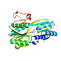
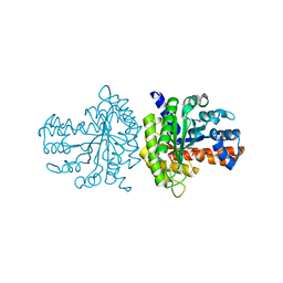
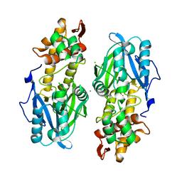
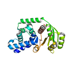
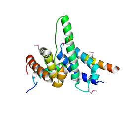
 | | Crystal structure of Cryptococcus neoformans H99 Acetyl-CoA Synthetase in complex with Ac-AMS | | Descriptor: | 1,2-ETHANEDIOL, 5'-O-(acetylsulfamoyl)adenosine, Acetyl-coenzyme A synthetase, ... | | Authors: | Seattle Structural Genomics Center for Infectious Disease (SSGCID), Fox III, D, Edwards, T.E, Potts, K.T, Taylor, B.M. | | Deposit date: | 2016-11-30 | | Release date: | 2017-12-06 | | Last modified: | 2023-10-04 | | Method: | X-RAY DIFFRACTION (2.5 Å) | | Cite: | Crystal structure of Cryptococcus neoformans H99 Acetyl-CoA Synthetase in complex with Ac-AMS
To Be Published
|
|
3SIB
 | |
3QRH
 | |
5U8O
 | |
5UXV
 | |
5UXX
 | |
5UXW
 | |
5U2A
 | |
4FKX
 | |
4FKY
 | |
4FI5
 | |
6D46
 | |
7SBC
 | |
7S5E
 | |
6DEG
 | |
6D47
 | |
5VMK
 | |
5VK4
 | |
5VMB
 | |


