Loading
PDBj
MenuPDBj@FacebookPDBj@TwitterPDBj@YouTubewwPDB FoundationwwPDB
RCSB PDBPDBeBMRBAdv. SearchSearch help
Search by PDB author
2ZW7
DownloadVisualize
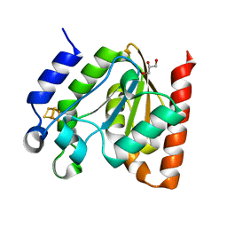
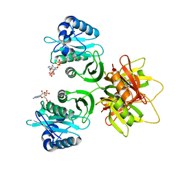
BU of 2zw7 by Molmil
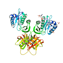
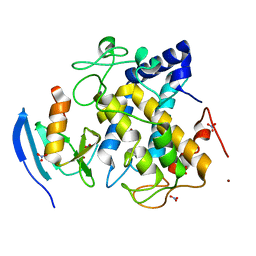
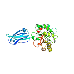
Crystal structure of bleomycin N-acetyltransferase complexed with bleomycin A2 and coenzyme A
Descriptor: BLEOMYCIN A2, Bleomycin acetyltransferase, COENZYME A
Authors:Oda, K, Matoba, Y, Sugiyama, M.
Deposit date:2008-12-01
Release date:2009-11-03
Last modified:2023-11-15
Method:X-RAY DIFFRACTION (2.8 Å)
Cite:Catalytic mechanism of bleomycin N-acetyltransferase proposed on the basis of its crystal structure.
J.Biol.Chem., 285, 2010
2ZW5
DownloadVisualize
BU of 2zw5 by Molmil
Crystal structure of bleomycin N-acetyltransferase complexed with coenzyme A in the trigonal crystal
Descriptor: Bleomycin acetyltransferase, COENZYME A
Authors:Oda, K, Matoba, Y, Sugiyama, M.
Deposit date:2008-12-01
Release date:2009-11-03
Last modified:2023-11-01
Method:X-RAY DIFFRACTION (2.4 Å)
Cite:Catalytic mechanism of bleomycin N-acetyltransferase proposed on the basis of its crystal structure.
J.Biol.Chem., 285, 2010
2ZW4
DownloadVisualize
BU of 2zw4 by Molmil
Crystal structure of bleomycin N-acetyltransferase complexed with coenzyme A in the orthorhombic crystal
Descriptor: Bleomycin acetyltransferase, COENZYME A, SULFATE ION
Authors:Oda, K, Matoba, Y, Sugiyama, M.
Deposit date:2008-12-01
Release date:2009-11-03
Last modified:2011-07-13
Method:X-RAY DIFFRACTION (2.7 Å)
Cite:Catalytic mechanism of bleomycin N-acetyltransferase proposed on the basis of its crystal structure.
J.Biol.Chem., 285, 2010
3WSW
DownloadVisualize
BU of 3wsw by Molmil
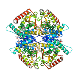
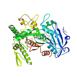
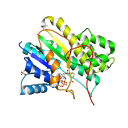
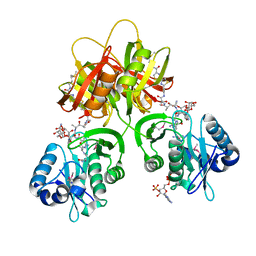
Crystal structure of minor L-lactate dehydrogenase from Enterococcus mundtii in the ligands-bound form
Descriptor: 1,6-di-O-phosphono-beta-D-fructofuranose, GLYCEROL, L-lactate dehydrogenase, ...
Authors:Matoba, Y, Sugiyama, M.
Deposit date:2014-03-27
Release date:2014-09-10
Last modified:2023-11-08
Method:X-RAY DIFFRACTION (2.3 Å)
Cite:An alternative allosteric regulation mechanism of an acidophilic l-lactate dehydrogenase from Enterococcus mundtii 15-1A.
Febs Open Bio, 4, 2014
3WSV
DownloadVisualize
BU of 3wsv by Molmil
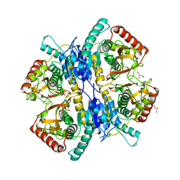
Crystal structure of minor L-lactate dehydrogenase from Enterococcus mundtii in the ligands-unbound form
Descriptor: GLYCEROL, L-lactate dehydrogenase
Authors:Matoba, Y, Sugiyama, M.
Deposit date:2014-03-27
Release date:2014-09-10
Last modified:2023-11-08
Method:X-RAY DIFFRACTION (2.38 Å)
Cite:An alternative allosteric regulation mechanism of an acidophilic l-lactate dehydrogenase from Enterococcus mundtii 15-1A.
Febs Open Bio, 4, 2014
3WA6
DownloadVisualize
BU of 3wa6 by Molmil
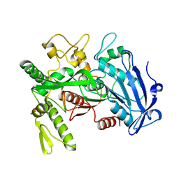
Crystal structure of tannase from Lactobacillus plantarum in the orthorhombic crystal
Descriptor: SULFATE ION, tannase
Authors:Matoba, Y, Tanaka, N, Sugiyama, M.
Deposit date:2013-04-27
Release date:2013-07-24
Last modified:2024-03-20
Method:X-RAY DIFFRACTION (1.8 Å)
Cite:Crystallographic and mutational analyses of tannase from Lactobacillus plantarum
Proteins, 81, 2013
3WA7
DownloadVisualize
BU of 3wa7 by Molmil
Crystal structure of selenomethionine-labeled tannase from Lactobacillus plantarum in the orthorhombic crystal
Descriptor: ACETATE ION, GLYCEROL, SULFATE ION, ...
Authors:Matoba, Y, Tanaka, N, Sugiyama, M.
Deposit date:2013-04-27
Release date:2013-07-24
Last modified:2013-11-06
Method:X-RAY DIFFRACTION (1.7 Å)
Cite:Crystallographic and mutational analyses of tannase from Lactobacillus plantarum.
Proteins, 81, 2013
3WCK
DownloadVisualize
BU of 3wck by Molmil
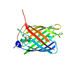
Crystal structure of monomeric photosensitizing fluorescent protein, Supernova
Descriptor: Monomeric photosenitizing fluorescent protein supernova
Authors:Sakai, N, Matsuda, T, Takemoto, K, Nagai, T.
Deposit date:2013-05-27
Release date:2013-10-02
Last modified:2023-11-15
Method:X-RAY DIFFRACTION (2.3 Å)
Cite:SuperNova, a monomeric photosensitizing fluorescent protein for chromophore-assisted light inactivation
Sci Rep, 3, 2013
3AL9
DownloadVisualize
BU of 3al9 by Molmil
Mouse Plexin A2 extracellular domain
Descriptor: 1,2-ETHANEDIOL, 2-acetamido-2-deoxy-beta-D-glucopyranose, Plexin-A2
Authors:Nogi, T, Yasui, N, Mihara, E, Takagi, J.
Deposit date:2010-07-28
Release date:2010-10-06
Last modified:2023-11-01
Method:X-RAY DIFFRACTION (2.1 Å)
Cite:Structural basis for semaphorin signalling through the plexin receptor.
Nature, 467, 2010
3AFC
DownloadVisualize
BU of 3afc by Molmil
Mouse Semaphorin 6A extracellular domain
Descriptor: 2-acetamido-2-deoxy-beta-D-glucopyranose, 2-acetamido-2-deoxy-beta-D-glucopyranose-(1-4)-2-acetamido-2-deoxy-beta-D-glucopyranose, Semaphorin-6A
Authors:Yasui, N, Nogi, T, Mihara, E, Takagi, J.
Deposit date:2010-02-26
Release date:2010-10-06
Last modified:2023-11-01
Method:X-RAY DIFFRACTION (2.5 Å)
Cite:Structural basis for semaphorin signalling through the plexin receptor.
Nature, 467, 2010
3AL8
DownloadVisualize
BU of 3al8 by Molmil
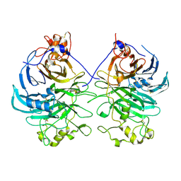
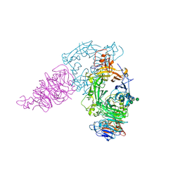
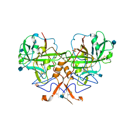
Plexin A2 / Semaphorin 6A complex
Descriptor: 2-acetamido-2-deoxy-beta-D-glucopyranose, 2-acetamido-2-deoxy-beta-D-glucopyranose-(1-4)-2-acetamido-2-deoxy-beta-D-glucopyranose, Plexin-A2, ...
Authors:Nogi, T, Yasui, N, Mihara, E, Takagi, J.
Deposit date:2010-07-28
Release date:2010-10-06
Last modified:2020-07-29
Method:X-RAY DIFFRACTION (3.6 Å)
Cite:Structural basis for semaphorin signalling through the plexin receptor.
Nature, 467, 2010
2ZMX
DownloadVisualize
BU of 2zmx by Molmil
Crystal structure of the met1-form of the copper-bound tyrosinase in complex with a caddie protein from Streptomyces castaneoglobisporus obtained by soaking in cupric sulfate solution for 36 hours
Descriptor: CADDIE, COPPER (II) ION, NITRATE ION, ...
Authors:Matoba, Y, Sugiyama, M.
Deposit date:2008-04-21
Release date:2009-04-07
Last modified:2023-11-01
Method:X-RAY DIFFRACTION (1.33 Å)
Cite:Crystallographic Evidence That the Dinuclear Copper Center of Tyrosinase Is Flexible during Catalysis
J.Biol.Chem., 281, 2006
1N5D
DownloadVisualize
BU of 1n5d by Molmil
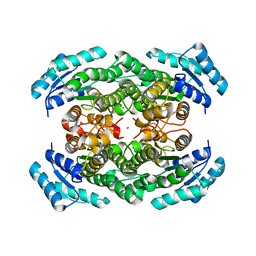
CRYSTAL STRUCTURE OF PORCINE TESTICULAR CARBONYL REDUCTASE/ 20BETA-HYDROXYSTEROID DEHYDROGENASE
Descriptor: CARBONYL REDUCTASE/20BETA-HYDROXYSTEROID DEHYDROGENASE, NADPH DIHYDRO-NICOTINAMIDE-ADENINE-DINUCLEOTIDE PHOSPHATE, SULFATE ION
Authors:Ghosh, D.
Deposit date:2002-11-05
Release date:2002-11-13
Last modified:2024-02-14
Method:X-RAY DIFFRACTION (2.3 Å)
Cite:Porcine Carbonyl Reductase: Structural Basis for a Functional Monomer in Short-Chain Dehydrogenases/Reductases
J.Biol.Chem., 276, 2001
3N4O
DownloadVisualize
BU of 3n4o by Molmil
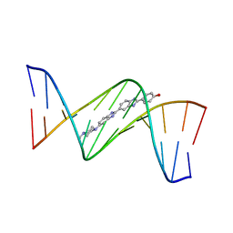
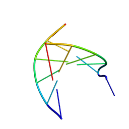
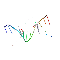
Insights into the stabilizing contributions of a bicyclic cytosine analogue: crystal structures of DNA duplexes containing 7,8-dihydropyrido[2,3-d]pyrimidin-2-one
Descriptor: 2'-(4-HYDROXYPHENYL)-5-(4-METHYL-1-PIPERAZINYL)-2,5'-BI-BENZIMIDAZOLE, 5'-D(*CP*GP*CP*GP*AP*AP*TP*(B7C)P*CP*GP*CP*G)-3'
Authors:Takenaka, A, Juan, E.C.M, Shimizu, S.
Deposit date:2010-05-22
Release date:2010-08-11
Last modified:2023-11-01
Method:X-RAY DIFFRACTION (2.9 Å)
Cite:Insights into the DNA stabilizing contributions of a bicyclic cytosine analogue: crystal structures of DNA duplexes containing 7,8-dihydropyrido [2,3-d]pyrimidin-2-one.
Nucleic Acids Res., 2010
3N4N
DownloadVisualize
BU of 3n4n by Molmil
Insights into the stabilizing contributions of a bicyclic cytosine analogue: crystal structures of DNA duplexes containing 7,8-dihydropyrido[2,3-d]pyrimidin-2-one
Descriptor: 5'-D(*CP*GP*CP*GP*AP*A)-3', 5'-D(P*TP*TP*(B7C)P*GP*CP*G)-3'
Authors:Takenaka, A, Juan, E.C.M, Shimizu, S.
Deposit date:2010-05-22
Release date:2010-08-11
Last modified:2023-11-01
Method:X-RAY DIFFRACTION (1.92 Å)
Cite:Insights into the DNA stabilizing contributions of a bicyclic cytosine analogue: crystal structures of DNA duplexes containing 7,8-dihydropyrido [2,3-d]pyrimidin-2-one.
Nucleic Acids Res., 2010
6GO1
DownloadVisualize
BU of 6go1 by Molmil
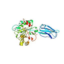
Crystal Structure of a Bacillus anthracis peptidoglycan deacetylase
Descriptor: 1,2-ETHANEDIOL, ACETATE ION, Polysaccharide deacetylase-like protein, ...
Authors:Giastas, P, Andreou, A, Eliopoulos, E.E.
Deposit date:2018-06-01
Release date:2019-04-10
Last modified:2024-01-17
Method:X-RAY DIFFRACTION (2.59 Å)
Cite:The putative polysaccharide deacetylase Ba0331: cloning, expression, crystallization and structure determination.
Acta Crystallogr.,Sect.F, 75, 2019
3AIX
DownloadVisualize
BU of 3aix by Molmil
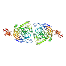
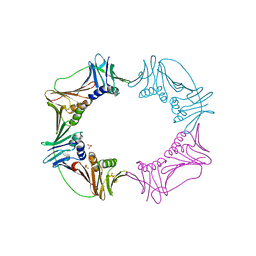
Crystal structure of PCNA2-PCNA3 complex from Sulfolobus tokodaii (I222)
Descriptor: DNA polymerase sliding clamp B, DNA polymerase sliding clamp C, SULFATE ION
Authors:Kawai, A, Higuchi, S, Miyamoto, S.
Deposit date:2010-05-18
Release date:2011-05-18
Last modified:2023-11-01
Method:X-RAY DIFFRACTION (2.9 Å)
Cite:A novel heterotetrameric structure of the crenarchaeal PCNA2-PCNA3 complex
J.Struct.Biol., 174, 2011
3AIZ
DownloadVisualize
BU of 3aiz by Molmil
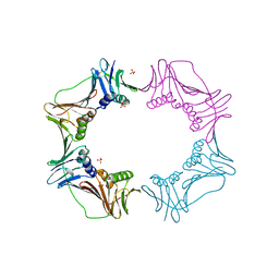
Crystal structure of PCNA2-PCNA3 complex from Sulfolobus tokodaii (P21212)
Descriptor: DNA polymerase sliding clamp B, DNA polymerase sliding clamp C, SULFATE ION
Authors:Kawai, A, Higuchi, S, Miyamoto, S.
Deposit date:2010-05-18
Release date:2011-05-18
Last modified:2023-11-01
Method:X-RAY DIFFRACTION (2.8 Å)
Cite:A novel heterotetrameric structure of the crenarchaeal PCNA2-PCNA3 complex
J.Struct.Biol., 174, 2011
4V33
DownloadVisualize
BU of 4v33 by Molmil
Crystal structure of the putative polysaccharide deacetylase BA0330 from bacillus anthracis
Descriptor: 1,2-ETHANEDIOL, ACETATE ION, POLYSACCHARIDE DEACETYLASE-LIKE PROTEIN, ...
Authors:Giastas, P, Andreou, A, Bouriotis, V, Eliopoulos, E.
Deposit date:2014-10-16
Release date:2015-04-08
Last modified:2024-01-10
Method:X-RAY DIFFRACTION (1.48 Å)
Cite:Two Putative Polysaccharide Deacetylases are Required for Osmotic Stability and Cell Shape Maintenance in Bacillus Anthracis.
J.Biol.Chem., 290, 2015
1I3T
DownloadVisualize
BU of 1i3t by Molmil
MOLECULAR AND CRYSTAL STRUCTURE OF D(CGCGAATT(MO4)CGCG): THE WATSON-CRICK TYPE AND WOBBLE N4-METHOXYCYTIDINE/GUANOSINE BASE PAIRS IN B-DNA
Descriptor: 5'-D(*CP*GP*CP*GP*AP*AP*TP*TP*(C45)P*GP*CP*GP)-3', MAGNESIUM ION
Authors:Hossain, M.T, Hikima, T, Chatake, T, Masaru, T, Sunami, T, Ueno, Y, Matsuda, A, Takenaka, A.
Deposit date:2001-02-16
Release date:2001-09-14
Last modified:2023-08-09
Method:X-RAY DIFFRACTION (1.6 Å)
Cite:Crystallographic studies on damaged DNAs: III. N(4)-methoxycytosine can form both Watson-Crick type and wobbled base pairs in a B-form duplex.
J.Biochem.(Tokyo), 130, 2001
4ZBY
DownloadVisualize
BU of 4zby by Molmil
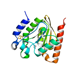
Family 4 uracil-DNA glycosylase from Sulfolobus tokodaii (uracil complex form)
Descriptor: 2-(N-MORPHOLINO)-ETHANESULFONIC ACID, IRON/SULFUR CLUSTER, URACIL, ...
Authors:Kawai, A, Miyamoto, S.
Deposit date:2015-04-15
Release date:2015-09-09
Last modified:2023-11-08
Method:X-RAY DIFFRACTION (1.7 Å)
Cite:Crystal structure of family 4 uracil-DNA glycosylase from Sulfolobus tokodaii and a function of tyrosine 170 in DNA binding
Febs Lett., 589, 2015
4ZBX
DownloadVisualize
BU of 4zbx by Molmil
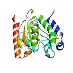
Family 4 uracil-DNA glycosylase from Sulfolobus tokodaii (free form, X-ray wavelength=0.9000)
Descriptor: 2-(N-MORPHOLINO)-ETHANESULFONIC ACID, IRON/SULFUR CLUSTER, Uracil-DNA glycosylase
Authors:Kawai, A, Miyamoto, S.
Deposit date:2015-04-15
Release date:2015-09-09
Last modified:2023-11-08
Method:X-RAY DIFFRACTION (1.7 Å)
Cite:Crystal structure of family 4 uracil-DNA glycosylase from Sulfolobus tokodaii and a function of tyrosine 170 in DNA binding
Febs Lett., 589, 2015
4ZBZ
DownloadVisualize
BU of 4zbz by Molmil
Family 4 uracil-DNA glycosylase from Sulfolobus tokodaii (free form, X-ray wavelength=1.5418)
Descriptor: 2-(N-MORPHOLINO)-ETHANESULFONIC ACID, GLYCEROL, IRON/SULFUR CLUSTER, ...
Authors:Kawai, A, Miyamoto, S.
Deposit date:2015-04-15
Release date:2015-09-09
Last modified:2023-11-08
Method:X-RAY DIFFRACTION (1.9 Å)
Cite:Crystal structure of family 4 uracil-DNA glycosylase from Sulfolobus tokodaii and a function of tyrosine 170 in DNA binding
Febs Lett., 589, 2015
3B1B
DownloadVisualize
BU of 3b1b by Molmil
The unique structure of wild type carbonic anhydrase alpha-CA1 from Chlamydomonas reinhardtii
Descriptor: 2-acetamido-2-deoxy-beta-D-glucopyranose, Carbonic anhydrase 1, SULFATE ION, ...
Authors:Shimizu, S, Takenaka, A.
Deposit date:2011-06-29
Release date:2011-11-16
Last modified:2020-07-29
Method:X-RAY DIFFRACTION (1.88 Å)
Cite:The unique structure of carbonic anhydrase alpha CA1 from Chlamydomonas reinhardtii
Acta Crystallogr.,Sect.D, 67, 2011
2YZ7
DownloadVisualize
BU of 2yz7 by Molmil
X-ray analyses of 3-hydroxybutyrate dehydrogenase from Alcaligenes faecalis
Descriptor: CALCIUM ION, CHLORIDE ION, D-3-hydroxybutyrate dehydrogenase
Authors:Hoque, M.M, Juan, E.C.M, Shimizu, S, Hossain, M.T, Takenaka, A.
Deposit date:2007-05-04
Release date:2008-04-22
Last modified:2023-10-25
Method:X-RAY DIFFRACTION (2.19 Å)
Cite:The structures of Alcaligenes faecalisD-3-hydroxybutyrate dehydrogenase before and after NAD(+) and acetate binding suggest a dynamical reaction mechanism as a member of the SDR family.
Acta Crystallogr.,Sect.D, 64, 2008

222926

건을2024-07-24부터공개중

PDB statisticsPDBj update infoContact PDBjnumon