Loading
PDBj
MenuPDBj@FacebookPDBj@TwitterPDBj@YouTubewwPDB FoundationwwPDB
RCSB PDBPDBeBMRBAdv. SearchSearch help
Search by PDB author
3RIZ
DownloadVisualize
BU of 3riz by Molmil
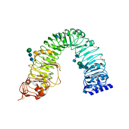
Crystal structure of the plant steroid receptor BRI1 ectodomain
Descriptor: 2-acetamido-2-deoxy-beta-D-glucopyranose, 2-acetamido-2-deoxy-beta-D-glucopyranose-(1-4)-2-acetamido-2-deoxy-beta-D-glucopyranose, Protein BRASSINOSTEROID INSENSITIVE 1, ...
Authors:Hothorn, M.
Deposit date:2011-04-14
Release date:2011-06-22
Last modified:2020-07-29
Method:X-RAY DIFFRACTION (2.523 Å)
Cite:Structural basis of steroid hormone perception by the receptor kinase BRI1.
Nature, 474, 2011
2OOK
DownloadVisualize
BU of 2ook by Molmil
Crystal structure of a protein with unknown function (YP_749275.1) from Shewanella Frigidimarina NCIMB 400 at 1.80 A resolution
Descriptor: 1,2-ETHANEDIOL, Hypothetical protein
Authors:Joint Center for Structural Genomics (JCSG)
Deposit date:2007-01-25
Release date:2007-02-06
Last modified:2023-01-25
Method:X-RAY DIFFRACTION (1.8 Å)
Cite:Open and closed conformations of two SpoIIAA-like proteins (YP_749275.1 and YP_001095227.1) provide insights into membrane association and ligand binding.
Acta Crystallogr.,Sect.F, 66, 2010
2OOC
DownloadVisualize
BU of 2ooc by Molmil
Crystal structure of Histidine Phosphotransferase ShpA (NP_419930.1) from Caulobacter crescentus at 1.52 A resolution
Descriptor: GLYCEROL, Histidine phosphotransferase, TETRAETHYLENE GLYCOL
Authors:Joint Center for Structural Genomics (JCSG)
Deposit date:2007-01-25
Release date:2007-02-06
Last modified:2023-01-25
Method:X-RAY DIFFRACTION (1.52 Å)
Cite:Crystal structure of histidine phosphotransfer protein ShpA, an essential regulator of stalk biogenesis in Caulobacter crescentus.
J.Mol.Biol., 390, 2009
2F58
DownloadVisualize
BU of 2f58 by Molmil
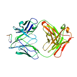
IGG1 FAB FRAGMENT (58.2) COMPLEX WITH 12-RESIDUE CYCLIC PEPTIDE (INCLUDING RESIDUES 315-324 OF HIV-1 GP120) (MN ISOLATE)
Descriptor: 1-IMINO-5-PENTANONE, PROTEIN (HIV-1 GP120), PROTEIN (IGG1 FAB 58.2 ANTIBODY (HEAVY CHAIN)), ...
Authors:Stanfield, R.L, Cabezas, E, Satterthwait, A.C, Stura, E.A, Profy, A.T, Wilson, I.A.
Deposit date:1998-10-23
Release date:1999-02-09
Last modified:2024-04-03
Method:X-RAY DIFFRACTION (2.8 Å)
Cite:Dual conformations for the HIV-1 gp120 V3 loop in complexes with different neutralizing fabs.
Structure Fold.Des., 7, 1999
1F58
DownloadVisualize
BU of 1f58 by Molmil
IGG1 FAB FRAGMENT (58.2) COMPLEX WITH 24-RESIDUE PEPTIDE (RESIDUES 308-333 OF HIV-1 GP120 (MN ISOLATE) WITH ALA TO AIB SUBSTITUTION AT POSITION 323
Descriptor: Envelope glycoprotein gp120, PROTEIN (IGG1 ANTIBODY 58.2 (HEAVY CHAIN)), PROTEIN (IGG1 ANTIBODY 58.2 (LIGHT CHAIN))
Authors:Stanfield, R.L, Cabezas, E, Satterthwait, A.C, Stura, E.A, Profy, A.T, Wilson, I.A.
Deposit date:1998-10-21
Release date:1999-02-02
Last modified:2024-04-03
Method:X-RAY DIFFRACTION (2 Å)
Cite:Dual conformations for the HIV-1 gp120 V3 loop in complexes with different neutralizing fabs.
Structure Fold.Des., 7, 1999
1HTM
DownloadVisualize
BU of 1htm by Molmil
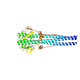
STRUCTURE OF INFLUENZA HAEMAGGLUTININ AT THE PH OF MEMBRANE FUSION
Descriptor: HEMAGGLUTININ HA1 CHAIN, HEMAGGLUTININ HA2 CHAIN
Authors:Bullough, P.A, Hughson, F.M, Skehel, J.J, Wiley, D.C.
Deposit date:1994-11-02
Release date:1995-02-14
Last modified:2024-06-05
Method:X-RAY DIFFRACTION (2.5 Å)
Cite:Structure of influenza haemagglutinin at the pH of membrane fusion.
Nature, 371, 1994
3BOS
DownloadVisualize
BU of 3bos by Molmil
Crystal structure of a putative dna replication regulator HDA (SAMA_1916) from Shewanella amazonensis sb2b at 1.75 A resolution
Descriptor: 1,2-ETHANEDIOL, CHLORIDE ION, CYTIDINE-5'-DIPHOSPHATE, ...
Authors:Joint Center for Structural Genomics (JCSG)
Deposit date:2007-12-17
Release date:2008-01-15
Last modified:2023-01-25
Method:X-RAY DIFFRACTION (1.75 Å)
Cite:A structural basis for the regulatory inactivation of DnaA.
J.Mol.Biol., 385, 2009
3CM1
DownloadVisualize
BU of 3cm1 by Molmil
Crystal structure of SsgA-like sporulation-specific cell division protein (YP_290167.1) from Thermobifida fusca YX-ER1 at 2.60 A resolution
Descriptor: SsgA-like sporulation-specific cell division protein
Authors:Joint Center for Structural Genomics (JCSG), Chruszcz, M, Minor, W, Wang, S.
Deposit date:2008-03-20
Release date:2008-04-01
Last modified:2023-02-01
Method:X-RAY DIFFRACTION (2.6 Å)
Cite:Structural and functional characterizations of SsgB, a conserved activator of developmental cell division in morphologically complex actinomycetes.
J.Biol.Chem., 284, 2009
3D00
DownloadVisualize
BU of 3d00 by Molmil
Crystal structure of a tungsten formylmethanofuran dehydrogenase subunit e (fmde)-like protein (syn_00638) from syntrophus aciditrophicus at 1.90 A resolution
Descriptor: CHLORIDE ION, Tungsten formylmethanofuran dehydrogenase subunit E, ZINC ION
Authors:Joint Center for Structural Genomics (JCSG)
Deposit date:2008-04-30
Release date:2008-05-20
Last modified:2023-02-01
Method:X-RAY DIFFRACTION (1.9 Å)
Cite:Structures of three members of Pfam PF02663 (FmdE) implicated in microbial methanogenesis reveal a conserved alpha+beta core domain and an auxiliary C-terminal treble-clef zinc finger.
Acta Crystallogr.,Sect.F, 66, 2010
3DEE
DownloadVisualize
BU of 3dee by Molmil
CRYSTAL STRUCTURE OF A PUTATIVE REGULATORY PROTEIN INVOLVED IN TRANSCRIPTION (NGO1945) FROM NEISSERIA GONORRHOEAE FA 1090 AT 2.25 A RESOLUTION
Descriptor: CHLORIDE ION, IMIDAZOLE, Putative regulatory protein
Authors:Joint Center for Structural Genomics (JCSG)
Deposit date:2008-06-09
Release date:2008-08-05
Last modified:2023-02-01
Method:X-RAY DIFFRACTION (2.1 Å)
Cite:The structure of the first representative of Pfam family PF09836 reveals a two-domain organization and suggests involvement in transcriptional regulation.
Acta Crystallogr.,Sect.F, 66, 2010
1PSH
DownloadVisualize
BU of 1psh by Molmil
CRYSTAL STRUCTURE OF PHOSPHOLIPASE A2 FROM INDIAN COBRA REVEALS A TRIMERIC ASSOCIATION
Descriptor: CALCIUM ION, PHOSPHOLIPASE A2
Authors:Fremont, D, Xuong, N.-H, Wilson, I.
Deposit date:1992-07-28
Release date:1994-01-31
Last modified:2024-06-05
Method:X-RAY DIFFRACTION (2.3 Å)
Cite:Crystal structure of phospholipase A2 from Indian cobra reveals a trimeric association.
Proc.Natl.Acad.Sci.USA, 90, 1993
3E0F
DownloadVisualize
BU of 3e0f by Molmil
Crystal structure of a putative metal-dependent phosphoesterase (bad_1165) from bifidobacterium adolescentis atcc 15703 at 2.40 A resolution
Descriptor: ACETATE ION, FE (III) ION, PHOSPHATE ION, ...
Authors:Joint Center for Structural Genomics (JCSG)
Deposit date:2008-07-31
Release date:2008-09-02
Last modified:2023-02-01
Method:X-RAY DIFFRACTION (2.4 Å)
Cite:Crystal structure of a metal-dependent phosphoesterase (YP_910028.1) from Bifidobacterium adolescentis: Computational prediction and experimental validation of phosphoesterase activity.
Proteins, 79, 2011
3DUE
DownloadVisualize
BU of 3due by Molmil
CRYSTAL STRUCTURE OF A PUTATIVE PERIPLASMIC PROTEIN FROM DUF2874 FAMILY (BVU_2987) FROM BACTEROIDES VULGATUS ATCC 8482 AT 1.85 A RESOLUTION
Descriptor: CACODYLATE ION, Putative periplasmic protein
Authors:Joint Center for Structural Genomics (JCSG)
Deposit date:2008-07-17
Release date:2008-08-05
Last modified:2023-02-01
Method:X-RAY DIFFRACTION (1.85 Å)
Cite:The structure of BVU2987 from Bacteroides vulgatus reveals a superfamily of bacterial periplasmic proteins with possible inhibitory function.
Acta Crystallogr.,Sect.F, 66, 2010
3B77
DownloadVisualize
BU of 3b77 by Molmil
Crystal structure of a ph domain containing bacterial protein (exig_2160) from exiguobacterium sibiricum 255-15 at 2.42 A resolution
Descriptor: Uncharacterized protein
Authors:Joint Center for Structural Genomics (JCSG)
Deposit date:2007-10-30
Release date:2007-11-13
Last modified:2023-01-25
Method:X-RAY DIFFRACTION (2.42 Å)
Cite:Bacterial pleckstrin homology domains: a prokaryotic origin for the PH domain.
J.Mol.Biol., 396, 2010
3BY7
DownloadVisualize
BU of 3by7 by Molmil
CRYSTAL STRUCTURE OF A PROTEIN STRUCTURALLY SIMILAR TO SM/LSM-LIKE RNA-BINDING PROTEINS (JCVI_PEP_1096686650277) FROM UNCULTURED MARINE ORGANISM AT 2.60 A RESOLUTION
Descriptor: uncharacterized protein
Authors:Joint Center for Structural Genomics (JCSG)
Deposit date:2008-01-15
Release date:2008-01-29
Last modified:2023-02-01
Method:X-RAY DIFFRACTION (2.6 Å)
Cite:Crystal structure of a novel Sm-like protein of putative cyanophage origin at 2.60 A resolution.
Proteins, 75, 2009
3BYQ
DownloadVisualize
BU of 3byq by Molmil
Crystal structure of a duf1185 family protein (bb2672) from bordetella bronchiseptica rb50 at 1.70 A resolution
Descriptor: 1,2-ETHANEDIOL, CHLORIDE ION, TETRAETHYLENE GLYCOL, ...
Authors:Joint Center for Structural Genomics (JCSG)
Deposit date:2008-01-16
Release date:2008-01-29
Last modified:2023-02-01
Method:X-RAY DIFFRACTION (1.7 Å)
Cite:Structures of the first representatives of Pfam family PF06684 (DUF1185) reveal a novel variant of the Bacillus chorismate mutase fold and suggest a role in amino-acid metabolism.
Acta Crystallogr.,Sect.F, 66, 2010
3DCX
DownloadVisualize
BU of 3dcx by Molmil
Crystal structure of a duf1696 family protein with a pleckstrin-homology domain (shew_0819) from shewanella loihica pv-4 at 2.00 A resolution
Descriptor: (4S)-2-METHYL-2,4-PENTANEDIOL, CHLORIDE ION, Protein of Unknown Function (DUF1696) with Pleckstrin-homology Domains
Authors:Joint Center for Structural Genomics (JCSG)
Deposit date:2008-06-04
Release date:2008-07-29
Last modified:2023-02-01
Method:X-RAY DIFFRACTION (2 Å)
Cite:Bacterial pleckstrin homology domains: a prokaryotic origin for the PH domain.
J.Mol.Biol., 396, 2010
3DL1
DownloadVisualize
BU of 3dl1 by Molmil
Crystal structure of a Putative Metal-dependent Hydrolase (YP_001336084.1) from Klebsiella pneumoniae subsp. pneumoniae MGH 78578 at 2.20 A resolution
Descriptor: CHLORIDE ION, Putative Metal-dependent Hydrolase
Authors:Joint Center for Structural Genomics (JCSG)
Deposit date:2008-06-26
Release date:2008-08-26
Last modified:2023-02-01
Method:X-RAY DIFFRACTION (2.2 Å)
Cite:The Structure of Mlc Titration Factor A (MtfA/YeeI) Reveals a Prototypical Zinc Metallopeptidase Related to Anthrax Lethal Factor.
J.Bacteriol., 194, 2012
3CGH
DownloadVisualize
BU of 3cgh by Molmil
Crystal structure of a susd homolog (bt_3984) from bacteroides thetaiotaomicron vpi-5482 at 1.70 A resolution
Descriptor: ACETATE ION, SusD homolog, ZINC ION
Authors:Joint Center for Structural Genomics (JCSG)
Deposit date:2008-03-05
Release date:2008-03-18
Last modified:2023-02-01
Method:X-RAY DIFFRACTION (1.7 Å)
Cite:Structure of BT_3984, a member of the SusD/RagB family of nutrient-binding molecules.
Acta Crystallogr.,Sect.F, 66, 2010
3EQX
DownloadVisualize
BU of 3eqx by Molmil
CRYSTAL STRUCTURE OF A FIC FAMILY PROTEIN (SO_4266) FROM SHEWANELLA ONEIDENSIS AT 1.6 A RESOLUTION
Descriptor: FIC DOMAIN CONTAINING TRANSCRIPTIONAL REGULATOR, TRIETHYLENE GLYCOL
Authors:Joint Center for Structural Genomics (JCSG)
Deposit date:2008-10-01
Release date:2008-10-14
Last modified:2023-02-01
Method:X-RAY DIFFRACTION (1.6 Å)
Cite:Crystal structure of the Fic (Filamentation induced by cAMP) family protein SO4266 (gi|24375750) from Shewanella oneidensis MR-1 at 1.6 A resolution.
Proteins, 75, 2009
3ETN
DownloadVisualize
BU of 3etn by Molmil
Crystal structure of putative phosphosugar isomerase involved in capsule formation (YP_209877.1) from Bacteroides fragilis NCTC 9343 at 1.70 A resolution
Descriptor: 1,2-ETHANEDIOL, CYTIDINE 5'-MONOPHOSPHATE 3-DEOXY-BETA-D-GULO-OCT-2-ULO-PYRANOSONIC ACID, putative phosphosugar isomerase involved in capsule formation
Authors:Joint Center for Structural Genomics (JCSG)
Deposit date:2008-10-08
Release date:2008-10-21
Last modified:2023-02-01
Method:X-RAY DIFFRACTION (1.7 Å)
Cite:Structural analysis of arabinose-5-phosphate isomerase from Bacteroides fragilis and functional implications.
Acta Crystallogr.,Sect.D, 70, 2014
3G23
DownloadVisualize
BU of 3g23 by Molmil
Crystal structure of a ld-carboxypeptidase a (saro_1426) from novosphingobium aromaticivorans dsm at 1.89 A resolution
Descriptor: GLYCEROL, LD-carboxypeptidase A, SULFATE ION, ...
Authors:Joint Center for Structural Genomics (JCSG)
Deposit date:2009-01-30
Release date:2009-02-10
Last modified:2023-02-01
Method:X-RAY DIFFRACTION (1.89 Å)
Cite:Structure and function of a novel LD-carboxypeptidase a involved in peptidoglycan recycling.
J.Bacteriol., 195, 2013
3G0T
DownloadVisualize
BU of 3g0t by Molmil
Crystal structure of putative aspartate aminotransferase (NP_905498.1) from Porphyromonas gingivalis W83 at 1.75 A resolution
Descriptor: 1,2-ETHANEDIOL, 2-{2-[2-(2-{2-[2-(2-ETHOXY-ETHOXY)-ETHOXY]-ETHOXY}-ETHOXY)-ETHOXY]-ETHOXY}-ETHANOL, CHLORIDE ION, ...
Authors:Joint Center for Structural Genomics (JCSG)
Deposit date:2009-01-28
Release date:2009-02-10
Last modified:2023-02-01
Method:X-RAY DIFFRACTION (1.75 Å)
Cite:Molecular characterization of novel pyridoxal-5'-phosphate-dependent enzymes from the human microbiome.
Protein Sci., 23, 2014
3FKQ
DownloadVisualize
BU of 3fkq by Molmil
Crystal structure of NtrC-like two-domain protein (RER070207001320) from Eubacterium rectale at 2.10 A resolution
Descriptor: ADENOSINE-5'-TRIPHOSPHATE, CHLORIDE ION, GLYCEROL, ...
Authors:Joint Center for Structural Genomics (JCSG)
Deposit date:2008-12-17
Release date:2009-01-06
Last modified:2019-07-24
Method:X-RAY DIFFRACTION (2.1 Å)
Cite:Structure of the pilus assembly protein TadZ from Eubacterium rectale: implications for polar localization.
Mol.Microbiol., 83, 2012
3HN7
DownloadVisualize
BU of 3hn7 by Molmil
Crystal structure of a murein peptide ligase mpl (psyc_0032) from psychrobacter arcticus 273-4 at 1.65 A resolution
Descriptor: UDP-N-acetylmuramate-L-alanine ligase
Authors:Joint Center for Structural Genomics (JCSG)
Deposit date:2009-05-29
Release date:2009-06-16
Last modified:2023-02-01
Method:X-RAY DIFFRACTION (1.65 Å)
Cite:Structure and function of the first full-length murein Peptide ligase (mpl) cell wall recycling protein.
Plos One, 6, 2011

224201

건을2024-08-28부터공개중

PDB statisticsPDBj update infoContact PDBjnumon