Loading
PDBj
MenuPDBj@FacebookPDBj@TwitterPDBj@YouTubewwPDB FoundationwwPDB
RCSB PDBPDBeBMRBAdv. SearchSearch help
Search by PDB author
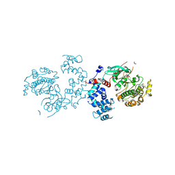
2GGZ
DownloadVisualize
BU of 2ggz by Molmil
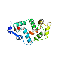
Crystal Structure of Human Guanylate Cyclase Activating Protein-3
Descriptor: CALCIUM ION, Guanylyl cyclase-activating protein 3
Authors:Stephen, R.
Deposit date:2006-03-24
Release date:2006-05-23
Last modified:2024-02-14
Method:X-RAY DIFFRACTION (3 Å)
Cite:The Crystal Structure of GCAP3 Suggests Molecular Mechanism of GCAP-linked Cone Dystrophies.
J.Mol.Biol., 359, 2006
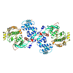
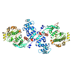
3C4W
DownloadVisualize
BU of 3c4w by Molmil
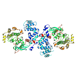
Crystal Structure of G protein coupled receptor kinase 1 bound to ATP and magnesium chloride at 2.7A
Descriptor: ADENOSINE-5'-TRIPHOSPHATE, CHLORIDE ION, MAGNESIUM ION, ...
Authors:Singh, P, Tesmer, J.J.G.
Deposit date:2008-01-30
Release date:2008-03-11
Last modified:2023-08-30
Method:X-RAY DIFFRACTION (2.7 Å)
Cite:Structures of rhodopsin kinase in different ligand states reveal key elements involved in G protein-coupled receptor kinase activation.
J.Biol.Chem., 283, 2008
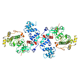
3C4Z
DownloadVisualize
BU of 3c4z by Molmil
Crystal structure of G protein coupled receptor kinase 1 bound to ADP and magnesium chloride at 1.84A
Descriptor: ADENOSINE-5'-DIPHOSPHATE, CHLORIDE ION, MAGNESIUM ION, ...
Authors:Singh, P, Tesmer, J.J.G.
Deposit date:2008-01-30
Release date:2008-03-11
Last modified:2023-08-30
Method:X-RAY DIFFRACTION (1.84 Å)
Cite:Structures of rhodopsin kinase in different ligand states reveal key elements involved in G protein-coupled receptor kinase activation.
J.Biol.Chem., 283, 2008
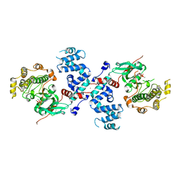
3C50
DownloadVisualize
BU of 3c50 by Molmil
Crystal Structure of G protein coupled receptor kinase 1 bound to ADP and magnesium chloride at 2.6A
Descriptor: 2-(N-MORPHOLINO)-ETHANESULFONIC ACID, ADENOSINE-5'-DIPHOSPHATE, CHLORIDE ION, ...
Authors:Singh, P, Tesmer, J.J.G.
Deposit date:2008-01-30
Release date:2008-03-11
Last modified:2023-08-30
Method:X-RAY DIFFRACTION (2.6 Å)
Cite:Structures of rhodopsin kinase in different ligand states reveal key elements involved in G protein-coupled receptor kinase activation.
J.Biol.Chem., 283, 2008
3C4Y
DownloadVisualize
BU of 3c4y by Molmil
Crystal Structure of Apo form of G protein coupled receptor kinase 1 at 7.51A
Descriptor: Rhodopsin kinase
Authors:Singh, P, Tesmer, J.J.G.
Deposit date:2008-01-30
Release date:2008-03-11
Last modified:2023-08-30
Method:X-RAY DIFFRACTION (7.509 Å)
Cite:Structures of rhodopsin kinase in different ligand states reveal key elements involved in G protein-coupled receptor kinase activation.
J.Biol.Chem., 283, 2008
3C51
DownloadVisualize
BU of 3c51 by Molmil
Crystal structure of G protein coupled receptor kinase 1 bound to ADP and magnesium chloride at 3.55A
Descriptor: ADENOSINE-5'-DIPHOSPHATE, BROMIDE ION, MAGNESIUM ION, ...
Authors:Singh, P, Tesmer, J.J.G.
Deposit date:2008-01-30
Release date:2008-03-11
Last modified:2023-08-30
Method:X-RAY DIFFRACTION (3.55 Å)
Cite:Structures of rhodopsin kinase in different ligand states reveal key elements involved in G protein-coupled receptor kinase activation.
J.Biol.Chem., 283, 2008
3C4X
DownloadVisualize
BU of 3c4x by Molmil
Crystal Structure of G protein coupled receptor kinase 1 bound to ATP and magnesium chloride at 2.9A
Descriptor: ADENOSINE-5'-TRIPHOSPHATE, CHLORIDE ION, MAGNESIUM ION, ...
Authors:Singh, P, Tesmer, J.J.G.
Deposit date:2008-01-30
Release date:2008-03-11
Last modified:2023-08-30
Method:X-RAY DIFFRACTION (2.9 Å)
Cite:Structures of rhodopsin kinase in different ligand states reveal key elements involved in G protein-coupled receptor kinase activation.
J.Biol.Chem., 283, 2008
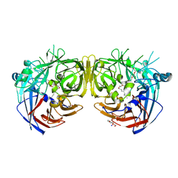
7K88
DownloadVisualize
BU of 7k88 by Molmil
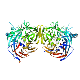
Crystal structure of bovine RPE65 in complex with hexaethylene glycol monooctyl ether
Descriptor: 3,6,9,12,15,18-hexaoxahexacosan-1-ol, FE (II) ION, Retinoid isomerohydrolase, ...
Authors:Kiser, P.D.
Deposit date:2020-09-26
Release date:2021-06-02
Last modified:2023-10-18
Method:X-RAY DIFFRACTION (2.1 Å)
Cite:Rational Alteration of Pharmacokinetics of Chiral Fluorinated and Deuterated Derivatives of Emixustat for Retinal Therapy.
J.Med.Chem., 64, 2021
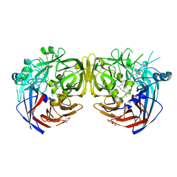
7K89
DownloadVisualize
BU of 7k89 by Molmil
Crystal structure of bovine RPE65 in complex with 4-fluoro-emixustat and palmitate
Descriptor: (1R)-3-amino-1-[3-(cyclohexylmethoxy)-4-fluorophenyl]propan-1-ol, FE (II) ION, PALMITIC ACID, ...
Authors:Kiser, P.D.
Deposit date:2020-09-26
Release date:2021-06-02
Last modified:2023-10-18
Method:X-RAY DIFFRACTION (2.15 Å)
Cite:Rational Alteration of Pharmacokinetics of Chiral Fluorinated and Deuterated Derivatives of Emixustat for Retinal Therapy.
J.Med.Chem., 64, 2021
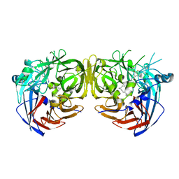
7K8G
DownloadVisualize
BU of 7k8g by Molmil
Crystal structure of bovine RPE65 in complex with 4-fluoro-MB-004 and palmitate
Descriptor: (1R)-3-amino-1-{4-fluoro-3-[(2-propylpentyl)oxy]phenyl}propan-1-ol, FE (II) ION, PALMITIC ACID, ...
Authors:Kiser, P.D.
Deposit date:2020-09-27
Release date:2021-06-02
Last modified:2023-10-18
Method:X-RAY DIFFRACTION (1.95 Å)
Cite:Rational Alteration of Pharmacokinetics of Chiral Fluorinated and Deuterated Derivatives of Emixustat for Retinal Therapy.
J.Med.Chem., 64, 2021
7L0E
DownloadVisualize
BU of 7l0e by Molmil
Crystal structure of bovine RPE65 in complex with gem-difluoro emixustat and palmitate
Descriptor: (1R)-3-amino-1-{3-[(4,4-difluorocyclohexyl)methoxy]phenyl}propan-1-ol, FE (II) ION, PALMITIC ACID, ...
Authors:Kiser, P.D.
Deposit date:2020-12-11
Release date:2021-06-02
Last modified:2023-10-18
Method:X-RAY DIFFRACTION (1.9 Å)
Cite:Rational Alteration of Pharmacokinetics of Chiral Fluorinated and Deuterated Derivatives of Emixustat for Retinal Therapy.
J.Med.Chem., 64, 2021
<1234


224572

건을2024-09-04부터공개중

PDB statisticsPDBj update infoContact PDBjnumon