Loading
PDBj
MenuPDBj@FacebookPDBj@TwitterPDBj@YouTubewwPDB FoundationwwPDB
RCSB PDBPDBeBMRBAdv. SearchSearch help
Search by PDB author
5LYB
DownloadVisualize
BU of 5lyb by Molmil
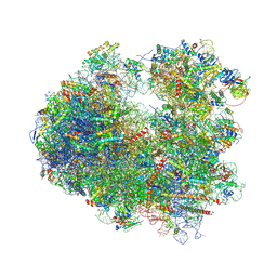
Crystal structure of the S.cerevisiae 80S ribosome in complex with the A-site bound aminoacyl-tRNA analog ACCPmn
Descriptor: 18S rRNA, 25S rRNA, 40S Ribosomal Protein S12, ...
Authors:Melnikov, S, Mailliot, J.
Deposit date:2016-09-26
Release date:2016-11-23
Last modified:2024-10-16
Method:X-RAY DIFFRACTION (3.25 Å)
Cite:Molecular insights into protein synthesis with proline residues.
EMBO Rep., 17, 2016
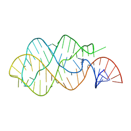
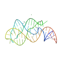
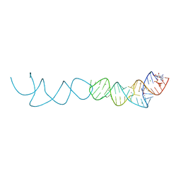
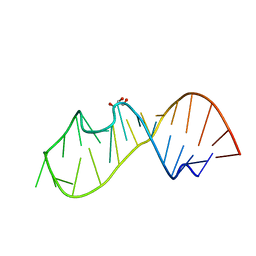
6QIT
DownloadVisualize
BU of 6qit by Molmil
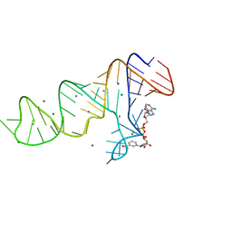
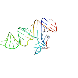
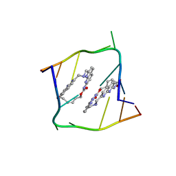
Crystal structure of CAG repeats with synthetic CMBL3b compound
Descriptor: CMBL3a, RNA (5'-R(*GP*CP*AP*GP*CP*AP*GP*C)-3')
Authors:Kiliszek, A, Blaszczyk, L, Rypniewski, W, Nakatani, K.
Deposit date:2019-01-21
Release date:2019-09-25
Last modified:2024-05-15
Method:X-RAY DIFFRACTION (1.501 Å)
Cite:Structural insights into synthetic ligands targeting A-A pairs in disease-related CAG RNA repeats.
Nucleic Acids Res., 47, 2019
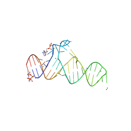
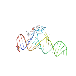
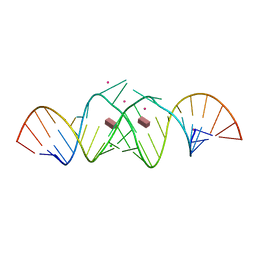
1Y27
DownloadVisualize
BU of 1y27 by Molmil
G-riboswitch-guanine complex
Descriptor: Bacillus subtilis xpt, GUANINE
Authors:Serganov, A, Yuan, Y.R, Patel, D.J.
Deposit date:2004-11-20
Release date:2004-12-28
Last modified:2023-08-23
Method:X-RAY DIFFRACTION (2.4 Å)
Cite:Structural Basis for Discriminative Regulation of Gene Expression by Adenine- and Guanine-Sensing mRNAs
Chem.Biol., 11, 2004
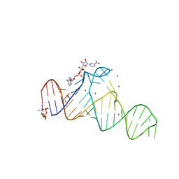
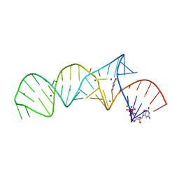
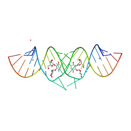
1Y26
DownloadVisualize
BU of 1y26 by Molmil
A-riboswitch-adenine complex
Descriptor: ADENINE, MAGNESIUM ION, Vibrio vulnificus A-riboswitch
Authors:Serganov, A, Yuan, Y.R, Patel, D.J.
Deposit date:2004-11-20
Release date:2004-12-28
Last modified:2024-02-14
Method:X-RAY DIFFRACTION (2.1 Å)
Cite:Structural Basis for Discriminative Regulation of Gene Expression by Adenine- and Guanine-Sensing mRNAs
Chem.Biol., 11, 2004
7D7X
DownloadVisualize
BU of 7d7x by Molmil
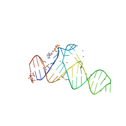
Crystal Structure of the Domain1 of NAD+ Riboswitch with adenosine diphosphate (ADP)
Descriptor: 18GAAA(52-MER), ADENOSINE-5'-DIPHOSPHATE, GUANOSINE-5'-TRIPHOSPHATE, ...
Authors:Chen, H, Ren, A.M.
Deposit date:2020-10-06
Release date:2020-11-25
Last modified:2023-11-29
Method:X-RAY DIFFRACTION (2.631 Å)
Cite:Structural distinctions between NAD+ riboswitch domains 1 and 2 determine differential folding and ligand binding.
Nucleic Acids Res., 48, 2020
7D7W
DownloadVisualize
BU of 7d7w by Molmil
Crystal Structure of the Domain1 of NAD+ Riboswitch with nicotinamide adenine dinucleotide (NAD+)
Descriptor: 18GAAA (52-MER), GUANOSINE-5'-TRIPHOSPHATE, MAGNESIUM ION, ...
Authors:Chen, H, Ren, A.M.
Deposit date:2020-10-06
Release date:2020-11-25
Last modified:2023-11-29
Method:X-RAY DIFFRACTION (2.391 Å)
Cite:Structural distinctions between NAD+ riboswitch domains 1 and 2 determine differential folding and ligand binding.
Nucleic Acids Res., 48, 2020
7D82
DownloadVisualize
BU of 7d82 by Molmil
Crystal Structure of the Domain2 of NAD+ Riboswitch with nicotinamide adenine dinucleotide (NAD+), soaked in Mn2+
Descriptor: 832GAAA (50-MER), MAGNESIUM ION, MANGANESE (II) ION, ...
Authors:Chen, H, Ren, A.M.
Deposit date:2020-10-06
Release date:2020-11-25
Last modified:2023-11-29
Method:X-RAY DIFFRACTION (2.489 Å)
Cite:Structural distinctions between NAD+ riboswitch domains 1 and 2 determine differential folding and ligand binding.
Nucleic Acids Res., 48, 2020
7D81
DownloadVisualize
BU of 7d81 by Molmil
Crystal Structure of the Domain2 of NAD+ Riboswitch with nicotinamide adenine dinucleotide (NAD+)
Descriptor: 832GAAA (50-MER), MAGNESIUM ION, NICOTINAMIDE-ADENINE-DINUCLEOTIDE
Authors:Chen, H, Ren, A.M.
Deposit date:2020-10-06
Release date:2020-11-25
Last modified:2023-11-29
Method:X-RAY DIFFRACTION (2.1 Å)
Cite:Structural distinctions between NAD+ riboswitch domains 1 and 2 determine differential folding and ligand binding.
Nucleic Acids Res., 48, 2020
7D7Y
DownloadVisualize
BU of 7d7y by Molmil
Crystal Structure of the Domain1 of NAD+ Riboswitch with adenosine triphosphate (ATP)
Descriptor: 18GAAA (52-MER), ADENOSINE-5'-TRIPHOSPHATE, GUANOSINE-5'-TRIPHOSPHATE, ...
Authors:Chen, H, Ren, A.M.
Deposit date:2020-10-06
Release date:2020-11-25
Last modified:2023-11-29
Method:X-RAY DIFFRACTION (2.8 Å)
Cite:Structural distinctions between NAD+ riboswitch domains 1 and 2 determine differential folding and ligand binding.
Nucleic Acids Res., 48, 2020
7D7V
DownloadVisualize
BU of 7d7v by Molmil
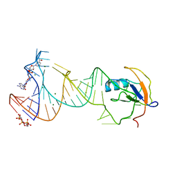
Crystal Structure of the Domain1 of NAD+ Riboswitch with nicotinamide adenine dinucleotide (NAD+) and U1A protein
Descriptor: 17delU1A (58-MER), GUANOSINE-5'-TRIPHOSPHATE, MAGNESIUM ION, ...
Authors:Chen, H, Ren, A.M.
Deposit date:2020-10-06
Release date:2020-11-25
Last modified:2024-03-27
Method:X-RAY DIFFRACTION (2.8 Å)
Cite:Structural distinctions between NAD+ riboswitch domains 1 and 2 determine differential folding and ligand binding.
Nucleic Acids Res., 48, 2020
7D7Z
DownloadVisualize
BU of 7d7z by Molmil
Crystal Structure of the Domain1 of NAD+ Riboswitch with nicotinamide adenine dinucleotide (NAD+), soaked in Mn2+
Descriptor: 18GAAA(52-MER), GUANOSINE-5'-TRIPHOSPHATE, MAGNESIUM ION, ...
Authors:Chen, H, Ren, A.M.
Deposit date:2020-10-06
Release date:2020-11-25
Last modified:2023-11-29
Method:X-RAY DIFFRACTION (2.6 Å)
Cite:Structural distinctions between NAD+ riboswitch domains 1 and 2 determine differential folding and ligand binding.
Nucleic Acids Res., 48, 2020
7ELR
DownloadVisualize
BU of 7elr by Molmil
Crystal structure of xanthine riboswitch with xanthine
Descriptor: GUANOSINE-5'-TRIPHOSPHATE, MAGNESIUM ION, NMT1 (46-MER), ...
Authors:Xu, X.C, Ren, A.M.
Deposit date:2021-04-12
Release date:2021-06-30
Last modified:2023-11-29
Method:X-RAY DIFFRACTION (2.66 Å)
Cite:Insights into xanthine riboswitch structure and metal ion-mediated ligand recognition.
Nucleic Acids Res., 49, 2021
7ELS
DownloadVisualize
BU of 7els by Molmil
Crystal structure of xanthine riboswitch with 8-azaxanthine
Descriptor: 8-AZAXANTHINE, GUANOSINE-5'-TRIPHOSPHATE, MAGNESIUM ION, ...
Authors:Xu, X.C, Ren, A.M.
Deposit date:2021-04-12
Release date:2021-06-30
Last modified:2023-11-29
Method:X-RAY DIFFRACTION (3 Å)
Cite:Insights into xanthine riboswitch structure and metal ion-mediated ligand recognition.
Nucleic Acids Res., 49, 2021
7ELP
DownloadVisualize
BU of 7elp by Molmil
Crystal structure of xanthine riboswitch with xanthine, iridium hexammine soak
Descriptor: GUANOSINE-5'-TRIPHOSPHATE, IRIDIUM ION, MAGNESIUM ION, ...
Authors:Xu, X.C, Ren, A.M.
Deposit date:2021-04-12
Release date:2021-06-30
Last modified:2024-05-29
Method:X-RAY DIFFRACTION (2.79 Å)
Cite:Insights into xanthine riboswitch structure and metal ion-mediated ligand recognition.
Nucleic Acids Res., 49, 2021
7ELQ
DownloadVisualize
BU of 7elq by Molmil
Crystal structure of xanthine riboswitch with xanthine, manganese saok
Descriptor: GUANOSINE-5'-TRIPHOSPHATE, MAGNESIUM ION, MANGANESE (II) ION, ...
Authors:Xu, X.C, Ren, A.M.
Deposit date:2021-04-12
Release date:2021-06-30
Last modified:2023-11-29
Method:X-RAY DIFFRACTION (2.6 Å)
Cite:Insights into xanthine riboswitch structure and metal ion-mediated ligand recognition.
Nucleic Acids Res., 49, 2021
5D99
DownloadVisualize
BU of 5d99 by Molmil
3DW4 redetermined by direct methods starting from random phase angles
Descriptor: GLYCEROL, RNA (27-MER) hairpin from sarcin-ricin domain of E. coli 23S rRNA
Authors:Mooers, B.H.M.
Deposit date:2015-08-18
Release date:2016-04-13
Last modified:2024-03-06
Method:X-RAY DIFFRACTION (0.97 Å)
Cite:Direct-methods structure determination of a trypanosome RNA-editing substrate fragment with translational pseudosymmetry.
Acta Crystallogr D Struct Biol, 72, 2016
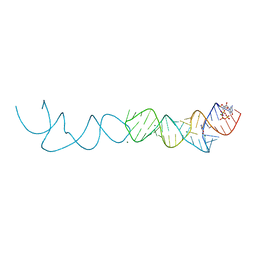
3C44
DownloadVisualize
BU of 3c44 by Molmil
Crystal structure of HIV-1 subtype F DIS extended duplex RNA bound to paromomycin
Descriptor: CHLORIDE ION, HIV-1 subtype F genomic RNA, PAROMOMYCIN, ...
Authors:Freisz, S, Ennifar, E, Dumas, P.
Deposit date:2008-01-29
Release date:2008-05-06
Last modified:2023-11-01
Method:X-RAY DIFFRACTION (2 Å)
Cite:Binding of aminoglycoside antibiotics to the duplex form of the HIV-1 genomic RNA dimerization initiation site.
Angew.Chem.Int.Ed.Engl., 47, 2008
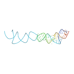
3DVV
DownloadVisualize
BU of 3dvv by Molmil
Crystal structure of HIV-1 subtype F DIS extended duplex RNA bound to ribostamycin (U267OMe)
Descriptor: CHLORIDE ION, HIV-1 genomic RNA, POTASSIUM ION, ...
Authors:Ennifar, E, Dumas, P, Olieric, V.
Deposit date:2008-07-21
Release date:2008-08-12
Last modified:2023-11-01
Method:X-RAY DIFFRACTION (2 Å)
Cite:A fast strategy for Selenium derivatization and phasing of RNA structures
To be Published
<1234


227111

건을2024-11-06부터공개중

PDB statisticsPDBj update infoContact PDBjnumon