Loading
PDBj
MenuPDBj@FacebookPDBj@TwitterPDBj@YouTubewwPDB FoundationwwPDB
RCSB PDBPDBeBMRBAdv. SearchSearch help
Search by PDB author
1VH8
DownloadVisualize
BU of 1vh8 by Molmil
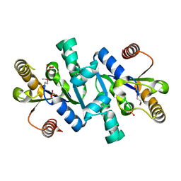
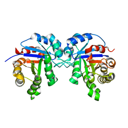
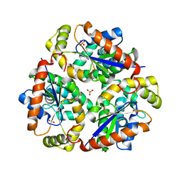
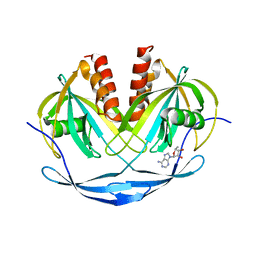
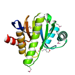
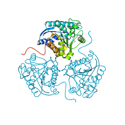
Crystal structure of a 2C-methyl-D-erythritol 2,4-cyclodiphosphate synthase
Descriptor: 2-C-methyl-D-erythritol 2,4-cyclodiphosphate synthase, ACETIC ACID, PYROPHOSPHATE 2-
Authors:Structural GenomiX
Deposit date:2003-12-01
Release date:2003-12-30
Last modified:2023-12-27
Method:X-RAY DIFFRACTION (2.35 Å)
Cite:Structural analysis of a set of proteins resulting from a bacterial genomics project
Proteins, 60, 2005
1VI3
DownloadVisualize
BU of 1vi3 by Molmil
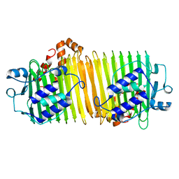
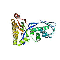
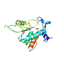
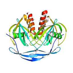
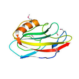
Crystal structure of an hypothetical protein
Descriptor: ACETATE ION, Protein ybhB
Authors:Structural GenomiX
Deposit date:2003-12-01
Release date:2003-12-30
Last modified:2023-12-27
Method:X-RAY DIFFRACTION (1.76 Å)
Cite:Structural analysis of a set of proteins resulting from a bacterial genomics project
Proteins, 60, 2005
1VIA
DownloadVisualize
BU of 1via by Molmil
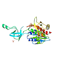
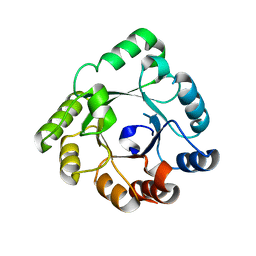
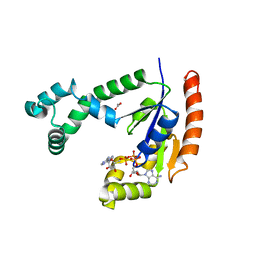
Crystal structure of shikimate kinase
Descriptor: SULFATE ION, shikimate kinase
Authors:Structural GenomiX
Deposit date:2003-12-01
Release date:2003-12-30
Last modified:2023-12-27
Method:X-RAY DIFFRACTION (1.57 Å)
Cite:Structural analysis of a set of proteins resulting from a bacterial genomics project
Proteins, 60, 2005
1VR2
DownloadVisualize
BU of 1vr2 by Molmil
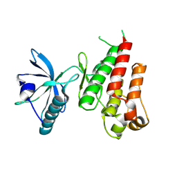
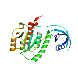
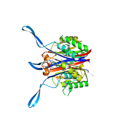
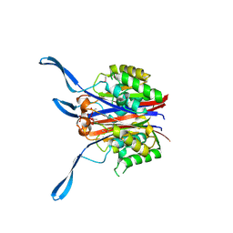
HUMAN VASCULAR ENDOTHELIAL GROWTH FACTOR RECEPTOR 2 (KDR) KINASE DOMAIN
Descriptor: PROTEIN (VASCULAR ENDOTHELIAL GROWTH FACTOR RECEPTOR KINASE)
Authors:Mctigue, M, Wickersham, J, Pinko, C, Showalter, R, Parast, C, Tempczyk-Russell, A, Gehring, M, Mroczkowski, B, Kan, C, Villafranca, J, Appelt, K.
Deposit date:1998-12-03
Release date:2000-03-15
Last modified:2023-11-15
Method:X-RAY DIFFRACTION (2.4 Å)
Cite:Crystal structure of the kinase domain of human vascular endothelial growth factor receptor 2: a key enzyme in angiogenesis.
Structure Fold.Des., 7, 1999
1VIQ
DownloadVisualize
BU of 1viq by Molmil
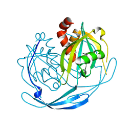
Crystal structure of putative ADP ribose pyrophosphatase
Descriptor: ADP-ribose pyrophosphatase
Authors:Structural GenomiX
Deposit date:2003-12-01
Release date:2003-12-30
Last modified:2023-12-27
Method:X-RAY DIFFRACTION (2.4 Å)
Cite:Structural analysis of a set of proteins resulting from a bacterial genomics project
Proteins, 60, 2005
3OHH
DownloadVisualize
BU of 3ohh by Molmil
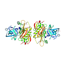
Crystal structure of beta-site app-cleaving enzyme 1 (bace-wt) complex with bms-681889 aka n~1~-butyl-5-cyano- n~3~-((1s,2r)-1-(3,5-difluorobenzyl)-2-hydroxy-3-((3- methoxybenzyl)amino)propyl)-n~1~-methyl-1h-indole-1,3- dicarboxamide
Descriptor: Beta-secretase 1, GLYCEROL, N~1~-butyl-5-cyano-N~3~-{(1S,2R)-1-(3,5-difluorobenzyl)-2-hydroxy-3-[(3-methoxybenzyl)amino]propyl}-N~1~-methyl-1H-indole-1,3-dicarboxamide, ...
Authors:Muckelbauer, J.K.
Deposit date:2010-08-17
Release date:2011-04-06
Last modified:2017-11-08
Method:X-RAY DIFFRACTION (2.01 Å)
Cite:Synthesis and SAR of indole-and 7-azaindole-1,3-dicarboxamide hydroxyethylamine inhibitors of BACE-1.
Bioorg.Med.Chem.Lett., 21, 2011
1HTI
DownloadVisualize
BU of 1hti by Molmil
CRYSTAL STRUCTURE OF RECOMBINANT HUMAN TRIOSEPHOSPHATE ISOMERASE AT 2.8 ANGSTROMS RESOLUTION. TRIOSEPHOSPHATE ISOMERASE RELATED HUMAN GENETIC DISORDERS AND COMPARISON WITH THE TRYPANOSOMAL ENZYME
Descriptor: 2-PHOSPHOGLYCOLIC ACID, TRIOSEPHOSPHATE ISOMERASE
Authors:Mande, S.C, Hol, W.G.J.
Deposit date:1994-10-12
Release date:1995-01-26
Last modified:2024-02-07
Method:X-RAY DIFFRACTION (2.8 Å)
Cite:Crystal structure of recombinant human triosephosphate isomerase at 2.8 A resolution. Triosephosphate isomerase-related human genetic disorders and comparison with the trypanosomal enzyme.
Protein Sci., 3, 1994
1VH4
DownloadVisualize
BU of 1vh4 by Molmil
Crystal structure of a stabilizer of iron transporter
Descriptor: SufD protein
Authors:Structural GenomiX
Deposit date:2003-12-01
Release date:2003-12-30
Last modified:2023-12-27
Method:X-RAY DIFFRACTION (1.75 Å)
Cite:Structural analysis of a set of proteins resulting from a bacterial genomics project
Proteins, 60, 2005
1VHO
DownloadVisualize
BU of 1vho by Molmil
Crystal structure of a putative peptidase/endoglucanase
Descriptor: CACODYLATE ION, SULFATE ION, endoglucanase
Authors:Structural GenomiX
Deposit date:2003-12-01
Release date:2003-12-30
Last modified:2023-12-27
Method:X-RAY DIFFRACTION (1.86 Å)
Cite:Structural analysis of a set of proteins resulting from a bacterial genomics project
Proteins, 60, 2005
1VHY
DownloadVisualize
BU of 1vhy by Molmil
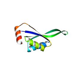
Crystal structure of Haemophilus influenzae protein HI0303, Pfam DUF558
Descriptor: Hypothetical protein HI0303
Authors:Structural GenomiX, Burley, S.K, New York SGX Research Center for Structural Genomics (NYSGXRC)
Deposit date:2003-12-01
Release date:2003-12-30
Last modified:2023-12-27
Method:X-RAY DIFFRACTION (1.9 Å)
Cite:Structural analysis of a set of proteins resulting from a bacterial genomics project
Proteins, 60, 2005
1VIY
DownloadVisualize
BU of 1viy by Molmil
Crystal structure of dephospho-CoA kinase
Descriptor: Dephospho-CoA kinase, SULFATE ION
Authors:Structural GenomiX
Deposit date:2003-12-01
Release date:2003-12-30
Last modified:2023-12-27
Method:X-RAY DIFFRACTION (1.89 Å)
Cite:Structural analysis of a set of proteins resulting from a bacterial genomics project
Proteins, 60, 2005
1VER
DownloadVisualize
BU of 1ver by Molmil
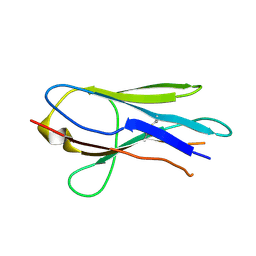
Structure of New Antigen Receptor variable domain from sharks
Descriptor: New Antigen Receptor variable domain
Authors:Streltsov, V.A.
Deposit date:2004-04-05
Release date:2004-08-24
Last modified:2023-12-27
Method:X-RAY DIFFRACTION (2.82 Å)
Cite:Structural evidence for evolution of shark Ig new antigen receptor variable domain antibodies from a cell-surface receptor
Proc.Natl.Acad.Sci.USA, 101, 2004
1VH7
DownloadVisualize
BU of 1vh7 by Molmil
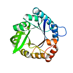
Crystal structure of a cyclase subunit of imidazolglycerolphosphate synthase
Descriptor: Imidazole glycerol phosphate synthase subunit hisF, PHOSPHATE ION
Authors:Structural GenomiX
Deposit date:2003-12-01
Release date:2003-12-30
Last modified:2023-12-27
Method:X-RAY DIFFRACTION (1.9 Å)
Cite:Structural analysis of a set of proteins resulting from a bacterial genomics project
Proteins, 60, 2005
1VHF
DownloadVisualize
BU of 1vhf by Molmil
Crystal structure of periplasmic divalent cation tolerance protein
Descriptor: periplasmic divalent cation tolerance protein
Authors:Structural GenomiX
Deposit date:2003-12-01
Release date:2003-12-30
Last modified:2023-12-27
Method:X-RAY DIFFRACTION (1.54 Å)
Cite:Structural analysis of a set of proteins resulting from a bacterial genomics project
Proteins, 60, 2005
1VHZ
DownloadVisualize
BU of 1vhz by Molmil
Crystal structure of ADP compounds hydrolase
Descriptor: ADENOSINE-5-DIPHOSPHORIBOSE, ADP compounds hydrolase nudE
Authors:Structural GenomiX
Deposit date:2003-12-01
Release date:2003-12-30
Last modified:2023-12-27
Method:X-RAY DIFFRACTION (2.32 Å)
Cite:Structural analysis of a set of proteins resulting from a bacterial genomics project
Proteins, 60, 2005
1VIS
DownloadVisualize
BU of 1vis by Molmil
Crystal structure of mevalonate kinase
Descriptor: 1,4-DIETHYLENE DIOXIDE, Mevalonate kinase
Authors:Structural GenomiX
Deposit date:2003-12-01
Release date:2003-12-30
Last modified:2023-12-27
Method:X-RAY DIFFRACTION (2.69 Å)
Cite:Structural analysis of a set of proteins resulting from a bacterial genomics project
Proteins, 60, 2005
1VIZ
DownloadVisualize
BU of 1viz by Molmil
Crystal structure of an hypothetical protein
Descriptor: PcrB protein homolog, SODIUM ION
Authors:Structural GenomiX
Deposit date:2003-12-01
Release date:2003-12-30
Last modified:2023-12-27
Method:X-RAY DIFFRACTION (1.85 Å)
Cite:Structural analysis of a set of proteins resulting from a bacterial genomics project
Proteins, 60, 2005
1VHA
DownloadVisualize
BU of 1vha by Molmil
Crystal structure of 2C-methyl-D-erythritol 2,4-cyclodiphosphate synthase
Descriptor: 2-C-methyl-D-erythritol 2,4-cyclodiphosphate synthase, ACETIC ACID, MANGANESE (II) ION, ...
Authors:Structural GenomiX
Deposit date:2003-12-01
Release date:2003-12-30
Last modified:2023-12-27
Method:X-RAY DIFFRACTION (2.35 Å)
Cite:Structural analysis of a set of proteins resulting from a bacterial genomics project
Proteins, 60, 2005
1VHU
DownloadVisualize
BU of 1vhu by Molmil
Crystal structure of a putative phosphoesterase
Descriptor: 2-(N-MORPHOLINO)-ETHANESULFONIC ACID, Hypothetical protein AF1521
Authors:Structural GenomiX
Deposit date:2003-12-01
Release date:2003-12-30
Last modified:2023-12-27
Method:X-RAY DIFFRACTION (1.34 Å)
Cite:Structural analysis of a set of proteins resulting from a bacterial genomics project
Proteins, 60, 2005
1VI5
DownloadVisualize
BU of 1vi5 by Molmil
Crystal structure of ribosomal protein S2P
Descriptor: 30S ribosomal protein S2P
Authors:Structural GenomiX
Deposit date:2003-12-01
Release date:2003-12-30
Last modified:2023-12-27
Method:X-RAY DIFFRACTION (2.65 Å)
Cite:Structural analysis of a set of proteins resulting from a bacterial genomics project
Proteins, 60, 2005
1GP2
DownloadVisualize
BU of 1gp2 by Molmil
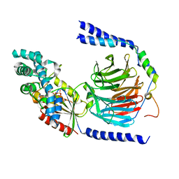
G PROTEIN HETEROTRIMER GI_ALPHA_1 BETA_1 GAMMA_2 WITH GDP BOUND
Descriptor: G PROTEIN GI ALPHA 1, G PROTEIN GI BETA 1, G PROTEIN GI GAMMA 2, ...
Authors:Wall, M.A, Sprang, S.R.
Deposit date:1996-11-13
Release date:1997-02-12
Last modified:2024-02-07
Method:X-RAY DIFFRACTION (2.3 Å)
Cite:The structure of the G protein heterotrimer Gi alpha 1 beta 1 gamma 2.
Cell(Cambridge,Mass.), 83, 1995
3M0C
DownloadVisualize
BU of 3m0c by Molmil
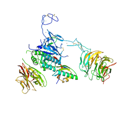
The X-ray Crystal Structure of PCSK9 in Complex with the LDL receptor
Descriptor: CALCIUM ION, Low-density lipoprotein receptor, Proprotein convertase subtilisin/kexin type 9
Authors:Spraggon, G, Hampton, E.N.
Deposit date:2010-03-02
Release date:2011-03-16
Last modified:2023-09-06
Method:X-RAY DIFFRACTION (7.01 Å)
Cite:The X-ray Crystal Structure of PCSK9 in Complex with the LDL receptor
To be Published
3MJL
DownloadVisualize
BU of 3mjl by Molmil
Crystal structure of human arginase I in complex with 2-aminoimidazole. Resolution 1.90 A.
Descriptor: 1H-imidazol-2-amine, Arginase-1, MANGANESE (II) ION
Authors:Di Costanzo, L, Christianson, D.W.
Deposit date:2010-04-13
Release date:2010-05-05
Last modified:2023-09-06
Method:X-RAY DIFFRACTION (1.9 Å)
Cite:Crystal structure of human arginase I in complex with 2-aminoimidazole. Resolution 1.90 A.
To be Published
1VHG
DownloadVisualize
BU of 1vhg by Molmil
Crystal structure of ADP compounds hydrolase
Descriptor: ADP compounds hydrolase nudE
Authors:Structural GenomiX
Deposit date:2003-12-01
Release date:2003-12-30
Last modified:2023-12-27
Method:X-RAY DIFFRACTION (2.7 Å)
Cite:Structural analysis of a set of proteins resulting from a bacterial genomics project
Proteins, 60, 2005
1VHT
DownloadVisualize
BU of 1vht by Molmil
Crystal structure of dephospho-coA kinase with bis(adenosine)-5'-triphosphate
Descriptor: ACETATE ION, BIS(ADENOSINE)-5'-TRIPHOSPHATE, Dephospho-CoA kinase
Authors:Structural GenomiX
Deposit date:2003-12-01
Release date:2003-12-30
Last modified:2023-12-27
Method:X-RAY DIFFRACTION (1.59 Å)
Cite:Structural analysis of a set of proteins resulting from a bacterial genomics project
Proteins, 60, 2005

225399

건을2024-09-25부터공개중

PDB statisticsPDBj update infoContact PDBjnumon