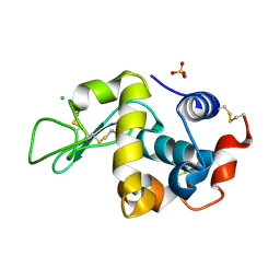
6F1O
 
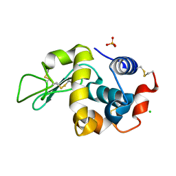 | | Orthorhombic Lysozyme crystallized at 298 K and pH 4.5 | | Descriptor: | CHLORIDE ION, Lysozyme C, PHOSPHATE ION | | Authors: | Camara-Artigas, A. | | Deposit date: | 2017-11-22 | | Release date: | 2018-05-09 | | Last modified: | 2024-01-17 | | Method: | X-RAY DIFFRACTION (0.96 Å) | | Cite: | Orthorhombic lysozyme crystallization at acidic pH values driven by phosphate binding.
Acta Crystallogr D Struct Biol, 74, 2018
|
|
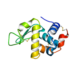
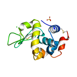
6F1M
 | |
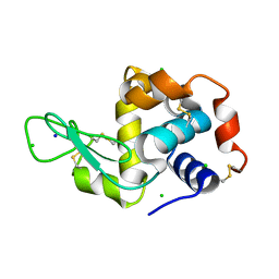
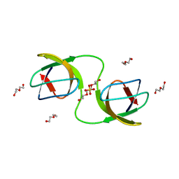
6F1R
 | |
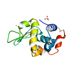
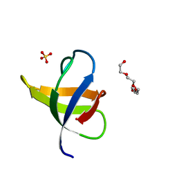
6FA0
 | |
6F9Y
 | |
6F9X
 | |
7NET
 | |
7NER
 | |
7NES
 | |
5DK8
 | |
8AH7
 | |
8AH5
 | |
8AH6
 | |
8AH8
 | |
8AH4
 | |


