Loading
PDBj
MenuPDBj@FacebookPDBj@TwitterPDBj@YouTubewwPDB FoundationwwPDB
RCSB PDBPDBeBMRBAdv. SearchSearch help
Search by PDB author
6OFS
DownloadVisualize
BU of 6ofs by Molmil
The crystal structure of the periplasmic protease PqqL from Escherichia coli
Descriptor: CHLORIDE ION, Probable zinc protease PqqL, ZINC ION
Authors:Grinter, R.
Deposit date:2019-04-01
Release date:2019-10-02
Last modified:2024-04-03
Method:X-RAY DIFFRACTION (2.6 Å)
Cite:Protease-associated import systems are widespread in Gram-negative bacteria.
Plos Genet., 15, 2019
6OFT
DownloadVisualize
BU of 6oft by Molmil
The crystal structure of the first half of the periplasmic protease PqqL from Escherichia coli
Descriptor: CHLORIDE ION, GLYCEROL, PHOSPHATE ION, ...
Authors:Grinter, R.
Deposit date:2019-04-01
Release date:2019-10-02
Last modified:2023-10-11
Method:X-RAY DIFFRACTION (2 Å)
Cite:Protease-associated import systems are widespread in Gram-negative bacteria.
Plos Genet., 15, 2019
6OFR
DownloadVisualize
BU of 6ofr by Molmil
The crystal structure of the outer membrane transporter YddB from Escherichia coli
Descriptor: GLYCEROL, MAGNESIUM ION, TonB-dependent outer membrane receptor, ...
Authors:Grinter, R.
Deposit date:2019-04-01
Release date:2019-10-02
Last modified:2023-10-11
Method:X-RAY DIFFRACTION (2.4 Å)
Cite:Protease-associated import systems are widespread in Gram-negative bacteria.
Plos Genet., 15, 2019
6V4V
DownloadVisualize
BU of 6v4v by Molmil
The crystal structure of BonA from Acinetobacter baumannii
Descriptor: BON domain protein, ZINC ION
Authors:Grinter, R.
Deposit date:2019-12-02
Release date:2021-06-02
Last modified:2021-08-25
Method:X-RAY DIFFRACTION (1.65 Å)
Cite:BonA from Acinetobacter baumannii Forms a Divisome-Localized Decamer That Supports Outer Envelope Function.
Mbio, 2021
6WQQ
DownloadVisualize
BU of 6wqq by Molmil
Structure of the 50S subunit of the ribosome from Methicillin Resistant Staphylococcus aureus in complex with the antibiotic, radezolid
Descriptor: 23S rRNA, 50S ribosomal protein L13, 50S ribosomal protein L14, ...
Authors:Belousoff, M.J.
Deposit date:2020-04-29
Release date:2020-06-03
Last modified:2020-12-16
Method:ELECTRON MICROSCOPY (3.1 Å)
Cite:Characterization of the Core Ribosomal Binding Region for the Oxazolidone Family of Antibiotics Using Cryo-EM.
Acs Pharmacol Transl Sci, 3, 2020
6V81
DownloadVisualize
BU of 6v81 by Molmil
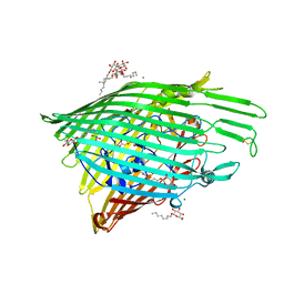
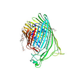
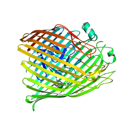
The crystal structure of the outer-membrane transporter YncD
Descriptor: CALCIUM ION, Probable TonB-dependent receptor YncD, octyl beta-D-glucopyranoside
Authors:Grinter, R.
Deposit date:2019-12-10
Release date:2020-05-06
Last modified:2020-07-29
Method:X-RAY DIFFRACTION (2.957 Å)
Cite:The crystal structure of the TonB-dependent transporter YncD reveals a positively charged substrate-binding site.
Acta Crystallogr D Struct Biol, 76, 2020
6WRU
DownloadVisualize
BU of 6wru by Molmil
Structure of the 50S subunit of the ribosome from Methicillin Resistant Staphylococcus aureus in complex with an isomer of the tedizolid
Descriptor: 23S rRNA, 50S ribosomal protein L13, 50S ribosomal protein L14, ...
Authors:Belousoff, M.J.
Deposit date:2020-04-30
Release date:2020-06-03
Last modified:2020-12-16
Method:ELECTRON MICROSCOPY (3.1 Å)
Cite:Characterization of the Core Ribosomal Binding Region for the Oxazolidone Family of Antibiotics Using Cryo-EM.
Acs Pharmacol Transl Sci, 3, 2020
6B05
DownloadVisualize
BU of 6b05 by Molmil
The Crystal Structure of the Ferredoxin Protease FusC E83A mutant in complex with Arabidopsis Ferredoxin
Descriptor: Ferredoxin-2, chloroplastic, Putative zinc protease, ...
Authors:Grinter, R.
Deposit date:2017-09-13
Release date:2018-06-20
Last modified:2024-03-13
Method:X-RAY DIFFRACTION (1.9 Å)
Cite:FusC, a member of the M16 protease family acquired by bacteria for iron piracy against plants.
PLoS Biol., 16, 2018
6B03
DownloadVisualize
BU of 6b03 by Molmil
The crystal structure of the ferredoxin protease FusC in complex with its substrate plant ferredoxin
Descriptor: Ferredoxin-2, chloroplastic, Putative zinc protease
Authors:Grinter, R.
Deposit date:2017-09-13
Release date:2018-06-20
Last modified:2024-03-13
Method:X-RAY DIFFRACTION (2.7 Å)
Cite:FusC, a member of the M16 protease family acquired by bacteria for iron piracy against plants.
PLoS Biol., 16, 2018
6BRS
DownloadVisualize
BU of 6brs by Molmil
The Crystal Structure of the Ferredoxin Protease FusC in complex with Arabidopsis Ferredoxin, Ethylmercury phosphate soaked dataset
Descriptor: Ferredoxin-2, chloroplastic, MERCURY (II) ION, ...
Authors:Grinter, R.
Deposit date:2017-11-30
Release date:2018-06-20
Last modified:2024-03-13
Method:X-RAY DIFFRACTION (2.3 Å)
Cite:FusC, a member of the M16 protease family acquired by bacteria for iron piracy against plants.
PLoS Biol., 16, 2018
6BPN
DownloadVisualize
BU of 6bpn by Molmil
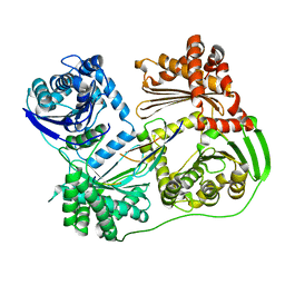
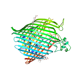
The crystal structure of the Ferric-Catecholate import receptor Fiu from E. coli K12: Open form (C2221)
Descriptor: (20S)-2,5,8,11,14,17-HEXAMETHYL-3,6,9,12,15,18-HEXAOXAHENICOSANE-1,20-DIOL, 2-AMINO-2-HYDROXYMETHYL-PROPANE-1,3-DIOL, CHLORIDE ION, ...
Authors:Grinter, R.
Deposit date:2017-11-23
Release date:2018-11-28
Last modified:2023-10-04
Method:X-RAY DIFFRACTION (2.1 Å)
Cite:The structure of the bacterial iron-catecholate transporter Fiu suggests that it imports substrates via a two-step mechanism.
J.Biol.Chem., 2019
6BPM
DownloadVisualize
BU of 6bpm by Molmil
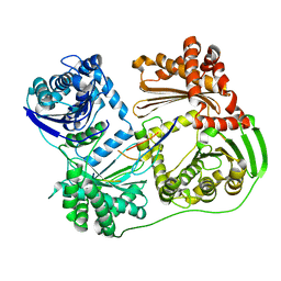
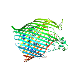
The crystal structure of the Ferric-Catecholate import receptor Fiu from K12 E. coli: Closed form (C21)
Descriptor: (20S)-2,5,8,11,14,17-HEXAMETHYL-3,6,9,12,15,18-HEXAOXAHENICOSANE-1,20-DIOL, Catecholate siderophore receptor Fiu, octyl beta-D-glucopyranoside
Authors:Grinter, R.
Deposit date:2017-11-23
Release date:2018-11-28
Last modified:2023-10-04
Method:X-RAY DIFFRACTION (2.5 Å)
Cite:The structure of the bacterial iron-catecholate transporter Fiu suggests that it imports substrates via a two-step mechanism.
J.Biol.Chem., 2019
6BPO
DownloadVisualize
BU of 6bpo by Molmil
The crystal structure of the Ferric-Catecholate import receptor Fiu from K12 E. coli: Closed form (P1)
Descriptor: Catecholate siderophore receptor Fiu, octyl beta-D-glucopyranoside
Authors:Grinter, R.
Deposit date:2017-11-23
Release date:2018-11-28
Last modified:2023-10-04
Method:X-RAY DIFFRACTION (2.9 Å)
Cite:The structure of the bacterial iron-catecholate transporter Fiu suggests that it imports substrates via a two-step mechanism.
J.Biol.Chem., 2019
<12


224004

건을2024-08-21부터공개중

PDB statisticsPDBj update infoContact PDBjnumon