1RPA
 
 | |
1RPT
 | |
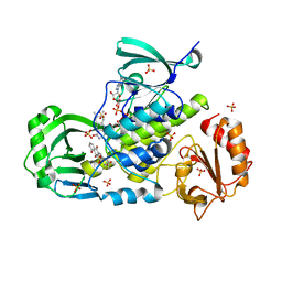
1QHW
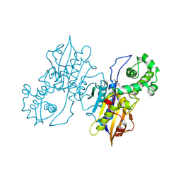
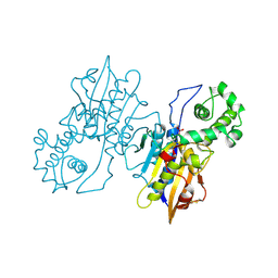
 | | PURPLE ACID PHOSPHATASE FROM RAT BONE | | Descriptor: | 2-acetamido-2-deoxy-beta-D-glucopyranose-(1-4)-2-acetamido-2-deoxy-beta-D-glucopyranose, FE (III) ION, PROTEIN (PURPLE ACID PHOSPHATASE), ... | | Authors: | Lindqvist, Y, Johansson, E, Kaija, H, Vihko, P, Schneider, G. | | Deposit date: | 1999-03-26 | | Release date: | 1999-09-15 | | Last modified: | 2024-11-13 | | Method: | X-RAY DIFFRACTION (2.2 Å) | | Cite: | Three-dimensional structure of a mammalian purple acid phosphatase at 2.2 A resolution with a mu-(hydr)oxo bridged di-iron center.
J.Mol.Biol., 291, 1999
|
|
1GOX
 | |
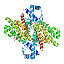
3IHG
 | | Crystal structure of a ternary complex of aklavinone-11 hydroxylase with FAD and aklavinone | | Descriptor: | FLAVIN-ADENINE DINUCLEOTIDE, RdmE, SULFATE ION, ... | | Authors: | Lindqvist, Y, Koskiniemi, H, Jansson, A, Sandalova, T, Schneider, G. | | Deposit date: | 2009-07-30 | | Release date: | 2009-09-29 | | Last modified: | 2024-02-21 | | Method: | X-RAY DIFFRACTION (2.49 Å) | | Cite: | Structural basis for substrate recognition and specificity in aklavinone-11-hydroxylase from rhodomycin biosynthesis.
J.Mol.Biol., 393, 2009
|
|
5DV5
 | |
5XGF
 | |
3U0J
 | |
7Y62
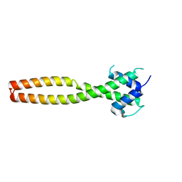 | | Crystal structure of human TFEB HLHLZ domain | | Descriptor: | Transcription factor EB | | Authors: | Yang, G, Li, P, Lin, Y, Liu, Z, Sun, H, Zhao, Z, Fang, P, Wang, J. | | Deposit date: | 2022-06-18 | | Release date: | 2023-03-22 | | Last modified: | 2023-11-29 | | Method: | X-RAY DIFFRACTION (2 Å) | | Cite: | A small-molecule drug inhibits autophagy gene expression through the central regulator TFEB.
Proc.Natl.Acad.Sci.USA, 120, 2023
|
|
1I7K
 | |
1TRK
 | |
1GYL
 | |
1I2A
 | |
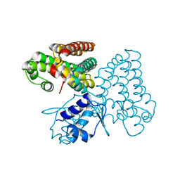
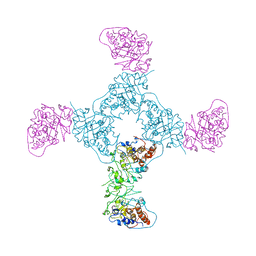
4KPR
 | | Tetrameric form of rat selenoprotein thioredoxin reductase 1 | | Descriptor: | (4S)-2-METHYL-2,4-PENTANEDIOL, FLAVIN-ADENINE DINUCLEOTIDE, SULFITE ION, ... | | Authors: | Lindqvist, Y, Sandalova, T, Xu, J, Arner, E. | | Deposit date: | 2013-05-14 | | Release date: | 2014-05-14 | | Last modified: | 2023-09-20 | | Method: | X-RAY DIFFRACTION (2.4 Å) | | Cite: | The Trp114 residue of thioredoxin reductase 1 is an electron relay sensor for oxidative stress
To be Published, 2013
|
|
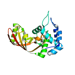
3SHB
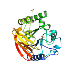
 | | Crystal Structure of PHD Domain of UHRF1 | | Descriptor: | E3 ubiquitin-protein ligase UHRF1, Histone H3 peptide, ZINC ION | | Authors: | Hu, L, Li, Z, Wang, P, Lin, Y, Xu, Y. | | Deposit date: | 2011-06-16 | | Release date: | 2011-08-24 | | Last modified: | 2024-03-20 | | Method: | X-RAY DIFFRACTION (1.8 Å) | | Cite: | Crystal structure of PHD domain of UHRF1 and insights into recognition of unmodified histone H3 arginine residue 2.
Cell Res., 2011
|
|
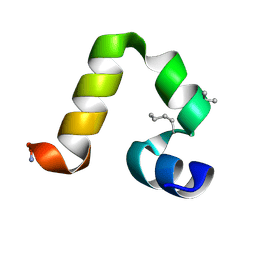
9MFB
 | | Backbone alpha-Methylation in the Villin Headpiece Miniprotein: HP35 with Calpha-methyl-Lys at Position 30 | | Descriptor: | Villin-1 | | Authors: | Harmon, T.W, Lin, Y, Sutton, R.T, Osborne, S, Horne, W.S. | | Deposit date: | 2024-12-09 | | Release date: | 2025-01-22 | | Last modified: | 2025-03-26 | | Method: | SOLUTION NMR | | Cite: | Interplay between C alpha Methylation and C alpha Stereochemistry in the Folding Energetics of a Helix-Rich Miniprotein.
Chembiochem, 26, 2025
|
|
9MF9
 | | Backbone alpha-Methylation in the Villin Headpiece Miniprotein: HP35 with Calpha-methyl-Ser at Position 15 | | Descriptor: | Villin-1 | | Authors: | Harmon, T.W, Lin, Y, Sutton, R.T, Osborne, S, Horne, W.S. | | Deposit date: | 2024-12-09 | | Release date: | 2025-01-22 | | Last modified: | 2025-03-26 | | Method: | SOLUTION NMR | | Cite: | Interplay between C alpha Methylation and C alpha Stereochemistry in the Folding Energetics of a Helix-Rich Miniprotein.
Chembiochem, 26, 2025
|
|
9MF8
 | | Backbone alpha-Methylation in the Villin Headpiece Miniprotein: HP35 with Aib at Position 15 | | Descriptor: | Villin-1 | | Authors: | Harmon, T.W, Lin, Y, Sutton, R.T, Osborne, S, Horne, W.S. | | Deposit date: | 2024-12-09 | | Release date: | 2025-01-22 | | Last modified: | 2025-03-26 | | Method: | SOLUTION NMR | | Cite: | Interplay between C alpha Methylation and C alpha Stereochemistry in the Folding Energetics of a Helix-Rich Miniprotein.
Chembiochem, 26, 2025
|
|
9MFA
 | | Backbone alpha-Methylation in the Villin Headpiece Miniprotein: HP35 with Aib at Position 30 | | Descriptor: | Villin-1 | | Authors: | Harmon, T.W, Lin, Y, Sutton, R.T, Osborne, S, Horne, W.S. | | Deposit date: | 2024-12-09 | | Release date: | 2025-01-22 | | Last modified: | 2025-03-26 | | Method: | SOLUTION NMR | | Cite: | Interplay between C alpha Methylation and C alpha Stereochemistry in the Folding Energetics of a Helix-Rich Miniprotein.
Chembiochem, 26, 2025
|
|
9MF7
 | | Backbone alpha-Methylation in the Villin Headpiece Miniprotein: Prototype HP35 | | Descriptor: | Villin-1 | | Authors: | Harmon, T.W, Lin, Y, Sutton, R.T, Osborne, S, Horne, W.S. | | Deposit date: | 2024-12-09 | | Release date: | 2025-01-22 | | Last modified: | 2025-03-26 | | Method: | SOLUTION NMR | | Cite: | Interplay between C alpha Methylation and C alpha Stereochemistry in the Folding Energetics of a Helix-Rich Miniprotein.
Chembiochem, 26, 2025
|
|
4V0J
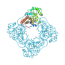
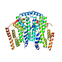 | | The channel-block Ser202Glu, Thr104Lys double mutant of Stearoyl-ACP- Desaturase from Castor bean (Ricinus communis) | | Descriptor: | ACYL-[ACYL-CARRIER-PROTEIN] DESATURASE, CHLOROPLASTIC, FE (II) ION, ... | | Authors: | Moche, M, Guy, J, Whittle, E, Lindqvist, Y, Shanklin, J. | | Deposit date: | 2014-09-17 | | Release date: | 2015-08-26 | | Last modified: | 2024-01-10 | | Method: | X-RAY DIFFRACTION (2.8 Å) | | Cite: | Half-of-the-Sites Reactivity of the Castor Delta9-18:0-Acp Desaturase.
Plant Physiol., 169, 2015
|
|
1KO5
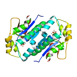 | | Crystal structure of gluconate kinase | | Descriptor: | ADENOSINE-5'-TRIPHOSPHATE, Gluconate kinase, MAGNESIUM ION | | Authors: | Kraft, L, Sprenger, G.A, Lindqvist, Y. | | Deposit date: | 2001-12-20 | | Release date: | 2002-05-29 | | Last modified: | 2024-03-13 | | Method: | X-RAY DIFFRACTION (2.28 Å) | | Cite: | Conformational changes during the catalytic cycle of gluconate kinase as revealed by X-ray crystallography.
J.Mol.Biol., 318, 2002
|
|
1KO8
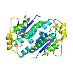 | | Crystal structure of gluconate kinase | | Descriptor: | 6-PHOSPHOGLUCONIC ACID, Gluconate kinase, MAGNESIUM ION | | Authors: | Kraft, L, Sprenger, G.A, Lindqvist, Y. | | Deposit date: | 2001-12-20 | | Release date: | 2002-05-29 | | Last modified: | 2024-03-13 | | Method: | X-RAY DIFFRACTION (2.4 Å) | | Cite: | Conformational changes during the catalytic cycle of gluconate kinase as revealed by X-ray crystallography.
J.Mol.Biol., 318, 2002
|
|
1KNQ
 | | Crystal structure of gluconate kinase | | Descriptor: | CHLORIDE ION, Gluconate kinase | | Authors: | Kraft, L, Sprenger, G.A, Lindqvist, Y. | | Deposit date: | 2001-12-19 | | Release date: | 2002-05-29 | | Last modified: | 2024-03-13 | | Method: | X-RAY DIFFRACTION (2 Å) | | Cite: | Conformational changes during the catalytic cycle of gluconate kinase as revealed by X-ray crystallography.
J.Mol.Biol., 318, 2002
|
|
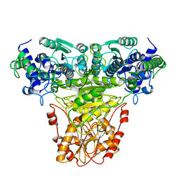
1KAS
 | | BETA-KETOACYL-ACP SYNTHASE II FROM ESCHERICHIA COLI | | Descriptor: | BETA-KETOACYL ACP SYNTHASE II | | Authors: | Huang, W, Jia, J, Edwards, P, Dehesh, K, Schneider, G, Lindqvist, Y. | | Deposit date: | 1997-12-22 | | Release date: | 1999-03-02 | | Last modified: | 2024-02-07 | | Method: | X-RAY DIFFRACTION (2.4 Å) | | Cite: | Crystal structure of beta-ketoacyl-acyl carrier protein synthase II from E.coli reveals the molecular architecture of condensing enzymes.
EMBO J., 17, 1998
|
|


