5KHA
 
 | |
5KOI
 | |
4XK1
 | |
6P81
 | |
4Y0V
 | |
5ELO
 | |
6OHZ
 | |
3EOM
 | |
6OJM
 | |
3EON
 | |
3EZ4
 | |
6OMZ
 | |
6O4N
 | |
3EZN
 | |
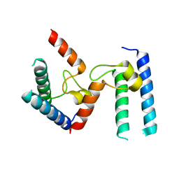
3FDZ
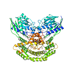
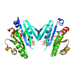
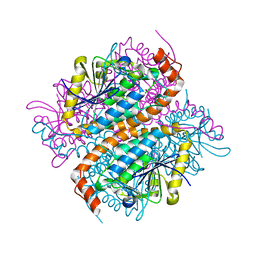
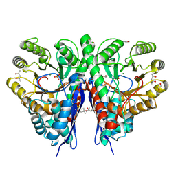
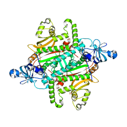
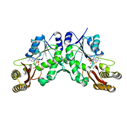
 | | Crystal structure of phosphoglyceromutase from burkholderia pseudomallei 1710b with bound 2,3-diphosphoglyceric acid and 3-phosphoglyceric acid | | Descriptor: | (2R)-2,3-diphosphoglyceric acid, 2,3-bisphosphoglycerate-dependent phosphoglycerate mutase, 3-PHOSPHOGLYCERIC ACID, ... | | Authors: | Seattle Structural Genomics Center for Infectious Disease (SSGCID) | | Deposit date: | 2008-11-26 | | Release date: | 2009-01-13 | | Last modified: | 2023-12-27 | | Method: | X-RAY DIFFRACTION (2.25 Å) | | Cite: | An ensemble of structures of Burkholderia pseudomallei 2,3-bisphosphoglycerate-dependent phosphoglycerate mutase.
Acta Crystallogr.,Sect.F, 67, 2011
|
|
4YS8
 | |
8F87
 | |
4WXT
 | |
6PQH
 | |
4XXP
 | |
6PBL
 | |
4XWI
 | |
3LUZ
 | |
5KWV
 | |
6PTG
 | |


