Loading
PDBj
メニューPDBj@FacebookPDBj@TwitterPDBj@YouTubewwPDB FoundationwwPDB
RCSB PDBPDBeBMRBAdv. SearchSearch help
Search by PDB author
4EV8
DownloadVisualize
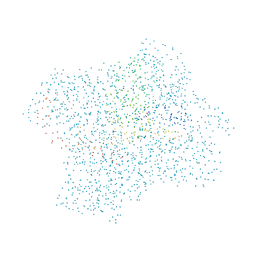
BU of 4ev8 by Molmil
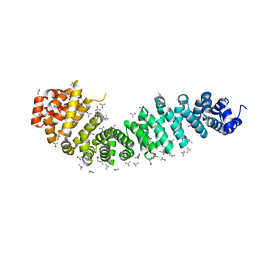
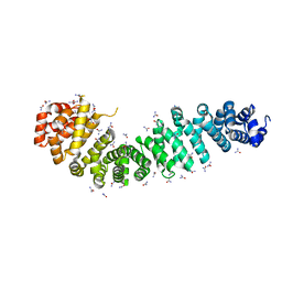
Crystal structure of mouse catenin beta-59 in 2.4M urea.
分子名称: Catenin beta-1, UREA
著者Wang, C, Zhang, G.Y.
登録日2012-04-26
公開日2013-05-01
最終更新日2023-09-13
実験手法X-RAY DIFFRACTION (1.9 Å)
主引用文献Crystal structure of mouse catenin beta-59 in 2.4M urea.
TO BE PUBLISHED
4EVA
DownloadVisualize
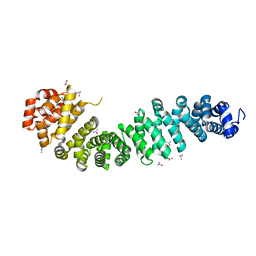
BU of 4eva by Molmil
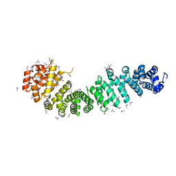
Crystal Structure of Mouse Catenin beta-59 in 5.6M urea
分子名称: Catenin beta-1, UREA
著者Wang, C, Zhang, G.Y.
登録日2012-04-26
公開日2013-05-01
最終更新日2024-02-28
実験手法X-RAY DIFFRACTION (2.003 Å)
主引用文献Crystal Structure of Mouse Catenin beta-59 in 5.6M urea
TO BE PUBLISHED
4QU1
DownloadVisualize
BU of 4qu1 by Molmil
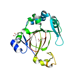
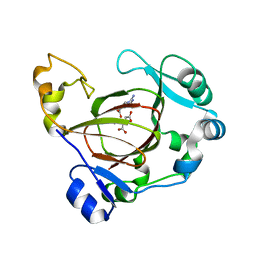
Crystal structure of human JMJD5 jmj-c domain
分子名称: 2-OXOGLUTARIC ACID, GLYCEROL, Lysine-specific demethylase 8, ...
著者Liu, H.L, Wang, C, Zhang, G.Y.
登録日2014-07-10
公開日2015-07-15
最終更新日2024-02-28
実験手法X-RAY DIFFRACTION (1.57 Å)
主引用文献The Histone Arginine Demethylase JMJD5
To be Published
4QU2
DownloadVisualize
BU of 4qu2 by Molmil
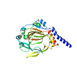
The structure of JMJD7 with alpha-KG
分子名称: 2-OXOGLUTARIC ACID, JmjC domain-containing protein 7, NICKEL (II) ION
著者Liu, H.L, Wang, C, Zhang, G.Y.
登録日2014-07-10
公開日2015-07-15
最終更新日2024-02-28
実験手法X-RAY DIFFRACTION (2.7 Å)
主引用文献The structure of JMJD7 with alpha-KG
To be Published
4EVT
DownloadVisualize
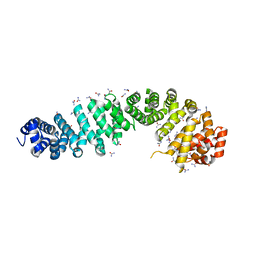
BU of 4evt by Molmil
Crystal Structure of Mouse Catenin beta-59 in 8.3M urea
分子名称: Catenin beta-1, UREA
著者Wang, C, Zhang, G.Y.
登録日2012-04-26
公開日2013-05-01
最終更新日2024-02-28
実験手法X-RAY DIFFRACTION (2.339 Å)
主引用文献Crystal Structure of Mouse Catenin beta-59 in 8.3M urea
TO BE PUBLISHED
4EV9
DownloadVisualize
BU of 4ev9 by Molmil
Crystal Structure of Mouse Catenin beta-59 in 4.0M urea
分子名称: Catenin beta-1, UREA
著者Wang, C, Zhang, G.Y.
登録日2012-04-26
公開日2013-05-01
最終更新日2024-02-28
実験手法X-RAY DIFFRACTION (2.1 Å)
主引用文献Crystal Structure of Mouse Catenin beta-59 in 4.0M urea
TO BE PUBLISHED
4EVP
DownloadVisualize
BU of 4evp by Molmil
Crystal Structure of Mouse Catenin beta-59 in 7.2M urea
分子名称: Catenin beta-1, UREA
著者Wang, C, Zhang, G.Y.
登録日2012-04-26
公開日2013-05-01
最終更新日2024-02-28
実験手法X-RAY DIFFRACTION (2.255 Å)
主引用文献Crystal Structure of Mouse Catenin beta-59 in 7.2M urea
TO BE PUBLISHED
5FBJ
DownloadVisualize
BU of 5fbj by Molmil
Complex structure of JMJD5 and substrate
分子名称: (2S)-2-amino-5-[(N-methylcarbamimidoyl)amino]pentanoic acid, 2-OXOGLUTARIC ACID, Lysine-specific demethylase 8, ...
著者Liu, H.L, Wang, Y, Wang, C, Zhang, G.Y.
登録日2015-12-14
公開日2016-12-14
最終更新日2023-09-27
実験手法X-RAY DIFFRACTION (2.42 Å)
主引用文献to be published
To Be Published
8U77
DownloadVisualize
BU of 8u77 by Molmil
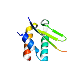
Crystal structure of Taf14 in complex with Yng1
分子名称: Protein YNG1, Transcription initiation factor TFIID subunit 14
著者Nguyen, M.C, Wei, P.C, Zhang, G.Y, Kutateladze, T.G.
登録日2023-09-14
公開日2024-08-21
実験手法X-RAY DIFFRACTION (1.93 Å)
主引用文献Molecular insight into interactions between the Taf14, Yng1 and Sas3 subunits of the NuA3 complex.
Nat Commun, 15, 2024
4QSZ
DownloadVisualize
BU of 4qsz by Molmil
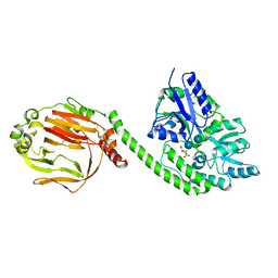
Crystal structure of mouse JMJd7 fused with maltose-binding protein
分子名称: CITRATE ANION, Maltose-binding periplasmic protein, JmjC domain-containing protein 7 chimera, ...
著者Liu, H, Wang, C, Zhang, G.Y.
登録日2014-07-06
公開日2015-07-08
最終更新日2020-07-29
実験手法X-RAY DIFFRACTION (2.86 Å)
主引用文献Crystal structure of mouse JMJd7 fused with maltose-binding protein
To be Published
1CLK
DownloadVisualize
BU of 1clk by Molmil
CRYSTAL STRUCTURE OF STREPTOMYCES DIASTATICUS NO.7 STRAIN M1033 XYLOSE ISOMERASE AT 1.9 A RESOLUTION WITH PSEUDO-I222 SPACE GROUP
分子名称: COBALT (II) ION, MAGNESIUM ION, XYLOSE ISOMERASE
著者Niu, L, Teng, M, Zhu, X, Gong, W.
登録日1999-04-29
公開日2000-05-03
最終更新日2023-08-09
実験手法X-RAY DIFFRACTION (1.9 Å)
主引用文献Structure of xylose isomerase from Streptomyces diastaticus no. 7 strain M1033 at 1.85 A resolution.
Acta Crystallogr.,Sect.D, 56, 2000

225946

件を2024-10-09に公開中

PDB statisticsPDBj update infoContact PDBjnumon