4DNR
 
 | |
7N6B
 | |
5D19
 | |
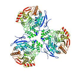
5D9R
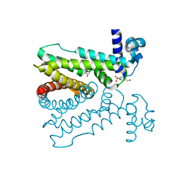
 | | Crystal structure of a conserved domain in the intermembrane space region of the plastid division protein ARC6 | | 分子名称: | Protein ACCUMULATION AND REPLICATION OF CHLOROPLASTS 6, chloroplastic | | 著者 | Radhakrishnan, A, Kumar, N, Su, C.-C, Chou, T.-H, Yu, E. | | 登録日 | 2015-08-18 | | 公開日 | 2015-11-11 | | 最終更新日 | 2024-03-06 | | 実験手法 | X-RAY DIFFRACTION (2.052 Å) | | 主引用文献 | Crystal structure of a conserved domain in the intermembrane space region of the plastid division protein ARC6.
Protein Sci., 25, 2016
|
|
5D18
 | | Crystal structure of Mycobacterium tuberculosis Rv0302, form I | | 分子名称: | 4-(2-HYDROXYETHYL)-1-PIPERAZINE ETHANESULFONIC ACID, ISOPROPYL ALCOHOL, SODIUM ION, ... | | 著者 | Chou, T.-H, Delmar, J, Su, C.-C, Yu, E. | | 登録日 | 2015-08-04 | | 公開日 | 2015-10-07 | | 最終更新日 | 2024-03-06 | | 実験手法 | X-RAY DIFFRACTION (2.04 Å) | | 主引用文献 | Crystal structure of the Mycobacterium tuberculosis transcriptional regulator Rv0302.
Protein Sci., 2015
|
|
5D1R
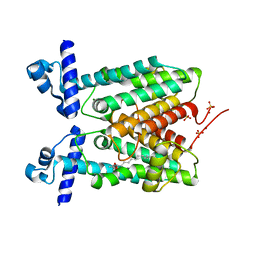 | | Crystal structure of Mycobacterium tuberculosis Rv1816 transcriptional regulator. | | 分子名称: | MAGNESIUM ION, NICKEL (II) ION, Rv1816 transcriptional regulator, ... | | 著者 | Chou, T.-H, Delmar, J, Su, C.-C, Yu, E. | | 登録日 | 2015-08-04 | | 公開日 | 2015-09-30 | | 最終更新日 | 2024-03-06 | | 実験手法 | X-RAY DIFFRACTION (2 Å) | | 主引用文献 | Structural Basis for the Regulation of the MmpL Transporters of Mycobacterium tuberculosis.
J.Biol.Chem., 290, 2015
|
|
5D1W
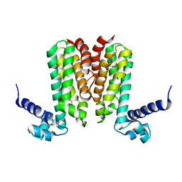 | | Crystal structure of Mycobacterium tuberculosis Rv3249c transcriptional regulator. | | 分子名称: | PALMITIC ACID, Rv3249c transcriptional regulator | | 著者 | Chou, T.-H, Delmar, J, Su, C.-C, Yu, E. | | 登録日 | 2015-08-04 | | 公開日 | 2015-09-30 | | 最終更新日 | 2024-03-06 | | 実験手法 | X-RAY DIFFRACTION (3.59 Å) | | 主引用文献 | Structural Basis for the Regulation of the MmpL Transporters of Mycobacterium tuberculosis.
J.Biol.Chem., 290, 2015
|
|
8EL9
 | | Cryo-EM structure of human catalase | | 分子名称: | Catalase, NADPH DIHYDRO-NICOTINAMIDE-ADENINE-DINUCLEOTIDE PHOSPHATE, PROTOPORPHYRIN IX CONTAINING FE | | 著者 | Su, C.C. | | 登録日 | 2022-09-23 | | 公開日 | 2023-10-04 | | 実験手法 | ELECTRON MICROSCOPY (2.27 Å) | | 主引用文献 | Cryo-EM structure of human catalase
To Be Published
|
|
8EOR
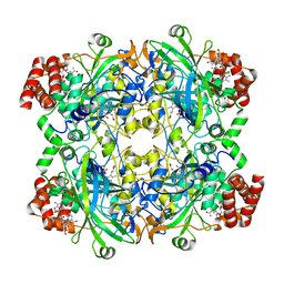
 | | Liver carboxylesterase 1 | | 分子名称: | ETHYL ACETATE, Liver carboxylesterase 1, beta-D-mannopyranose-(1-4)-2-acetamido-2-deoxy-beta-D-glucopyranose-(1-4)-2-acetamido-2-deoxy-beta-D-glucopyranose | | 著者 | Zhang, Z, Yu, E. | | 登録日 | 2022-10-04 | | 公開日 | 2023-05-03 | | 最終更新日 | 2023-11-15 | | 実験手法 | ELECTRON MICROSCOPY (2.67 Å) | | 主引用文献 | High-resolution structural-omics of human liver enzymes.
Cell Rep, 42, 2023
|
|
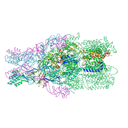
4DNT
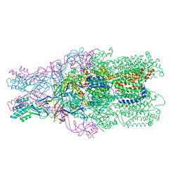 | | Crystal structure of the CusBA heavy-metal efflux complex from Escherichia coli, mutant | | 分子名称: | Cation efflux system protein CusA, Cation efflux system protein CusB | | 著者 | Su, C.-C, Long, F, Yu, E. | | 登録日 | 2012-02-09 | | 公開日 | 2012-06-20 | | 最終更新日 | 2024-02-28 | | 実験手法 | X-RAY DIFFRACTION (3.1 Å) | | 主引用文献 | Charged Amino Acids (R83, E567, D617, E625, R669, and K678) of CusA Are Required for Metal Ion Transport in the Cus Efflux System.
J.Mol.Biol., 422, 2012
|
|
4NN1
 | |
4DOP
 | | Crystal structure of the CusBA heavy-metal efflux complex from Escherichia coli, R mutant | | 分子名称: | Cation efflux system protein CusA, Cation efflux system protein CusB | | 著者 | Su, C.-C, Long, F, Yu, E. | | 登録日 | 2012-02-10 | | 公開日 | 2012-06-20 | | 最終更新日 | 2024-02-28 | | 実験手法 | X-RAY DIFFRACTION (4.2 Å) | | 主引用文献 | Charged Amino Acids (R83, E567, D617, E625, R669, and K678) of CusA Are Required for Metal Ion Transport in the Cus Efflux System.
J.Mol.Biol., 422, 2012
|
|


