2OSG
 
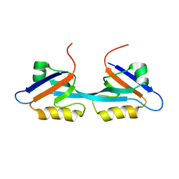 | | Solution Structure and Binding Property of the Domain-swapped Dimer of ZO2PDZ2 | | 分子名称: | Tight junction protein ZO-2 | | 著者 | Wu, J.W, Yang, Y.S, Zhang, J.H, Ji, P, Wu, J.H, Shi, Y.Y. | | 登録日 | 2007-02-05 | | 公開日 | 2007-09-25 | | 最終更新日 | 2024-05-29 | | 実験手法 | SOLUTION NMR | | 主引用文献 | Domain-swapped Dimerization of the Second PDZ Domain of ZO2 May Provide a Structural Basis for the Polymerization of Claudins
J.Biol.Chem., 282, 2007
|
|
1JD6
 
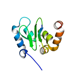 | | Crystal Structure of DIAP1-BIR2/Hid Complex | | 分子名称: | APOPTOSIS 1 INHIBITOR, ZINC ION, head involution defective protein | | 著者 | Wu, J.W, Cocina, A.E, Chai, J, Hay, B.A, Shi, Y. | | 登録日 | 2001-06-12 | | 公開日 | 2001-12-05 | | 最終更新日 | 2024-02-07 | | 実験手法 | X-RAY DIFFRACTION (2.7 Å) | | 主引用文献 | Structural analysis of a functional DIAP1 fragment bound to grim and hid peptides.
Mol.Cell, 8, 2001
|
|
1JD4
 
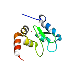 | | Crystal Structure of DIAP1-BIR2 | | 分子名称: | APOPTOSIS 1 INHIBITOR, ZINC ION | | 著者 | Wu, J.W, Cocina, A.E, Chai, J, Hay, B.A, Shi, Y. | | 登録日 | 2001-06-12 | | 公開日 | 2001-12-05 | | 最終更新日 | 2023-08-16 | | 実験手法 | X-RAY DIFFRACTION (2.7 Å) | | 主引用文献 | Structural analysis of a functional DIAP1 fragment bound to grim and hid peptides.
Mol.Cell, 8, 2001
|
|
1JD5
 
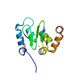 | | Crystal Structure of DIAP1-BIR2/GRIM | | 分子名称: | APOPTOSIS 1 INHIBITOR, ZINC ION, cell death protein GRIM | | 著者 | Wu, J.W, Cocina, A.E, Chai, J, Hay, B.A, Shi, Y. | | 登録日 | 2001-06-12 | | 公開日 | 2001-12-05 | | 最終更新日 | 2024-02-07 | | 実験手法 | X-RAY DIFFRACTION (1.9 Å) | | 主引用文献 | Structural analysis of a functional DIAP1 fragment bound to grim and hid peptides.
Mol.Cell, 8, 2001
|
|
3GMJ
 
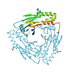 | | Crystal structure of MAD MH2 domain | | 分子名称: | Protein mothers against dpp | | 著者 | Wu, J.W, Wang, C. | | 登録日 | 2009-03-14 | | 公開日 | 2009-12-15 | | 最終更新日 | 2024-03-20 | | 実験手法 | X-RAY DIFFRACTION (2.8 Å) | | 主引用文献 | Crystal structure of the MH2 domain of Drosophila Mad
SCI.CHINA, SER.C: LIFE SCI., 52, 2009
|
|
4MJS
 
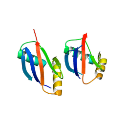 | | crystal structure of a PB1 complex | | 分子名称: | 1,2-ETHANEDIOL, Protein kinase C zeta type, Sequestosome-1 | | 著者 | Ren, J, Wang, Z.X, Wu, J.W. | | 登録日 | 2013-09-04 | | 公開日 | 2014-08-27 | | 最終更新日 | 2023-11-08 | | 実験手法 | X-RAY DIFFRACTION (2.5 Å) | | 主引用文献 | Structural and biochemical insights into the homotypic PB1-PB1 complex between PKC zeta and p62
Sci China Life Sci, 57, 2014
|
|
7XBR
 
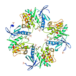 | |
4YNZ
 
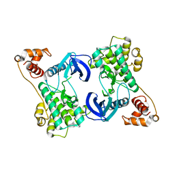 | | Structure of the N-terminal domain of SAD | | 分子名称: | Serine/threonine-protein kinase BRSK2 | | 著者 | Wu, J.X, Wang, J, Chen, L, Wang, Z.X, Wu, J.W. | | 登録日 | 2015-03-11 | | 公開日 | 2015-12-16 | | 最終更新日 | 2023-11-08 | | 実験手法 | X-RAY DIFFRACTION (2 Å) | | 主引用文献 | Structural insight into the mechanism of synergistic autoinhibition of SAD kinases
Nat Commun, 6, 2015
|
|
4YOM
 
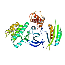 | | Structure of SAD kinase | | 分子名称: | 1,2-ETHANEDIOL, Serine/threonine-protein kinase BRSK2 | | 著者 | Wu, J.X, Wang, J, Chen, L, Wang, Z.X, Wu, J.W. | | 登録日 | 2015-03-12 | | 公開日 | 2015-12-16 | | 最終更新日 | 2023-11-08 | | 実験手法 | X-RAY DIFFRACTION (2.49 Å) | | 主引用文献 | Structural insight into the mechanism of synergistic autoinhibition of SAD kinases
Nat Commun, 6, 2015
|
|
7Y11
 
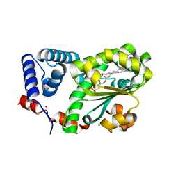 | | Crystal structure of AtSFH5-Sec14 in complex with egg PA | | 分子名称: | (2R)-1-(hexadecanoyloxy)-3-(phosphonooxy)propan-2-yl (9Z)-octadec-9-enoate, CHLORIDE ION, NICKEL (II) ION, ... | | 著者 | Lu, Y.Q, Wang, X.Q, Luo, Z.P, Wu, J.W. | | 登録日 | 2022-06-06 | | 公開日 | 2023-04-19 | | 最終更新日 | 2023-11-29 | | 実験手法 | X-RAY DIFFRACTION (1.95 Å) | | 主引用文献 | Arabidopsis Sec14 proteins (SFH5 and SFH7) mediate interorganelle transport of phosphatidic acid and regulate chloroplast development.
Proc.Natl.Acad.Sci.USA, 120, 2023
|
|
7Y10
 
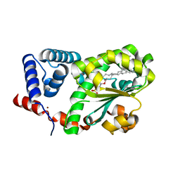 | | Crystal structure of AtSFH5-Sec14 in complex with DPPA | | 分子名称: | 1,2-DIPALMITOYL-SN-GLYCERO-3-PHOSPHATE, NICKEL (II) ION, Phosphatidylinositol/phosphatidylcholine transfer protein SFH5 | | 著者 | Lu, Y.Q, Wang, X.Q, Luo, Z.P, Wu, J.W. | | 登録日 | 2022-06-06 | | 公開日 | 2023-05-31 | | 最終更新日 | 2023-11-29 | | 実験手法 | X-RAY DIFFRACTION (2.1 Å) | | 主引用文献 | Arabidopsis Sec14 proteins (SFH5 and SFH7) mediate interorganelle transport of phosphatidic acid and regulate chloroplast development.
Proc.Natl.Acad.Sci.USA, 120, 2023
|
|
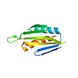
5IRI
 
 | |
4YR8
 
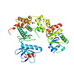 | | Crystal structure of JNK in complex with a regulator protein | | 分子名称: | CHLORIDE ION, Dual specificity protein phosphatase 16, Mitogen-activated protein kinase 8 | | 著者 | Liu, X, Wang, J, Wu, J.W, Wang, Z.X. | | 登録日 | 2015-03-14 | | 公開日 | 2016-03-16 | | 最終更新日 | 2023-11-08 | | 実験手法 | X-RAY DIFFRACTION (2.4 Å) | | 主引用文献 | A conserved motif in JNK/p38-specific MAPK phosphatases as a determinant for JNK1 recognition and inactivation.
Nat Commun, 7, 2016
|
|
4IXP
 
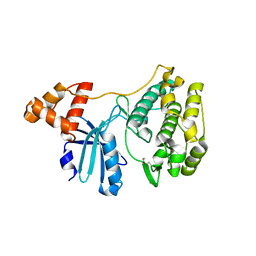 | |
4KA3
 
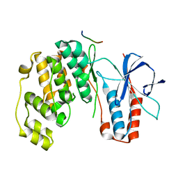 | | Structure of MAP kinase in complex with a docking peptide | | 分子名称: | Mitogen-activated protein kinase 14, TGF-beta-activated kinase 1 and MAP3K7-binding protein 1 | | 著者 | Xin, F.J, Wu, J.W. | | 登録日 | 2013-04-22 | | 公開日 | 2013-06-26 | | 最終更新日 | 2023-11-08 | | 実験手法 | X-RAY DIFFRACTION (2.707 Å) | | 主引用文献 | Crystal structure of the p38 alpha MAP kinase in complex with a docking peptide from TAB1
Sci China Life Sci, 56, 2013
|
|
6KRX
 
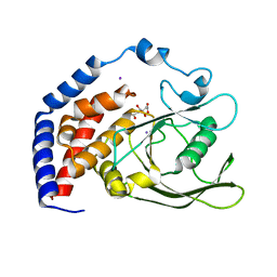 | |
6KRW
 
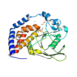 | | Crystal Structure of AtPTP1 at 1.4 angstrom | | 分子名称: | CITRATE ANION, DI(HYDROXYETHYL)ETHER, IODIDE ION, ... | | 著者 | Zhao, Y.Y, Luo, Z.P, Wang, J, Wu, J.W. | | 登録日 | 2019-08-22 | | 公開日 | 2020-08-26 | | 最終更新日 | 2023-11-22 | | 実験手法 | X-RAY DIFFRACTION (1.4 Å) | | 主引用文献 | Crystal structure of AtPTP1 at 1.4 Angstroms
To Be Published
|
|
3PZ7
 
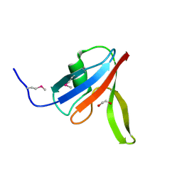 | |
3PZ8
 
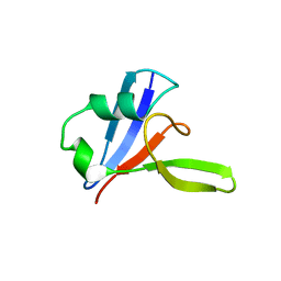 | |
2JWE
 
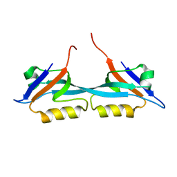 | | Solution structure of the second PDZ domain from human zonula occludens-1: A dimeric form with 3D domain swapping | | 分子名称: | Tight junction protein ZO-1 | | 著者 | Ji, P, Wu, J.W, Zhang, J.H, Yang, Y.S, Wu, J.H, Shi, Y.Y. | | 登録日 | 2007-10-10 | | 公開日 | 2007-10-30 | | 最終更新日 | 2024-05-01 | | 実験手法 | SOLUTION NMR | | 主引用文献 | Solution structure of the second PDZ domain of Zonula Occludens 1
Proteins, 79, 2011
|
|
1SE0
 
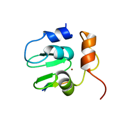 | | Crystal structure of DIAP1 BIR1 bound to a Grim peptide | | 分子名称: | Apoptosis 1 inhibitor, Cell death protein Grim, ZINC ION | | 著者 | Yan, N, Wu, J.W, Shi, Y. | | 登録日 | 2004-02-15 | | 公開日 | 2004-04-27 | | 最終更新日 | 2024-02-14 | | 実験手法 | X-RAY DIFFRACTION (1.75 Å) | | 主引用文献 | Molecular mechanisms of DrICE inhibition by DIAP1 and removal of inhibition by Reaper, Hid and Grim.
Nat.Struct.Mol.Biol., 11, 2004
|
|
1LCY
 
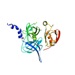 | | Crystal Structure of the Mitochondrial Serine Protease HtrA2 | | 分子名称: | HtrA2 serine protease | | 著者 | Li, W, Srinivasula, S.M, Chai, J, Li, P, Wu, J.W, Zhang, Z, Alnemri, E.S, Shi, Y. | | 登録日 | 2002-04-07 | | 公開日 | 2002-05-22 | | 最終更新日 | 2024-02-14 | | 実験手法 | X-RAY DIFFRACTION (2 Å) | | 主引用文献 | Structural insights into the pro-apoptotic function of mitochondrial serine protease HtrA2/Omi.
Nat.Struct.Biol., 9, 2002
|
|
1SDZ
 
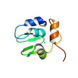 | | Crystal structure of DIAP1 BIR1 bound to a Reaper peptide | | 分子名称: | Apoptosis 1 inhibitor, Reaper, ZINC ION | | 著者 | Yan, N, Wu, J.W, Shi, Y. | | 登録日 | 2004-02-15 | | 公開日 | 2004-04-27 | | 最終更新日 | 2024-02-14 | | 実験手法 | X-RAY DIFFRACTION (1.78 Å) | | 主引用文献 | Molecular mechanisms of DrICE inhibition by DIAP1 and removal of inhibition by Reaper, Hid and Grim.
Nat.Struct.Mol.Biol., 11, 2004
|
|
3DIT
 
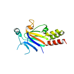 | |
5YYS
 
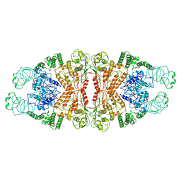 | | Cryo-EM structure of L-fucokinase, GDP-fucose pyrophosphorylase (FKP)in Bacteroides fragilis | | 分子名称: | L-fucokinase, L-fucose-1-P guanylyltransferase | | 著者 | Wang, J, Hu, H, Liu, Y, Zhou, Q, Wu, P, Yan, N, Wang, H.W, Wu, J.W, Sun, L. | | 登録日 | 2017-12-11 | | 公開日 | 2018-12-12 | | 最終更新日 | 2024-03-27 | | 実験手法 | ELECTRON MICROSCOPY (4.2 Å) | | 主引用文献 | Cryo-EM structure of L-fucokinase/GDP-fucose pyrophosphorylase (FKP) in Bacteroides fragilis.
Protein Cell, 10, 2019
|
|
