Loading
PDBj
メニューPDBj@FacebookPDBj@TwitterPDBj@YouTubewwPDB FoundationwwPDB
RCSB PDBPDBeBMRBAdv. SearchSearch help
Search by PDB author
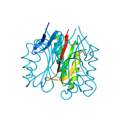
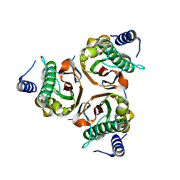
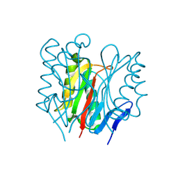
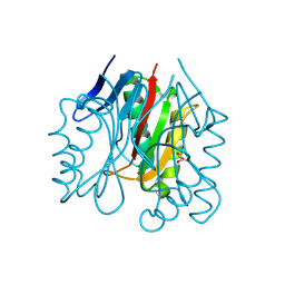
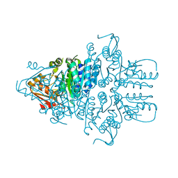
4WBX
DownloadVisualize
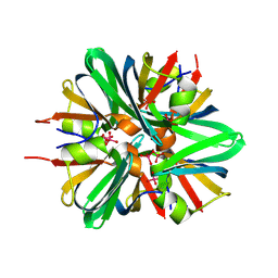
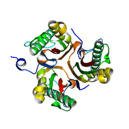
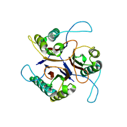
BU of 4wbx by Molmil
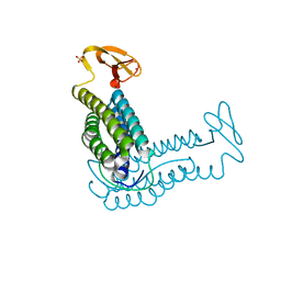
Conserved hypothetical protein PF1771 from Pyrococcus furiosus solved by sulfur SAD using Swiss Light Source data
分子名称: 2-keto acid:ferredoxin oxidoreductase subunit alpha
著者Weinert, T, Waltersperger, S, Olieric, V, Panepucci, E, Chen, L, Rose, J.P, Wang, M, Wang, B.C, Southeast Collaboratory for Structural Genomics (SECSG)
登録日2014-09-04
公開日2014-12-10
最終更新日2023-12-27
実験手法X-RAY DIFFRACTION (2.301 Å)
主引用文献Fast native-SAD phasing for routine macromolecular structure determination.
Nat.Methods, 12, 2015
1QV0
DownloadVisualize
BU of 1qv0 by Molmil
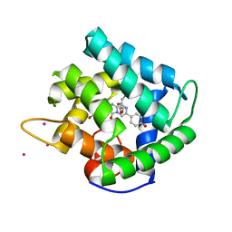
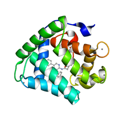
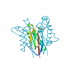
Atomic resolution structure of obelin from Obelia longissima
分子名称: C2-HYDROPEROXY-COELENTERAZINE, COBALT (II) ION, GLYCEROL, ...
著者Liu, Z.J, Vysotski, E.S, Deng, L, Lee, J, Rose, J, Wang, B.C.
登録日2003-08-26
公開日2003-11-11
最終更新日2023-08-16
実験手法X-RAY DIFFRACTION (1.1 Å)
主引用文献Atomic resolution structure of obelin: soaking with calcium enhances electron density of the second oxygen atom substituted at the C2-position of coelenterazine.
Biochem.Biophys.Res.Commun., 311, 2003
6EB0
DownloadVisualize
BU of 6eb0 by Molmil
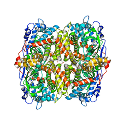
STRUCTURE OF 4-HYDROXYPHENYLACETATE 3-MONOOXYGENASE (HPAB), OXYGENASE COMPONENT FROM ESCHERICHIA COLI
分子名称: 4-hydroxyphenylacetate 3-monooxygenase, oxygenase subunit, ACETATE ION
著者Zhou, D, Kandavelu, P, Zhang, H, Wang, B.C, Yan, Y, Rose, J.
登録日2018-08-03
公開日2019-05-22
最終更新日2023-10-11
実験手法X-RAY DIFFRACTION (2.37 Å)
主引用文献Structural Insights into Catalytic Versatility of the Flavin-dependent Hydroxylase (HpaB) from Escherichia coli.
Sci Rep, 9, 2019
1QYB
DownloadVisualize
BU of 1qyb by Molmil
X-ray crystal structure of Desulfovibrio vulgaris rubrerythrin with zinc substituted into the [Fe(SCys)4] site and alternative diiron site structures
分子名称: FE (III) ION, Rubrerythrin, SULFATE ION, ...
著者Jin, S, Kurtz, D.M, Liu, Z.J, Rose, J, Wang, B.C.
登録日2003-09-10
公開日2004-03-30
最終更新日2023-08-23
実験手法X-RAY DIFFRACTION (1.75 Å)
主引用文献X-ray Crystal Structure of Desulfovibrio vulgaris Rubrerythrin with Zinc Substituted into the [Fe(SCys)(4)] Site and Alternative Diiron Site Structures.
Biochemistry, 43, 2004
2QUF
DownloadVisualize
BU of 2quf by Molmil
Crystal Structure of Transcription Factor AXXA-PF0095 from Pyrococcus furiosus
分子名称: GLYCEROL, Transcription Factor PF0095
著者Yang, H, Lipscomb, G.L, Scott, R.A, Rose, J.P, Wang, B.C.
登録日2007-08-05
公開日2008-08-05
最終更新日2023-08-30
実験手法X-RAY DIFFRACTION (2.8 Å)
主引用文献Crystal Structure of Transcription Factor AXXA-PF0095 from Pyrococcus furiosus
To be Published
2F8P
DownloadVisualize
BU of 2f8p by Molmil
Crystal structure of obelin following Ca2+ triggered bioluminescence suggests neutral coelenteramide as the primary excited state
分子名称: CALCIUM ION, N-[3-BENZYL-5-(4-HYDROXYPHENYL)PYRAZIN-2-YL]-2-(4-HYDROXYPHENYL)ACETAMIDE, Obelin
著者Liu, Z.J, Stepanyuk, G.A, Vysotski, E.S, Lee, J, Wang, B.C, Southeast Collaboratory for Structural Genomics (SECSG)
登録日2005-12-03
公開日2006-02-14
最終更新日2023-08-30
実験手法X-RAY DIFFRACTION (1.93 Å)
主引用文献Crystal structure of obelin after Ca2+-triggered bioluminescence suggests neutral coelenteramide as the primary excited state.
Proc.Natl.Acad.Sci.Usa, 103, 2006
2BN2
DownloadVisualize
BU of 2bn2 by Molmil
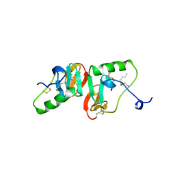
CRYSTAL STRUCTURE OF BOVINE NEUROPHYSIN II COMPLEXED WITH THE VASOPRESSIN ANALOGUE PHE-TYR AMIDE
分子名称: NEUROPHYSIN II, PHENYLALANINE, TYROSINE
著者Rose, J.P, Wang, B.C.
登録日1998-12-18
公開日1999-02-16
最終更新日2024-10-30
実験手法X-RAY DIFFRACTION (2.8 Å)
主引用文献Crystal structure of a bovine neurophysin II dipeptide complex at 2.8 A determined from the single-wavelength anomalous scattering signal of an incorporated iodine atom.
Proc.Natl.Acad.Sci.USA, 88, 1991
4RNP
DownloadVisualize
BU of 4rnp by Molmil
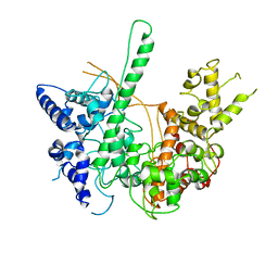
BACTERIOPHAGE T7 RNA POLYMERASE, HIGH SALT CRYSTAL FORM, LOW TEMPERATURE DATA, ALPHA-CARBONS ONLY
分子名称: RNA POLYMERASE
著者Liu, Z.J, Sousa, R, Rose, J.P, Wang, B.C.
登録日1997-09-11
公開日1997-12-03
最終更新日2024-02-28
実験手法X-RAY DIFFRACTION (3 Å)
主引用文献Crystal structure of bacteriophage T7 RNA polymerase at 3.3 A resolution.
Nature, 364, 1993
9BED
DownloadVisualize
BU of 9bed by Molmil
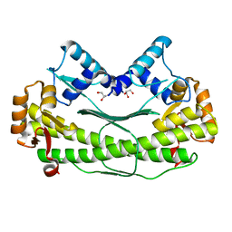
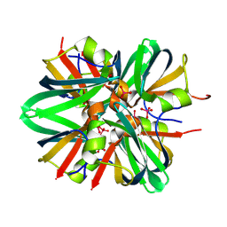
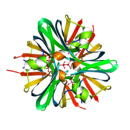
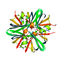
Tungstate binding protein (Tungbindin) from Eubacterium limosum with eight molybdates bound
分子名称: MOLYBDATE ION, Molybdenum-pterin-binding protein
著者Zhou, D, Rose, J.P, Chen, L, Wang, B.C.
登録日2024-04-15
公開日2024-09-04
実験手法X-RAY DIFFRACTION (2.02 Å)
主引用文献Tungstate binding protein (Tungbindin) from Eubacterium limosum with eight molybdates bound
To Be Published
9BOM
DownloadVisualize
BU of 9bom by Molmil
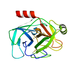
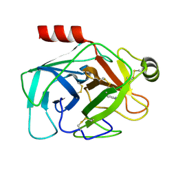
Crystal structure of reduced bovine trypsin, 25mM DTT-treated
分子名称: BENZAMIDINE, Cationic trypsin
著者Zhou, D, Chen, L, Rose, J.P, Wang, B.C.
登録日2024-05-03
公開日2024-08-07
最終更新日2024-10-23
実験手法X-RAY DIFFRACTION (1.35 Å)
主引用文献Crystal structure of reduced bovine trypsin, 25mM DTT-treated
To Be Published
9BEO
DownloadVisualize
BU of 9beo by Molmil
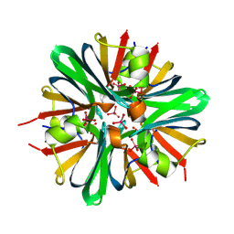
Tungstate binding protein (Tungbindin) from Eubacterium limosum with 7.5 tungstates bound
分子名称: Molybdenum-pterin binding domain-containing protein, TUNGSTATE(VI)ION
著者Zhou, D, Rose, J.P, Chen, L, Wang, B.C.
登録日2024-04-16
公開日2024-09-04
実験手法X-RAY DIFFRACTION (1.78 Å)
主引用文献Tungstate binding protein (Tungbindin) from Eubacterium limosum with 7.5 tungstates bound
To Be Published
9BEB
DownloadVisualize
BU of 9beb by Molmil
Tungstate binding protein (Tungbindin) from Eubacterium limosum with eight Tungstates bound
分子名称: Molybdenum-pterin binding domain-containing protein, TUNGSTATE(VI)ION
著者Zhou, D, Rose, J.P, Chen, L, Wang, B.C.
登録日2024-04-15
公開日2024-09-04
実験手法X-RAY DIFFRACTION (1.85 Å)
主引用文献Tungstate binding protein (Tungbindin) from Eubacterium limosum with eight Tungstates bound
To be published
9BJF
DownloadVisualize
BU of 9bjf by Molmil
Tungstate binding protein (Tungbindin) from Eubacterium limosum in apo form
分子名称: Molybdenum-pterin binding domain-containing protein, SULFATE ION
著者Zhou, D, Rose, J.P, Chen, L, Wang, B.C.
登録日2024-04-25
公開日2024-09-04
実験手法X-RAY DIFFRACTION (2.89 Å)
主引用文献Tungstate binding protein (Tungbindin) from Eubacterium limosum in apo form
To Be Published
9BOK
DownloadVisualize
BU of 9bok by Molmil
Crystal structure of reduced bovine trypsin, 50mM DTT-treated
分子名称: BENZAMIDINE, Cationic trypsin
著者Zhou, D, Chen, L, Rose, J.P, Wang, B.C.
登録日2024-05-03
公開日2024-08-07
最終更新日2024-10-16
実験手法X-RAY DIFFRACTION (1.35 Å)
主引用文献Crystal structure of reduced bovine trypsin, 50mM DTT-treated
To Be Published
9BEM
DownloadVisualize
BU of 9bem by Molmil
Tungstate binding protein (Tungbindin) from Eubacterium limosum with seven Tungstates bound
分子名称: Molybdenum-pterin-binding protein, SULFATE ION, TUNGSTATE(VI)ION
著者Zhou, D, Rose, J.P, Chen, L, Wang, B.C.
登録日2024-04-15
公開日2024-09-04
実験手法X-RAY DIFFRACTION (2.26 Å)
主引用文献Tungstate binding protein (Tungbindin) from Eubacterium limosum with seven Tungstates bound
To Be Published
9BKA
DownloadVisualize
BU of 9bka by Molmil
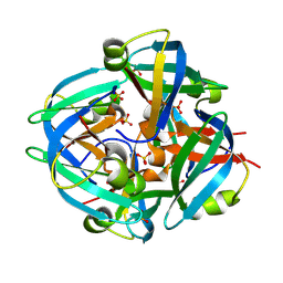
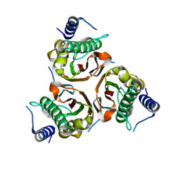
Crystal structure of Rid family protein PSPTO3006
分子名称: Rid family protein PSPTO3006
著者Zhou, D, Chen, L, Rose, J.P, Wang, B.C.
登録日2024-04-26
公開日2024-09-04
実験手法X-RAY DIFFRACTION (1.86 Å)
主引用文献Crystal structure of Rid family protein PSPTO3006
To Be Published
9BKB
DownloadVisualize
BU of 9bkb by Molmil
Crystal structure of Rid family protein ACIAD3089 from Acinetobacter baylyi
分子名称: Rid family protein ACIAD3089
著者Zhou, D, Chen, L, Rose, J.P, Wang, B.C.
登録日2024-04-26
公開日2024-09-04
実験手法X-RAY DIFFRACTION (2.1 Å)
主引用文献Crystal structure of Rid family protein ACIAD3089 from Acinetobacter baylyi
To Be Published
9BKH
DownloadVisualize
BU of 9bkh by Molmil
Crystal structure of RidA family protein PA5083 from Pseudomonas aeruginosa with Acetaldehyde bound
分子名称: ACETYL GROUP, RidA family protein PA5083
著者Zhou, D, Chen, L, Rose, J.P, Wang, B.C.
登録日2024-04-28
公開日2024-09-04
実験手法X-RAY DIFFRACTION (1.67 Å)
主引用文献Crystal structure of RidA family protein PA5083 from Pseudomonas aeruginosa with Acetaldehyde bound
To Be Published
9BKI
DownloadVisualize
BU of 9bki by Molmil
Crystal structure of Rid family protein ACIAD3089 from Acinetobacter baylyi in C2 space group
分子名称: Rid family protein ACIAD3089
著者Zhou, D, Chen, L, Rose, J.P, Wang, B.C.
登録日2024-04-28
公開日2024-09-04
最終更新日2024-10-09
実験手法X-RAY DIFFRACTION (2.38 Å)
主引用文献Crystal structure of Rid family protein ACIAD3089 from Acinetobacter baylyi in C2 space group
To Be Published
9BJV
DownloadVisualize
BU of 9bjv by Molmil
Crystal structure of hypothetical protein PA5083 from Pseudomonas aeruginosa Sulfur SAD phased
分子名称: RidA family protein PA5083
著者Zhou, D, Chen, L, Rose, J.P, Wang, B.C.
登録日2024-04-25
公開日2024-09-04
実験手法X-RAY DIFFRACTION (1.8 Å)
主引用文献Structure of the Rid protein PA5083 from Pseudomonas aeruginosa (Apo form) solved by Sulfur SAD
To Be Published
9BK4
DownloadVisualize
BU of 9bk4 by Molmil
Crystal structure of RidA family protein PA5083 from Pseudomonas aeruginosa with pyruvic acid bound
分子名称: PYRUVIC ACID, RidA family protein PA5083, SULFATE ION
著者Zhou, D, Chen, L, Rose, J.P, Wang, B.C.
登録日2024-04-26
公開日2024-09-04
実験手法X-RAY DIFFRACTION (2.08 Å)
主引用文献Crystal structure of RidA family protein PA5083 from Pseudomonas aeruginosa with pyruvic acid bound
To Be Published
9D2C
DownloadVisualize
BU of 9d2c by Molmil
Crystal Structure of Tungbindin Treated with Proteinase K
分子名称: MOLYBDATE ION, Molybdenum-pterin binding domain-containing protein
著者Zhou, D, Chen, L, Rose, J.P, Wang, B.C.
登録日2024-08-08
公開日2024-09-04
実験手法X-RAY DIFFRACTION (2.38 Å)
主引用文献Crystal structure of Tungbindin treated with proteinase K
To Be Published
9BK9
DownloadVisualize
BU of 9bk9 by Molmil
Crystal structure of Rid family protein PA0814 from Pseudomonas aeruginosa
分子名称: Rid family protein PA0814
著者Zhou, D, Chen, L, Rose, J.P, Wang, B.C.
登録日2024-04-26
公開日2024-09-04
実験手法X-RAY DIFFRACTION (1.78 Å)
主引用文献Crystal structure of Rid family protein PA0814 from Pseudomonas aeruginosa
To Be Published
9BKC
DownloadVisualize
BU of 9bkc by Molmil
Crystal structure of Rid family protein PFL1385 from Pseudomonas fluorescens
分子名称: Rid family protein PFL1385
著者Zhou, D, Chen, L, Rose, J.P, Wang, B.C.
登録日2024-04-27
公開日2024-09-04
実験手法X-RAY DIFFRACTION (2.49 Å)
主引用文献Crystal structure of Rid family protein PFL1385 from Pseudomonas fluorescens
To Be Published
9BKT
DownloadVisualize
BU of 9bkt by Molmil
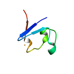
Crystal structure of rubredoxin from Pyrococcus furiosus reconstituted with FeSO4 solved by Fe/S-SAD
分子名称: FE (II) ION, Rubredoxin
著者Zhou, D, Chen, L, Rose, J.P, Wang, B.C.
登録日2024-04-29
公開日2024-06-05
実験手法X-RAY DIFFRACTION (1.8 Å)
主引用文献Crystal structure of rubredoxin from Pyrococcus furiosus reconstituted with FeSO4 solved by Fe/S-SAD
To Be Published

226707

件を2024-10-30に公開中

PDB statisticsPDBj update infoContact PDBjnumon