1NLA
 
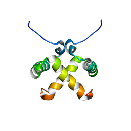 | | Solution Structure of Switch Arc, a Mutant with 3(10) Helices Replacing a Wild-Type Beta-Ribbon | | 分子名称: | Transcriptional repressor arc | | 著者 | Cordes, M.H, Walsh, N.P, McKnight, C.J, Sauer, R.T. | | 登録日 | 2003-01-06 | | 公開日 | 2003-03-18 | | 最終更新日 | 2024-05-22 | | 実験手法 | SOLUTION NMR | | 主引用文献 | Solution structure of Switch Arc, a mutant with 3(10) helices replacing a wild-type beta-ribbon
J.Mol.Biol., 326, 2003
|
|
1QTG
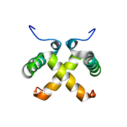 | | AVERAGED NMR MODEL OF SWITCH ARC, A DOUBLE MUTANT OF ARC REPRESSOR | | 分子名称: | Transcriptional repressor arc | | 著者 | Cordes, M.H.J, Walsh, N.P, McKnight, C.J, Sauer, R.T. | | 登録日 | 1999-06-27 | | 公開日 | 1999-07-12 | | 最終更新日 | 2024-05-22 | | 実験手法 | SOLUTION NMR | | 主引用文献 | Evolution of a protein fold in vitro.
Science, 284, 1999
|
|
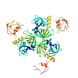
1SOT
 | | Crystal Structure of the DegS stress sensor | | 分子名称: | Protease degS | | 著者 | Wilken, C, Kitzing, K, Kurzbauer, R, Ehrmann, M, Clausen, T. | | 登録日 | 2004-03-15 | | 公開日 | 2004-06-08 | | 最終更新日 | 2023-11-15 | | 実験手法 | X-RAY DIFFRACTION (2.3 Å) | | 主引用文献 | Crystal structure of the DegS stress sensor: How a PDZ domain recognizes misfolded protein and activates a protease
Cell(Cambridge,Mass.), 117, 2004
|
|
1SOZ
 | | Crystal Structure of DegS protease in complex with an activating peptide | | 分子名称: | Protease degS, activating peptide | | 著者 | Wilken, C, Kitzing, K, Kurzbauer, R, Ehrmann, M, Clausen, T. | | 登録日 | 2004-03-16 | | 公開日 | 2004-06-08 | | 最終更新日 | 2024-04-03 | | 実験手法 | X-RAY DIFFRACTION (2.4 Å) | | 主引用文献 | Crystal structure of the DegS stress sensor: How a PDZ domain recognizes misfolded protein and activates a protease
Cell(Cambridge,Mass.), 117, 2004
|
|
1VCW
 | | Crystal structure of DegS after backsoaking the activating peptide | | 分子名称: | Protease degS | | 著者 | Wilken, C, Kitzing, K, Kurzbauer, R, Ehrmann, M, Clausen, T. | | 登録日 | 2004-03-16 | | 公開日 | 2004-06-08 | | 最終更新日 | 2023-12-27 | | 実験手法 | X-RAY DIFFRACTION (3.05 Å) | | 主引用文献 | Crystal structure of the DegS stress sensor: How a PDZ domain recognizes misfolded protein and activates a protease.
Cell(Cambridge,Mass.), 117, 2004
|
|


