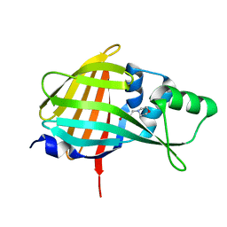
7EA4
 
 | |
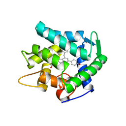
7EBY
 | |
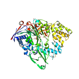
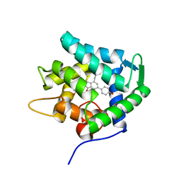
7FHT
 | | Crystal structure of DYRK1A in complex with RD0448 | | 分子名称: | (5~{Z})-5-[(3-ethynyl-4-methoxy-phenyl)methylidene]-2-sulfanylidene-1,3-thiazolidin-4-one, Dual specificity tyrosine-phosphorylation-regulated kinase 1A | | 著者 | Kikuchi, M, Sumida, Y, Hosoya, T, Kii, I, Umehara, T. | | 登録日 | 2021-07-30 | | 公開日 | 2022-03-23 | | 最終更新日 | 2023-11-29 | | 実験手法 | X-RAY DIFFRACTION (2.68 Å) | | 主引用文献 | Structure-activity relationship for the folding intermediate-selective inhibition of DYRK1A.
Eur.J.Med.Chem., 227, 2022
|
|
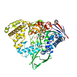
7FHS
 | | Crystal structure of DYRK1A in complex with RD0392 | | 分子名称: | (5~{Z})-5-[(3-ethoxy-4-oxidanyl-phenyl)methylidene]-2-sulfanylidene-1,3-thiazolidin-4-one, Dual specificity tyrosine-phosphorylation-regulated kinase 1A, GLYCEROL | | 著者 | Kikuchi, M, Sumida, T, Hosoya, T, Kii, I, Umehara, T. | | 登録日 | 2021-07-30 | | 公開日 | 2022-03-23 | | 最終更新日 | 2023-11-29 | | 実験手法 | X-RAY DIFFRACTION (2.42 Å) | | 主引用文献 | Structure-activity relationship for the folding intermediate-selective inhibition of DYRK1A.
Eur.J.Med.Chem., 227, 2022
|
|
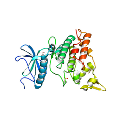
7VSX
 | | Crystal structure of QL-nanoKAZ (Reverse mutant of nanoKAZ with L18Q and V27L) | | 分子名称: | 2-(N-MORPHOLINO)-ETHANESULFONIC ACID, QLnK | | 著者 | Tomabechi, Y, Sekine, S, Shirouzu, M, Takamitsu, H, Satoshi, I. | | 登録日 | 2021-10-27 | | 公開日 | 2022-08-24 | | 最終更新日 | 2023-11-29 | | 実験手法 | X-RAY DIFFRACTION (1.698 Å) | | 主引用文献 | Reverse mutants of the catalytic 19 kDa mutant protein (nanoKAZ/nanoLuc) from Oplophorus luciferase with coelenterazine as preferred substrate.
Plos One, 17, 2022
|
|
7EG3
 | |
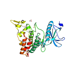
7EG2
 | | Crystal structure of the apoAequorin complex with (S)-daCTZ | | 分子名称: | (2~{S})-2-(hydroxymethyl)-6-(4-hydroxyphenyl)-2-[(4-hydroxyphenyl)methyl]-4-(phenylmethyl)-3~{H}-inden-1-one, Aequorin-2 | | 著者 | Tomabechi, Y, Shirouzu, M. | | 登録日 | 2021-03-24 | | 公開日 | 2021-06-23 | | 最終更新日 | 2023-11-29 | | 実験手法 | X-RAY DIFFRACTION (2.22 Å) | | 主引用文献 | Chiral deaza-coelenterazine analogs for probing a substrate-binding site in the Ca2+-binding photoprotein aequorin.
Plos One, 16, 2021
|
|


