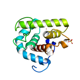
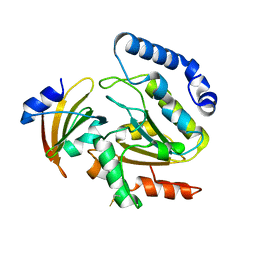
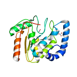
2TBS
 
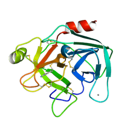 | |
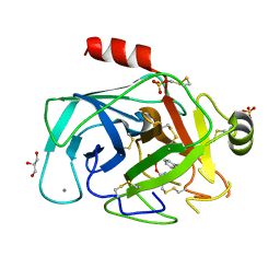
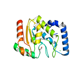
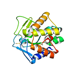
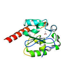
5AFD
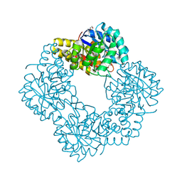 | | Native structure of N-acetylneuramininate lyase (sialic acid aldolase) from Aliivibrio salmonicida | | 分子名称: | 1,2-ETHANEDIOL, GLYCEROL, N-ACETYLNEURAMINATE LYASE | | 著者 | Gurung, M.K, Altermark, B, Rader, I.L.U, Helland, R, Smalas, A.O. | | 登録日 | 2015-01-21 | | 公開日 | 2016-03-02 | | 最終更新日 | 2024-01-10 | | 実験手法 | X-RAY DIFFRACTION (1.65 Å) | | 主引用文献 | Features and structure of a cold active N-acetylneuraminate lyase.
Plos One, 14, 2019
|
|
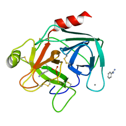
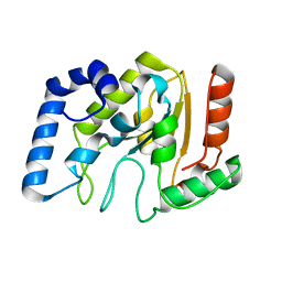
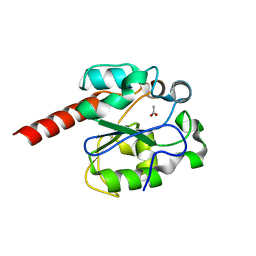
4UNF
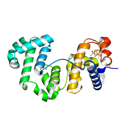 | | Crystal structure of Deinococcus radiodurans Endonuclease III-1 | | 分子名称: | ACETATE ION, ENDONUCLEASE III-1, IRON/SULFUR CLUSTER, ... | | 著者 | Sarre, A, Okvist, M, Klar, T, Hall, D, Smalas, A.O, McSweeney, S, Timmins, J, Moe, E. | | 登録日 | 2014-05-28 | | 公開日 | 2015-06-10 | | 最終更新日 | 2024-01-10 | | 実験手法 | X-RAY DIFFRACTION (2.15 Å) | | 主引用文献 | Structural and Functional Characterization of Two Unusual Endonuclease III Enzymes from Deinococcus Radiodurans.
J.Struct.Biol., 191, 2015
|
|
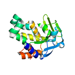
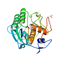
4UOB
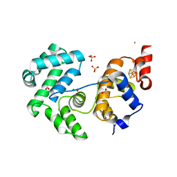 | | Crystal structure of Deinococcus radiodurans Endonuclease III-3 | | 分子名称: | ACETATE ION, COBALT (II) ION, ENDONUCLEASE III-3, ... | | 著者 | Sarre, A, Okvist, M, Klar, T, Hall, D, Smalas, A.O, McSweeney, S, Timmins, J, Moe, E. | | 登録日 | 2014-06-02 | | 公開日 | 2015-06-10 | | 最終更新日 | 2024-05-08 | | 実験手法 | X-RAY DIFFRACTION (1.31 Å) | | 主引用文献 | Structural and Functional Characterization of Two Unusual Endonuclease III Enzymes from Deinococcus Radiodurans.
J.Struct.Biol., 191, 2015
|
|
2STA
 | | ANIONIC SALMON TRYPSIN IN COMPLEX WITH SQUASH SEED INHIBITOR (CUCURBITA MAXIMA TRYPSIN INHIBITOR I) | | 分子名称: | CALCIUM ION, PROTEIN (TRYPSIN INHIBITOR), PROTEIN (TRYPSIN) | | 著者 | Helland, R, Berglund, G.I, Otlewski, J, Apostoluk, W, Andersen, O.A, Willassen, N.P, Smalas, A.O. | | 登録日 | 1998-12-10 | | 公開日 | 2000-01-19 | | 最終更新日 | 2023-08-30 | | 実験手法 | X-RAY DIFFRACTION (1.8 Å) | | 主引用文献 | High-resolution structures of three new trypsin-squash-inhibitor complexes: a detailed comparison with other trypsins and their complexes.
Acta Crystallogr.,Sect.D, 55, 1999
|
|
2STB
 | | ANIONIC SALMON TRYPSIN IN COMPLEX WITH SQUASH SEED INHIBITOR (CUCURBITA PEPO TRYPSIN INHIBITOR II) | | 分子名称: | CALCIUM ION, PROTEIN (TRYPSIN INHIBITOR), PROTEIN (TRYPSIN) | | 著者 | Helland, R, Berglund, G.I, Otlewski, J, Apostoluk, W, Andersen, O.A, Willassen, N.P, Smalas, A.O. | | 登録日 | 1998-12-11 | | 公開日 | 2000-01-19 | | 最終更新日 | 2023-08-30 | | 実験手法 | X-RAY DIFFRACTION (1.8 Å) | | 主引用文献 | High-resolution structures of three new trypsin-squash-inhibitor complexes: a detailed comparison with other trypsins and their complexes.
Acta Crystallogr.,Sect.D, 55, 1999
|
|
3MGW
 | | Thermodynamics and structure of a salmon cold-active goose-type lysozyme | | 分子名称: | COBALT (II) ION, Lysozyme g, SULFATE ION | | 著者 | Kyomuhendo, P, Myrnes, B, Brandsdal, B.O, Smalas, A.O, Nilsen, I.W, Helland, R. | | 登録日 | 2010-04-07 | | 公開日 | 2010-05-05 | | 最終更新日 | 2023-11-01 | | 実験手法 | X-RAY DIFFRACTION (1.75 Å) | | 主引用文献 | Thermodynamics and structure of a salmon cold active goose-type lysozyme
Comp.Biochem.Physiol. B: Biochem.Mol.Biol., 156, 2010
|
|
1HJ9
 | | Atomic resolution structures of trypsin provide insight into structural radiation damage | | 分子名称: | ANILINE, BETA-TRYPSIN, CALCIUM ION, ... | | 著者 | Leiros, H.-K.S, McSweeney, S.M, Smalas, A.O. | | 登録日 | 2001-01-10 | | 公開日 | 2002-01-04 | | 最終更新日 | 2011-07-13 | | 実験手法 | X-RAY DIFFRACTION (0.95 Å) | | 主引用文献 | Atomic Resolution Structure of Trypsin Provide Insight Into Structural Radiation Damage
Acta Crystallogr.,Sect.D, 57, 2001
|
|
1HJ8
 | | 1.00 AA Trypsin from Atlantic Salmon | | 分子名称: | BENZAMIDINE, CALCIUM ION, SULFATE ION, ... | | 著者 | Leiros, H.-K.S, Mcsweeney, S.M, Smalas, A.O. | | 登録日 | 2001-01-09 | | 公開日 | 2002-01-04 | | 最終更新日 | 2023-12-13 | | 実験手法 | X-RAY DIFFRACTION (1 Å) | | 主引用文献 | Atomic Resolution Structure of Trypsin Provide Insight Into Structural Radiation Damage
Acta Crystallogr.,Sect.D, 57, 2001
|
|
2VND
 | | The N69Q mutant of Vibrio cholerae endonuclease I | | 分子名称: | CHLORIDE ION, ENDONUCLEASE I, MAGNESIUM ION | | 著者 | Niiranen, L, Altermark, B, Brandsdal, B.O, Leiros, H.-K.S, Helland, R, Smalas, A.O, Willassen, N.P. | | 登録日 | 2008-02-04 | | 公開日 | 2008-02-12 | | 最終更新日 | 2023-12-13 | | 実験手法 | X-RAY DIFFRACTION (1.7 Å) | | 主引用文献 | Effects of Salt on the Kinetics and Thermodynamic Stability of Endonuclease I from Vibrio Salmonicida and Vibrio Cholerae.
FEBS J., 275, 2008
|
|
4LYL
 | | Crystal structure of uracil-DNA glycosylase from cod (Gadus morhua) in complex with the proteinaceous inhibitor UGI | | 分子名称: | Uracil-DNA glycosylase, Uracil-DNA glycosylase inhibitor | | 著者 | Assefa, N.G, Niiranen, L.M.K, Johnson, K.A, Leiros, H.-K.S, Smalas, A.O, Willassen, N.P, Moe, E. | | 登録日 | 2013-07-31 | | 公開日 | 2014-08-13 | | 実験手法 | X-RAY DIFFRACTION (1.93 Å) | | 主引用文献 | Structural and biophysical analysis of interactions between cod and human uracil-DNA N-glycosylase (UNG) and UNG inhibitor (Ugi).
Acta Crystallogr.,Sect.D, 70, 2014
|
|
1OKB
 | | crystal structure of Uracil-DNA glycosylase from Atlantic cod (Gadus morhua) | | 分子名称: | CHLORIDE ION, GLYCEROL, URACIL-DNA GLYCOSYLASE | | 著者 | Leiros, I, Moe, E, Lanes, O, Smalas, A.O, Willassen, N.P. | | 登録日 | 2003-07-21 | | 公開日 | 2004-04-05 | | 最終更新日 | 2023-12-13 | | 実験手法 | X-RAY DIFFRACTION (1.9 Å) | | 主引用文献 | The Crystal Structure of Uracil-DNA Glycosylase from Atlantic Cod (Gadus Morhua) Reveals Cold-Adaptation Features
Acta Crystallogr.,Sect.D, 59, 2003
|
|
1YUO
 | | Optimisation of the surface electrostatics as a strategy for cold adaptation of uracil-DNA N-glycosylase (UNG)from atlantic cod (Gadus morhua) | | 分子名称: | Uracil-DNA glycosylase | | 著者 | Moe, E, Leiros, I, Riise, E.K, Olufsen, M, Lanes, O, Smalas, A.O, Willassen, N.P. | | 登録日 | 2005-02-14 | | 公開日 | 2005-03-01 | | 最終更新日 | 2023-10-25 | | 実験手法 | X-RAY DIFFRACTION (1.95 Å) | | 主引用文献 | Optimisation of the surface electrostatics as a strategy for cold adaptation of uracil-DNA N-glycosylase (UNG) from Atlantic cod (Gadus morhua)
J.Mol.Biol., 343, 2004
|
|
2B6N
 | | The 1.8 A crystal structure of a Proteinase K like enzyme from a psychrotroph Serratia species | | 分子名称: | CALCIUM ION, SULFATE ION, TRIPEPTIDE, ... | | 著者 | Helland, R, Larsen, A.N, Smalas, A.O, Willassen, N.P. | | 登録日 | 2005-10-03 | | 公開日 | 2006-03-07 | | 最終更新日 | 2023-10-25 | | 実験手法 | X-RAY DIFFRACTION (1.8 Å) | | 主引用文献 | The 1.8 A crystal structure of a proteinase K-like enzyme from a psychrotroph Serratia species
Febs J., 273, 2006
|
|
2PU3
 | | Structural adaptation of endonuclease I from the cold-adapted and halophilic bacterium Vibrio salmonicida | | 分子名称: | CHLORIDE ION, Endonuclease I, MAGNESIUM ION | | 著者 | Altermark, B, Helland, R, Moe, E, Willassen, N.P, Smalas, A.O. | | 登録日 | 2007-05-08 | | 公開日 | 2008-03-18 | | 最終更新日 | 2023-08-30 | | 実験手法 | X-RAY DIFFRACTION (1.5 Å) | | 主引用文献 | Structural adaptation of endonuclease I from the cold-adapted and halophilic bacterium Vibrio salmonicida.
Acta Crystallogr.,Sect.D, 64, 2008
|
|
2ISA
 | | Crystal Structure of Vibrio salmonicida catalase | | 分子名称: | CHLORIDE ION, Catalase, GLYCEROL, ... | | 著者 | Riise, E.K, Lorentzen, M.S, Helland, R, Smalas, A.O, Leiros, H.K.S, Willassen, N.P. | | 登録日 | 2006-10-17 | | 公開日 | 2007-01-23 | | 最終更新日 | 2023-10-25 | | 実験手法 | X-RAY DIFFRACTION (1.97 Å) | | 主引用文献 | The first structure of a cold-active catalase from Vibrio salmonicida at 1.96A reveals structural aspects of cold adaptation
ACTA CRYSTALLOGR.,SECT.D, 63, 2007
|
|
2JHQ
 | | Crystal structure of Uracil DNA-glycosylase from Vibrio cholerae | | 分子名称: | CHLORIDE ION, URACIL DNA-GLYCOSYLASE | | 著者 | Raeder, I.L.U, Moe, E, Willassen, N.P, Smalas, A.O, Leiros, I. | | 登録日 | 2007-02-23 | | 公開日 | 2008-06-03 | | 最終更新日 | 2023-12-13 | | 実験手法 | X-RAY DIFFRACTION (1.5 Å) | | 主引用文献 | Structure of Uracil-DNA N-Glycosylase (Ung) from Vibrio Cholerae. Mapping Temperature Adaptation Through Structural and Mutational Analysis.
Acta Crystallogr.,Sect.F, 66, 2010
|
|
2BTC
 | | BOVINE TRYPSIN IN COMPLEX WITH SQUASH SEED INHIBITOR (CUCURBITA PEPO TRYPSIN INHIBITOR II) | | 分子名称: | CALCIUM ION, PROTEIN (TRYPSIN INHIBITOR), PROTEIN (TRYPSIN) | | 著者 | Helland, R, Berglund, G.I, Otlewski, J, Apostoluk, W, Andersen, O.A, Willassen, N.P, Smalas, A.O. | | 登録日 | 1998-12-11 | | 公開日 | 2000-01-19 | | 最終更新日 | 2023-08-23 | | 実験手法 | X-RAY DIFFRACTION (1.5 Å) | | 主引用文献 | High-resolution structures of three new trypsin-squash-inhibitor complexes: a detailed comparison with other trypsins and their complexes.
Acta Crystallogr.,Sect.D, 55, 1999
|
|
2BOO
 | | The crystal structure of Uracil-DNA N-Glycosylase (UNG) from Deinococcus radiodurans. | | 分子名称: | NITRATE ION, URACIL-DNA GLYCOSYLASE | | 著者 | Leiros, I, Moe, E, Smalas, A.O, McSweeney, S. | | 登録日 | 2005-04-13 | | 公開日 | 2005-07-27 | | 最終更新日 | 2023-12-13 | | 実験手法 | X-RAY DIFFRACTION (1.8 Å) | | 主引用文献 | Structure of the Uracil-DNA N-Glycosylase (Ung) from Deinococcus Radiodurans.
Acta Crystallogr.,Sect.D, 61, 2005
|
|
2C2Q
 | | The crystal structure of mismatch specific uracil-DNA glycosylase (MUG) from Deinococcus radiodurans. Inactive mutant Asp93Ala. | | 分子名称: | ACETATE ION, G/U MISMATCH-SPECIFIC DNA GLYCOSYLASE | | 著者 | Moe, E, Leiros, I, Smalas, A.O, McSweeney, S. | | 登録日 | 2005-09-29 | | 公開日 | 2005-10-18 | | 最終更新日 | 2023-12-13 | | 実験手法 | X-RAY DIFFRACTION (1.7 Å) | | 主引用文献 | The Crystal Structure of Mismatch Specific Uracil-DNA Glycosylase (Mug) from Deinococcus Radiodurans Reveals a Novel Catalytic Residue and Broad Substrate Specificity
J.Biol.Chem., 281, 2006
|
|
2C2P
 | | The crystal structure of mismatch specific uracil-DNA glycosylase (MUG) from Deinococcus radiodurans | | 分子名称: | ACETATE ION, G/U MISMATCH-SPECIFIC DNA GLYCOSYLASE | | 著者 | Moe, E, Leiros, I, Smalas, A.O, McSweeney, S. | | 登録日 | 2005-09-29 | | 公開日 | 2005-10-18 | | 最終更新日 | 2023-12-13 | | 実験手法 | X-RAY DIFFRACTION (1.75 Å) | | 主引用文献 | The Crystal Structure of Mismatch Specific Uracil-DNA Glycosylase (Mug) from Deinococcus Radiodurans Reveals a Novel Catalytic Residue and Broad Substrate Specificity
J.Biol.Chem., 281, 2006
|
|
1BZX
 | | THE CRYSTAL STRUCTURE OF ANIONIC SALMON TRYPSIN IN COMPLEX WITH BOVINE PANCREATIC TRYPSIN INHIBITOR | | 分子名称: | CALCIUM ION, PROTEIN (BOVINE PANCREATIC TRYPSIN INHIBITOR), PROTEIN (TRYPSIN) | | 著者 | Helland, R, Leiros, I, Berglund, G.I, Willassen, N.P, Smalas, A.O. | | 登録日 | 1998-11-05 | | 公開日 | 1998-11-11 | | 最終更新日 | 2023-08-09 | | 実験手法 | X-RAY DIFFRACTION (2.1 Å) | | 主引用文献 | The crystal structure of anionic salmon trypsin in complex with bovine pancreatic trypsin inhibitor.
Eur.J.Biochem., 256, 1998
|
|
1T8N
 | | CRYSTAL STRUCTURE OF THE P1 THR BPTI MUTANT- BOVINE CHYMOTRYPSIN COMPLEX | | 分子名称: | Chymotrypsin A, Pancreatic trypsin inhibitor, SULFATE ION | | 著者 | Czapinska, H, Helland, R, Otlewski, J, Smalas, A.O. | | 登録日 | 2004-05-13 | | 公開日 | 2005-03-08 | | 最終更新日 | 2023-08-23 | | 実験手法 | X-RAY DIFFRACTION (1.75 Å) | | 主引用文献 | Crystal structures of five bovine chymotrypsin complexes with P1 BPTI variants.
J.Mol.Biol., 344, 2004
|
|
1T8O
 | | CRYSTAL STRUCTURE OF THE P1 TRP BPTI MUTANT- BOVINE CHYMOTRYPSIN COMPLEX | | 分子名称: | Chymotrypsin A, Pancreatic trypsin inhibitor, SULFATE ION | | 著者 | Czapinska, H, Helland, R, Otlewski, J, Smalas, A.O. | | 登録日 | 2004-05-13 | | 公開日 | 2005-03-08 | | 最終更新日 | 2023-08-23 | | 実験手法 | X-RAY DIFFRACTION (1.7 Å) | | 主引用文献 | Crystal structures of five bovine chymotrypsin complexes with P1 BPTI variants.
J.Mol.Biol., 344, 2004
|
|
1T8M
 | | CRYSTAL STRUCTURE OF THE P1 HIS BPTI MUTANT- BOVINE CHYMOTRYPSIN COMPLEX | | 分子名称: | Chymotrypsin A, Pancreatic trypsin inhibitor, SULFATE ION | | 著者 | Czapinska, H, Helland, R, Otlewski, J, Smalas, A.O. | | 登録日 | 2004-05-13 | | 公開日 | 2005-03-08 | | 最終更新日 | 2023-08-23 | | 実験手法 | X-RAY DIFFRACTION (1.8 Å) | | 主引用文献 | Crystal structures of five bovine chymotrypsin complexes with P1 BPTI variants.
J.Mol.Biol., 344, 2004
|
|


