1ELT
 
 | |
1P2N
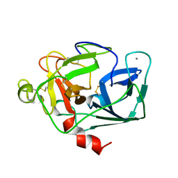
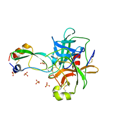
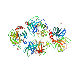
 | | Structural consequences of accommodation of four non-cognate amino-acid residues in the S1 pocket of bovine trypsin and chymotrypsin | | 分子名称: | Chymotrypsinogen A, Pancreatic trypsin inhibitor, SULFATE ION | | 著者 | Helland, R, Czapinska, H, Leiros, I, Olufsen, M, Otlewski, J, Smalaas, A.O. | | 登録日 | 2003-04-15 | | 公開日 | 2004-04-20 | | 最終更新日 | 2023-08-16 | | 実験手法 | X-RAY DIFFRACTION (1.8 Å) | | 主引用文献 | Structural consequences of accommodation of four non-cognate amino acid residues in the S1 pocket of bovine trypsin and chymotrypsin.
J.Mol.Biol., 333, 2003
|
|
1P2J
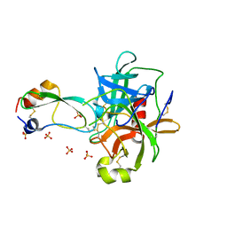
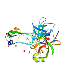
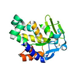
 | | Structural consequences of accommodation of four non-cognate amino-acid residues in the S1 pocket of bovine trypsin and chymotrypsin | | 分子名称: | CALCIUM ION, Pancreatic trypsin inhibitor, SULFATE ION, ... | | 著者 | Helland, R, Czapinska, H, Leiros, I, Olufsen, M, Otlewski, J, Smalaas, A.O. | | 登録日 | 2003-04-15 | | 公開日 | 2004-04-20 | | 最終更新日 | 2023-08-16 | | 実験手法 | X-RAY DIFFRACTION (1.35 Å) | | 主引用文献 | Structural consequences of accommodation of four non-cognate amino acid residues in the S1 pocket of bovine trypsin and chymotrypsin.
J.Mol.Biol., 333, 2003
|
|
1P2I
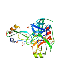
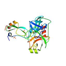
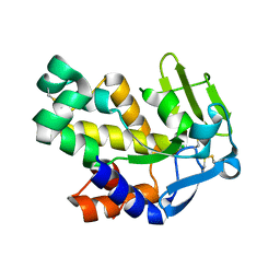
 | | Structural consequences of accommodation of four non-cognate amino-acid residues in the S1 pocket of bovine trypsin and chymotrypsin | | 分子名称: | CALCIUM ION, Pancreatic trypsin inhibitor, SULFATE ION, ... | | 著者 | Helland, R, Czapinska, H, Leiros, I, Olufsen, M, Otlewski, J, Smalaas, A.O. | | 登録日 | 2003-04-15 | | 公開日 | 2004-04-20 | | 最終更新日 | 2023-08-16 | | 実験手法 | X-RAY DIFFRACTION (1.65 Å) | | 主引用文献 | Structural consequences of accommodation of four non-cognate amino acid residues in the S1 pocket of bovine trypsin and chymotrypsin.
J.Mol.Biol., 333, 2003
|
|
1P2O
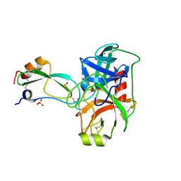
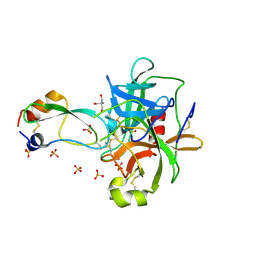
 | | Structural consequences of accommodation of four non-cognate amino-acid residues in the S1 pocket of bovine trypsin and chymotrypsin | | 分子名称: | Chymotrypsinogen A, Pancreatic trypsin inhibitor, SULFATE ION | | 著者 | Helland, R, Czapinska, H, Leiros, I, Olufsen, M, Otlewski, J, Smalaas, A.O. | | 登録日 | 2003-04-15 | | 公開日 | 2004-04-20 | | 最終更新日 | 2023-08-16 | | 実験手法 | X-RAY DIFFRACTION (2 Å) | | 主引用文献 | Structural consequences of accommodation of four non-cognate amino acid residues in the S1 pocket of bovine trypsin and chymotrypsin.
J.Mol.Biol., 333, 2003
|
|
1P2M
 | | Structural consequences of accommodation of four non-cognate amino-acid residues in the S1 pocket of bovine trypsin and chymotrypsin | | 分子名称: | Chymotrypsinogen A, Pancreatic trypsin inhibitor, SULFATE ION | | 著者 | Helland, R, Czapinska, H, Leiros, I, Olufsen, M, Otlewski, J, Smalaas, A.O. | | 登録日 | 2003-04-15 | | 公開日 | 2004-04-20 | | 最終更新日 | 2023-08-16 | | 実験手法 | X-RAY DIFFRACTION (1.75 Å) | | 主引用文献 | Structural consequences of accommodation of four non-cognate amino acid residues in the S1 pocket of bovine trypsin and chymotrypsin.
J.Mol.Biol., 333, 2003
|
|
1P2K
 | | Structural consequences of accommodation of four non-cognate amino-acid residues in the S1 pocket of bovine trypsin and chymotrypsin | | 分子名称: | CALCIUM ION, Pancreatic trypsin inhibitor, SULFATE ION, ... | | 著者 | Helland, R, Czapinska, H, Leiros, I, Olufsen, M, Otlewski, J, Smalaas, A.O. | | 登録日 | 2003-04-15 | | 公開日 | 2004-04-20 | | 最終更新日 | 2023-08-16 | | 実験手法 | X-RAY DIFFRACTION (1.6 Å) | | 主引用文献 | Structural consequences of accommodation of four non-cognate amino acid residues in the S1 pocket of bovine trypsin and chymotrypsin.
J.Mol.Biol., 333, 2003
|
|
1P2Q
 | | Structural consequences of accommodation of four non-cognate amino-acid residues in the S1 pocket of bovine trypsin and chymotrypsin | | 分子名称: | 2-AMINO-2-HYDROXYMETHYL-PROPANE-1,3-DIOL, Chymotrypsinogen A, Pancreatic trypsin inhibitor, ... | | 著者 | Helland, R, Czapinska, H, Leiros, I, Olufsen, M, Otlewski, J, Smalaas, A.O. | | 登録日 | 2003-04-15 | | 公開日 | 2004-04-20 | | 最終更新日 | 2023-08-16 | | 実験手法 | X-RAY DIFFRACTION (1.8 Å) | | 主引用文献 | Structural consequences of accommodation of four non-cognate amino acid residues in the S1 pocket of bovine trypsin and chymotrypsin.
J.Mol.Biol., 333, 2003
|
|
1A0J
 | | CRYSTAL STRUCTURE OF A NON-PSYCHROPHILIC TRYPSIN FROM A COLD-ADAPTED FISH SPECIES. | | 分子名称: | BENZAMIDINE, CALCIUM ION, SULFATE ION, ... | | 著者 | Schroeder, H.-K, Willassen, N.P, Smalaas, A.O. | | 登録日 | 1997-12-01 | | 公開日 | 1999-01-13 | | 最終更新日 | 2023-08-02 | | 実験手法 | X-RAY DIFFRACTION (1.7 Å) | | 主引用文献 | Structure of a non-psychrophilic trypsin from a cold-adapted fish species.
Acta Crystallogr.,Sect.D, 54, 1998
|
|
1T8L
 | | CRYSTAL STRUCTURE OF THE P1 MET BPTI MUTANT- BOVINE CHYMOTRYPSIN COMPLEX | | 分子名称: | Chymotrypsin A, Pancreatic trypsin inhibitor, SULFATE ION | | 著者 | Czapinska, H, Helland, R, Otlewski, J, Smalas, A.O. | | 登録日 | 2004-05-13 | | 公開日 | 2005-03-08 | | 最終更新日 | 2023-08-23 | | 実験手法 | X-RAY DIFFRACTION (1.75 Å) | | 主引用文献 | Crystal structures of five bovine chymotrypsin complexes with P1 BPTI variants.
J.Mol.Biol., 344, 2004
|
|
2G7F
 | | The 1.95 A crystal structure of Vibrio cholerae extracellular endonuclease I | | 分子名称: | CHLORIDE ION, Endonuclease I, MAGNESIUM ION | | 著者 | Altermark, B, Smalaas, A.O, Willassen, N.P, Helland, R. | | 登録日 | 2006-02-28 | | 公開日 | 2006-10-31 | | 最終更新日 | 2023-08-30 | | 実験手法 | X-RAY DIFFRACTION (1.95 Å) | | 主引用文献 | The structure of Vibrio cholerae extracellular endonuclease I reveals the presence of a buried chloride ion.
Acta Crystallogr.,Sect.D, 62, 2006
|
|
2G7E
 | | The 1.6 A crystal structure of Vibrio cholerae extracellular endonuclease I | | 分子名称: | CHLORIDE ION, Endonuclease I | | 著者 | Altermark, B, Smalaas, A.O, Willassen, N.P, Helland, R. | | 登録日 | 2006-02-28 | | 公開日 | 2006-10-31 | | 最終更新日 | 2023-08-30 | | 実験手法 | X-RAY DIFFRACTION (1.6 Å) | | 主引用文献 | The structure of Vibrio cholerae extracellular endonuclease I reveals the presence of a buried chloride ion.
Acta Crystallogr.,Sect.D, 62, 2006
|
|
1LMO
 | |
1LMC
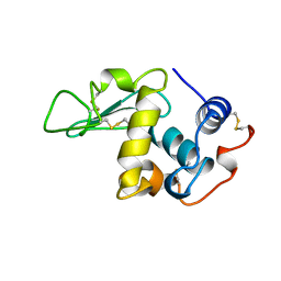
 | | THE CRYSTAL STRUCTURE OF A COMPLEX BETWEEN BULGECIN, A BACTERIAL METABOLITE, AND LYSOZYME FROM THE RAINBOW TROUT | | 分子名称: | BULGECIN A, LYSOZYME | | 著者 | Karlsen, S, Hough, E. | | 登録日 | 1994-11-14 | | 公開日 | 1996-01-01 | | 最終更新日 | 2023-07-26 | | 実験手法 | X-RAY DIFFRACTION (2 Å) | | 主引用文献 | Structure of a complex between bulgecin, a bacterial metabolite, and lysozyme from the rainbow trout.
Acta Crystallogr.,Sect.D, 52, 1996
|
|
1LMP
 | |
1LMQ
 | |


