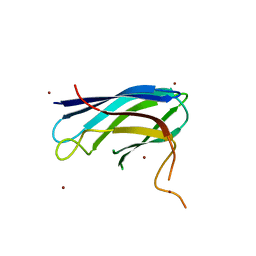
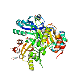
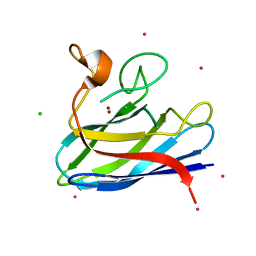
3ZSJ
 
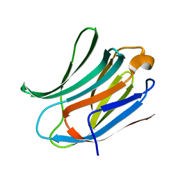 | | Crystal structure of Human Galectin-3 CRD in complex with Lactose at 0.86 angstrom resolution | | 分子名称: | GALECTIN-3, beta-D-galactopyranose-(1-4)-beta-D-glucopyranose | | 著者 | Saraboji, K, Hakansson, M, Diehl, C, Nilsson, U.J, Leffler, H, Akke, M, Logan, D.T. | | 登録日 | 2011-06-28 | | 公開日 | 2011-12-14 | | 最終更新日 | 2023-12-20 | | 実験手法 | X-RAY DIFFRACTION (0.86 Å) | | 主引用文献 | The Carbohydrate-Binding Site in Galectin-3 is Pre-Organized to Recognize a Sugar-Like Framework of Oxygens: Ultra-High Resolution Structures and Water Dynamics.
Biochemistry, 51, 2012
|
|
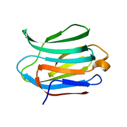
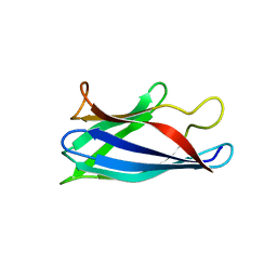
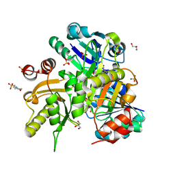
3ZSK
 | | Crystal structure of Human Galectin-3 CRD with glycerol bound at 0.90 angstrom resolution | | 分子名称: | GALECTIN-3, GLYCEROL | | 著者 | Saraboji, K, Hakansson, M, Diehl, C, Nilsson, U.J, Leffler, H, Akke, M, Logan, D.T. | | 登録日 | 2011-06-28 | | 公開日 | 2011-12-14 | | 最終更新日 | 2023-12-20 | | 実験手法 | X-RAY DIFFRACTION (0.9 Å) | | 主引用文献 | The Carbohydrate-Binding Site in Galectin-3 is Pre-Organized to Recognize a Sugar-Like Framework of Oxygens: Ultra-High Resolution Structures and Water Dynamics.
Biochemistry, 51, 2012
|
|
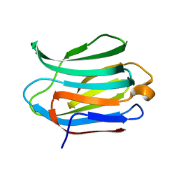
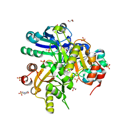
3ZSM
 | | Crystal structure of Apo Human Galectin-3 CRD at 1.25 angstrom resolution, at room temperature | | 分子名称: | GALECTIN-3 | | 著者 | Saraboji, K, Hakansson, M, Diehl, C, Nilsson, U.J, Leffler, H, Akke, M, Logan, D.T. | | 登録日 | 2011-06-28 | | 公開日 | 2011-12-14 | | 最終更新日 | 2023-12-20 | | 実験手法 | X-RAY DIFFRACTION (1.25 Å) | | 主引用文献 | The Carbohydrate-Binding Site in Galectin-3 is Pre-Organized to Recognize a Sugar-Like Framework of Oxygens: Ultra-High Resolution Structures and Water Dynamics.
Biochemistry, 51, 2012
|
|
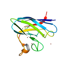
3ZSL
 | | Crystal structure of Apo Human Galectin-3 CRD at 1.08 angstrom resolution, at cryogenic temperature | | 分子名称: | GALECTIN-3 | | 著者 | Saraboji, K, Hakansson, M, Diehl, C, Nilsson, U.J, Leffler, H, Akke, M, Logan, D.T. | | 登録日 | 2011-06-28 | | 公開日 | 2011-12-14 | | 最終更新日 | 2023-12-20 | | 実験手法 | X-RAY DIFFRACTION (1.08 Å) | | 主引用文献 | The Carbohydrate-Binding Site in Galectin-3 is Pre-Organized to Recognize a Sugar-Like Framework of Oxygens: Ultra-High Resolution Structures and Water Dynamics.
Biochemistry, 51, 2012
|
|
2XJL
 | | Monomeric Human Cu,Zn Superoxide dismutase without Cu ligands | | 分子名称: | ACETATE ION, DI(HYDROXYETHYL)ETHER, SODIUM ION, ... | | 著者 | Saraboji, K, Leinartaite, L, Nordlund, A, Oliveberg, M, Logan, D.T. | | 登録日 | 2010-07-07 | | 公開日 | 2010-09-01 | | 最終更新日 | 2024-05-01 | | 実験手法 | X-RAY DIFFRACTION (1.55 Å) | | 主引用文献 | Folding Catalysis by Transient Coordination of Zn2+ to the Cu Ligands of the Als-Associated Enzyme Cu/Zn Superoxide Dismutase 1.
J.Am.Chem.Soc., 132, 2010
|
|
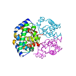
2XJK
 | | Monomeric Human Cu,Zn Superoxide dismutase | | 分子名称: | COPPER (II) ION, SUPEROXIDE DISMUTASE [CU-ZN], ZINC ION | | 著者 | Saraboji, K, Leinartaite, L, Nordlund, A, Oliveberg, M, Logan, D.T. | | 登録日 | 2010-07-07 | | 公開日 | 2010-09-01 | | 最終更新日 | 2023-12-20 | | 実験手法 | X-RAY DIFFRACTION (1.45 Å) | | 主引用文献 | Folding Catalysis by Transient Coordination of Zn2+ to the Cu Ligands of the Als-Associated Enzyme Cu/Zn Superoxide Dismutase 1.
J.Am.Chem.Soc., 132, 2010
|
|
3HFF
 | | Monomeric human Cu,Zn Superoxide dismutase without Zn ligands | | 分子名称: | Superoxide dismutase [Cu-Zn], ZINC ION | | 著者 | Saraboji, K, Nordlund, A, Leinartait, L, Oliveberg, M, Logan, D.T. | | 登録日 | 2009-05-11 | | 公開日 | 2009-06-16 | | 最終更新日 | 2024-05-29 | | 実験手法 | X-RAY DIFFRACTION (2.2 Å) | | 主引用文献 | Functional features cause misfolding of the ALS-provoking enzyme SOD1.
Proc.Natl.Acad.Sci.USA, 106, 2009
|
|
4BCZ
 | | Monomeric Human Cu,Zn Superoxide dismutase, loops IV and VII deleted, apo form. | | 分子名称: | SUPEROXIDE DISMUTASE [CU-ZN] | | 著者 | Saraboji, K, Awad, W, Danielsson, J, Lang, L, Kurnik, M, Marklund, S.L, Oliveberg, M, Logan, D.T. | | 登録日 | 2012-10-03 | | 公開日 | 2013-02-27 | | 最終更新日 | 2023-12-20 | | 実験手法 | X-RAY DIFFRACTION (1.93 Å) | | 主引用文献 | Global Structural Motions from the Strain of a Single Hydrogen Bond.
Proc.Natl.Acad.Sci.USA, 110, 2013
|
|
1S0H
 | |
2DSI
 | | Crystal structure of Glu171 to Arg mutant of Diphthine synthase | | 分子名称: | 2-(N-MORPHOLINO)-ETHANESULFONIC ACID, GLYCEROL, S-ADENOSYL-L-HOMOCYSTEINE, ... | | 著者 | Mizutani, H, Matsuura, Y, Saraboji, K, Malathy Sony, S.M, Ponnuswamy, M.N, Kumarevel, T.S, Kunishima, N, RIKEN Structural Genomics/Proteomics Initiative (RSGI) | | 登録日 | 2006-06-30 | | 公開日 | 2006-12-30 | | 最終更新日 | 2023-10-25 | | 実験手法 | X-RAY DIFFRACTION (2.2 Å) | | 主引用文献 | Crystal structure of diphthine synthase from Pyrococcus horikoshii OT3
To be Published
|
|
2DSG
 | | Crystal structure of Lys26 to Arg mutant of Diphthine synthase | | 分子名称: | 2-(N-MORPHOLINO)-ETHANESULFONIC ACID, GLYCEROL, S-ADENOSYL-L-HOMOCYSTEINE, ... | | 著者 | Mizutani, H, Matsuura, Y, Saraboji, K, Malathy Sony, S.M, Ponnuswamy, M.N, Kumarevel, T.S, Kunishima, N, RIKEN Structural Genomics/Proteomics Initiative (RSGI) | | 登録日 | 2006-06-30 | | 公開日 | 2006-12-30 | | 最終更新日 | 2023-10-25 | | 実験手法 | X-RAY DIFFRACTION (2 Å) | | 主引用文献 | Crystal structure of diphthine synthase from Pyrococcus horikoshii OT3
To be Published
|
|
2DV7
 | | Crystal structure of Lys187 to Arg mutant of Diphthine synthase | | 分子名称: | 2-(N-MORPHOLINO)-ETHANESULFONIC ACID, GLYCEROL, S-ADENOSYL-L-HOMOCYSTEINE, ... | | 著者 | Mizutani, H, Matsuura, Y, Saraboji, K, Malathy Sony, S.M, Ponnuswamy, M.N, Kumarevel, T.S, Kunishima, N, RIKEN Structural Genomics/Proteomics Initiative (RSGI) | | 登録日 | 2006-07-28 | | 公開日 | 2007-01-28 | | 最終更新日 | 2023-10-25 | | 実験手法 | X-RAY DIFFRACTION (2.3 Å) | | 主引用文献 | Crystal structure of diphthine synthase from Pyrococcus horikoshii OT3
To be Published
|
|
2DSH
 | | Crystal structure of Lys26 to Tyr mutant of Diphthine synthase | | 分子名称: | 2-(N-MORPHOLINO)-ETHANESULFONIC ACID, GLYCEROL, S-ADENOSYL-L-HOMOCYSTEINE, ... | | 著者 | Mizutani, H, Matsuura, Y, Saraboji, K, Malathy Sony, S.M, Ponnuswamy, M.N, Kumarevel, T.S, Kunishima, N, RIKEN Structural Genomics/Proteomics Initiative (RSGI) | | 登録日 | 2006-06-30 | | 公開日 | 2006-12-30 | | 最終更新日 | 2023-10-25 | | 実験手法 | X-RAY DIFFRACTION (2 Å) | | 主引用文献 | Crystal structure of diphthine synthase from Pyrococcus horikoshii OT3
To be Published
|
|
3WTG
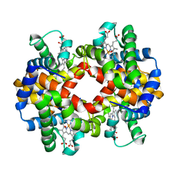 | | Crystal structure of Emu (dromaius novaehollandiae) hemoglobin at 2.3 angstrom resolution | | 分子名称: | Hemoglobin, Hemoglobin subunit alpha-A, OXYGEN MOLECULE, ... | | 著者 | Abubakkar, M.M, Maheshwaran, V, Ponnuswamy, M.N, Saraboji, K. | | 登録日 | 2014-04-11 | | 公開日 | 2014-07-02 | | 最終更新日 | 2023-11-08 | | 実験手法 | X-RAY DIFFRACTION (2.3 Å) | | 主引用文献 | Purification and preliminary structural studies of hemoglobin from high oxygen affinity species Emu (Dromaius novaehollandiae) at neutral pH
To be Published
|
|
4BD4
 | | Monomeric Human Cu,Zn Superoxide dismutase, loops IV and VII deleted, apo form, mutant H43F | | 分子名称: | GLYCEROL, SUPEROXIDE DISMUTASE [CU-ZN] | | 著者 | Awad, W, Saraboji, K, Danielsson, J, Lang, L, Kurnik, M, Marklund, S.L, Oliveberg, M, Logan, D.T. | | 登録日 | 2012-10-04 | | 公開日 | 2013-02-27 | | 最終更新日 | 2023-12-20 | | 実験手法 | X-RAY DIFFRACTION (2.78 Å) | | 主引用文献 | Global Structural Motions from the Strain of a Single Hydrogen Bond.
Proc.Natl.Acad.Sci.USA, 110, 2013
|
|
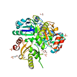
4BCY
 | | Monomeric Human Cu,Zn Superoxide dismutase, mutation H43F | | 分子名称: | CADMIUM ION, CHLORIDE ION, COPPER (II) ION, ... | | 著者 | Awad, W, Saraboji, K, Danielsson, J, Lang, L, Kurnik, M, Marklund, S.L, Oliveberg, M, Logan, D.T. | | 登録日 | 2012-10-03 | | 公開日 | 2013-02-27 | | 最終更新日 | 2023-12-20 | | 実験手法 | X-RAY DIFFRACTION (1.272 Å) | | 主引用文献 | Global Structural Motions from the Strain of a Single Hydrogen Bond.
Proc.Natl.Acad.Sci.USA, 110, 2013
|
|
3ZZP
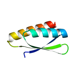 | | Circular permutant of ribosomal protein S6, lacking edge strand beta- 2 of wild-type S6. | | 分子名称: | RIBOSOMAL PROTEIN S6 | | 著者 | Saraboji, K, Haglund, E, Lindberg, M.O, Oliveberg, M, Logan, D.T. | | 登録日 | 2011-09-02 | | 公開日 | 2011-11-23 | | 最終更新日 | 2023-12-20 | | 実験手法 | X-RAY DIFFRACTION (0.96 Å) | | 主引用文献 | Trimming Down a Protein Structure to its Bare Foldons: Spatial Organization of the Cooperative Unit.
J.Biol.Chem., 287, 2012
|
|
2HUT
 | | Crystal structure of PH0725 from Pyrococcus horikoshii OT3 | | 分子名称: | Probable diphthine synthase, S-ADENOSYL-L-HOMOCYSTEINE | | 著者 | Sugahara, M, Saraboji, K, Malathy sony, S.M, Ponnuswamy, M.N, Kumarevel, T.S, Kunishima, N, RIKEN Structural Genomics/Proteomics Initiative (RSGI) | | 登録日 | 2006-07-27 | | 公開日 | 2007-01-27 | | 最終更新日 | 2023-10-25 | | 実験手法 | X-RAY DIFFRACTION (2.4 Å) | | 主引用文献 | Crystal structure of PH0725 from Pyrococcus horikoshii OT3
To be Published
|
|
2HUV
 | | Crystal structure of PH0725 from Pyrococcus horikoshii OT3 | | 分子名称: | PLATINUM (II) ION, Probable diphthine synthase, S-ADENOSYL-L-HOMOCYSTEINE, ... | | 著者 | Sugahara, M, Saraboji, K, Malathy sony, S.M, Ponnuswamy, M.N, Kumarevel, T.S, Kunishima, N, RIKEN Structural Genomics/Proteomics Initiative (RSGI) | | 登録日 | 2006-07-27 | | 公開日 | 2007-01-27 | | 最終更新日 | 2023-10-25 | | 実験手法 | X-RAY DIFFRACTION (2.1 Å) | | 主引用文献 | Crystal structure of PH0725 from Pyrococcus horikoshii OT3
To be Published
|
|
6EXY
 | | Neutron crystal structure of perdeuterated galectin-3C in complex with glycerol | | 分子名称: | GLYCEROL, Galectin-3 | | 著者 | Manzoni, F, Schrader, T.E, Ostermann, A, Oksanen, E, Logan, D.T. | | 登録日 | 2017-11-10 | | 公開日 | 2018-09-12 | | 最終更新日 | 2024-05-01 | | 実験手法 | NEUTRON DIFFRACTION (1.1 Å), X-RAY DIFFRACTION | | 主引用文献 | Elucidation of Hydrogen Bonding Patterns in Ligand-Free, Lactose- and Glycerol-Bound Galectin-3C by Neutron Crystallography to Guide Drug Design.
J. Med. Chem., 61, 2018
|
|
6EYM
 | | Neutron crystal structure of perdeuterated galectin-3C in complex with lactose | | 分子名称: | Galectin-3, beta-D-galactopyranose-(1-4)-beta-D-glucopyranose | | 著者 | Manzoni, F, Coates, L, Blakeley, M.P, Oksanen, E, Logan, D.T. | | 登録日 | 2017-11-13 | | 公開日 | 2018-09-12 | | 最終更新日 | 2024-05-01 | | 実験手法 | NEUTRON DIFFRACTION (1.7 Å), X-RAY DIFFRACTION | | 主引用文献 | Elucidation of Hydrogen Bonding Patterns in Ligand-Free, Lactose- and Glycerol-Bound Galectin-3C by Neutron Crystallography to Guide Drug Design.
J. Med. Chem., 61, 2018
|
|
6F2Q
 | | Neutron crystal structure of perdeuterated galectin-3C in the ligand-free form | | 分子名称: | Galectin-3 | | 著者 | Manzoni, F, Blakeley, M.P, Oksanen, E, Logan, D.T. | | 登録日 | 2017-11-27 | | 公開日 | 2018-05-02 | | 最終更新日 | 2024-05-01 | | 実験手法 | NEUTRON DIFFRACTION (1.03 Å), X-RAY DIFFRACTION | | 主引用文献 | Elucidation of Hydrogen Bonding Patterns in Ligand-Free, Lactose- and Glycerol-Bound Galectin-3C by Neutron Crystallography to Guide Drug Design.
J. Med. Chem., 61, 2018
|
|


