7BYK
 
 | |
5JE8
 | |
7CBV
 | |
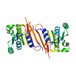
4POO
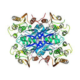
 | | The crystal structure of Bacillus subtilis YtqB in complex with SAM | | 分子名称: | Putative RNA methylase, S-ADENOSYLMETHIONINE | | 著者 | Park, S.C, Song, W.S, Yoon, S.I. | | 登録日 | 2014-02-26 | | 公開日 | 2014-04-02 | | 最終更新日 | 2023-11-08 | | 実験手法 | X-RAY DIFFRACTION (2.2 Å) | | 主引用文献 | Structural analysis of a putative SAM-dependent methyltransferase, YtqB, from Bacillus subtilis
Biochem.Biophys.Res.Commun., 446, 2014
|
|
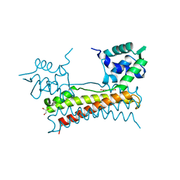
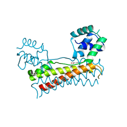
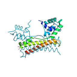
4PON
 | | The crystal structure of a putative SAM-dependent methyltransferase, YtqB, from Bacillus subtilis | | 分子名称: | Putative RNA methylase | | 著者 | Park, S.C, Song, W.S, Yoon, S.I. | | 登録日 | 2014-02-26 | | 公開日 | 2014-04-02 | | 最終更新日 | 2023-11-08 | | 実験手法 | X-RAY DIFFRACTION (1.9 Å) | | 主引用文献 | Structural analysis of a putative SAM-dependent methyltransferase, YtqB, from Bacillus subtilis
Biochem.Biophys.Res.Commun., 446, 2014
|
|
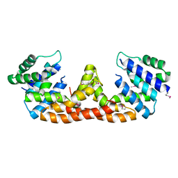
5X13
 | | Crystal structure of Bacillus subtilis PadR in complex with p-coumaric acid | | 分子名称: | 4'-HYDROXYCINNAMIC ACID, GLYCEROL, Transcriptional regulator | | 著者 | Park, S.C, Kwak, Y.M, Song, W.S, Hong, M, Yoon, S.I. | | 登録日 | 2017-01-24 | | 公開日 | 2017-11-22 | | 最終更新日 | 2023-11-22 | | 実験手法 | X-RAY DIFFRACTION (1.7 Å) | | 主引用文献 | Structural basis of effector and operator recognition by the phenolic acid-responsive transcriptional regulator PadR
Nucleic Acids Res., 45, 2017
|
|
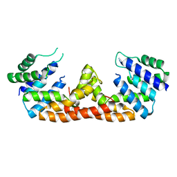
5Y8T
 | | Crystal structure of Bacillus subtilis PadR in complex with p-coumaric acid | | 分子名称: | 4'-HYDROXYCINNAMIC ACID, Transcriptional regulator | | 著者 | Park, S.C, Kwak, Y.M, Song, W.S, Hong, M, Yoon, S.I. | | 登録日 | 2017-08-21 | | 公開日 | 2017-11-29 | | 最終更新日 | 2023-11-22 | | 実験手法 | X-RAY DIFFRACTION (2 Å) | | 主引用文献 | Structural basis of effector and operator recognition by the phenolic acid-responsive transcriptional regulator PadR.
Nucleic Acids Res., 45, 2017
|
|
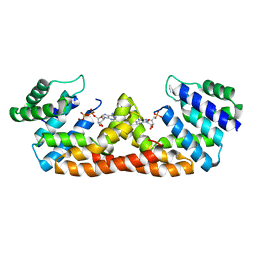
5X14
 | | Crystal structure of Bacillus subtilis PadR in complex with ferulic acid | | 分子名称: | 3-(4-HYDROXY-3-METHOXYPHENYL)-2-PROPENOIC ACID, GLYCEROL, Transcriptional regulator | | 著者 | Park, S.C, Kwak, Y.M, Song, W.S, Hong, M, Yoon, S.I. | | 登録日 | 2017-01-24 | | 公開日 | 2017-11-22 | | 最終更新日 | 2023-11-22 | | 実験手法 | X-RAY DIFFRACTION (1.68 Å) | | 主引用文献 | Structural basis of effector and operator recognition by the phenolic acid-responsive transcriptional regulator PadR
Nucleic Acids Res., 45, 2017
|
|
5X12
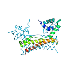 | | Crystal structure of Bacillus subtilis PadR | | 分子名称: | Transcriptional regulator | | 著者 | Park, S.C, Kwak, Y.M, Song, W.S, Hong, M, Yoon, S.I. | | 登録日 | 2017-01-24 | | 公開日 | 2017-11-22 | | 最終更新日 | 2024-03-20 | | 実験手法 | X-RAY DIFFRACTION (1.7 Å) | | 主引用文献 | Structural basis of effector and operator recognition by the phenolic acid-responsive transcriptional regulator PadR
Nucleic Acids Res., 45, 2017
|
|
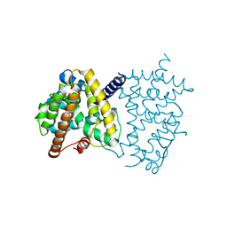
5X11
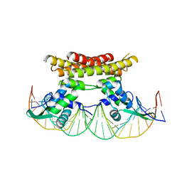 | | Crystal structure of Bacillus subtilis PadR in complex with operator DNA | | 分子名称: | DNA (28-MER), Transcriptional regulator | | 著者 | Park, S.C, Kwak, Y.M, Song, W.S, Hong, M, Yoon, S.I. | | 登録日 | 2017-01-24 | | 公開日 | 2017-11-22 | | 最終更新日 | 2023-11-22 | | 実験手法 | X-RAY DIFFRACTION (2.65 Å) | | 主引用文献 | Structural basis of effector and operator recognition by the phenolic acid-responsive transcriptional regulator PadR
Nucleic Acids Res., 45, 2017
|
|
8H50
 | |
8H4Z
 | |
8H52
 | | Crystal structure of Helicobacter pylori carboxyspermidine dehydrogenase in complex with NADP | | 分子名称: | NADP NICOTINAMIDE-ADENINE-DINUCLEOTIDE PHOSPHATE, Saccharopine dehydrogenase | | 著者 | Ko, K.Y, Park, S.C, Cho, S.Y, Yoon, S.I. | | 登録日 | 2022-10-11 | | 公開日 | 2022-11-09 | | 最終更新日 | 2023-10-25 | | 実験手法 | X-RAY DIFFRACTION (3.1 Å) | | 主引用文献 | Structural analysis of carboxyspermidine dehydrogenase from Helicobacter pylori.
Biochem.Biophys.Res.Commun., 635, 2022
|
|
6JYI
 | | Crystal structure of the PadR-like transcriptional regulator BC1756 from Bacillus cereus | | 分子名称: | Transcriptional repressor PadR | | 著者 | Kim, T.H, Park, S.C, Lee, K.C, Song, W.S, Yoon, S.I. | | 登録日 | 2019-04-26 | | 公開日 | 2019-06-26 | | 最終更新日 | 2019-07-10 | | 実験手法 | X-RAY DIFFRACTION (1.92 Å) | | 主引用文献 | Structural and DNA-binding studies of the PadR-like transcriptional regulator BC1756 from Bacillus cereus.
Biochem.Biophys.Res.Commun., 515, 2019
|
|
6JV6
 | | Crystal structure of the sirohydrochlorin chelatase SirB from Bacillus subtilis subspecies spizizenii in complex with cobalt | | 分子名称: | COBALT (II) ION, Sirohydrochlorin ferrochelatase | | 著者 | Nam, M.S, Song, W.S, Park, S.C, Yoon, S.I. | | 登録日 | 2019-04-16 | | 公開日 | 2019-06-12 | | 最終更新日 | 2023-11-22 | | 実験手法 | X-RAY DIFFRACTION (2.15 Å) | | 主引用文献 | Cobalt complex structure of the sirohydrochlorin chelatase SirB from Bacillus subtilis subsp. spizizenii.
KOREAN J MICROBIOL., 55, 2019
|
|
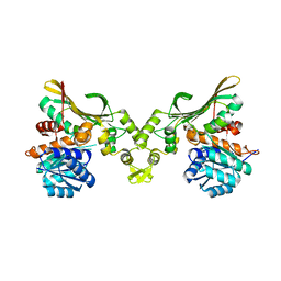
7X9R
 | | Crystal structure of the antirepressor GmaR | | 分子名称: | Glycosyl transferase family 2 | | 著者 | Cho, S.Y, Na, H.W, Oh, H.B, Kwak, Y.M, Song, W.S, Park, S.C, Yoon, S.I. | | 登録日 | 2022-03-16 | | 公開日 | 2022-11-09 | | 最終更新日 | 2024-05-29 | | 実験手法 | X-RAY DIFFRACTION (2.25 Å) | | 主引用文献 | Structural basis of flagellar motility regulation by the MogR repressor and the GmaR antirepressor in Listeria monocytogenes.
Nucleic Acids Res., 50, 2022
|
|
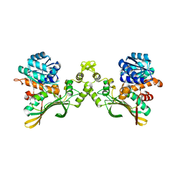
7X9S
 | | Crystal structure of a complex between the antirepressor GmaR and the transcriptional repressor MogR | | 分子名称: | GmaR, Motility gene repressor MogR | | 著者 | Cho, S.Y, Na, H.W, Oh, H.B, Kwak, Y.M, Song, W.S, Park, S.C, Yoon, S.I. | | 登録日 | 2022-03-16 | | 公開日 | 2022-11-23 | | 最終更新日 | 2023-11-29 | | 実験手法 | X-RAY DIFFRACTION (3.11 Å) | | 主引用文献 | Structural basis of flagellar motility regulation by the MogR repressor and the GmaR antirepressor in Listeria monocytogenes.
Nucleic Acids Res., 50, 2022
|
|
7W1F
 | | Crystal structure of the dNTP triphosphohydrolase PA1124 from Pseudomonas aeruginosa | | 分子名称: | NICKEL (II) ION, Probable deoxyguanosinetriphosphate triphosphohydrolase | | 著者 | Oh, H.B, Song, W.S, Lee, K.C, Park, S.C, Yoon, S.I. | | 登録日 | 2021-11-19 | | 公開日 | 2022-03-23 | | 最終更新日 | 2023-11-29 | | 実験手法 | X-RAY DIFFRACTION (2.9 Å) | | 主引用文献 | Structural analysis of the dNTP triphosphohydrolase PA1124 from Pseudomonas aeruginosa.
Biochem.Biophys.Res.Commun., 589, 2022
|
|
5Z7B
 | |
6L8B
 | | The ligand-free structure of human PPARgamma LBD | | 分子名称: | Peroxisome proliferator-activated receptor gamma | | 著者 | Jang, D.M, Han, B.W. | | 登録日 | 2019-11-05 | | 公開日 | 2020-09-16 | | 最終更新日 | 2023-11-22 | | 実験手法 | X-RAY DIFFRACTION (2.102 Å) | | 主引用文献 | Cyclin-Dependent Kinase 5 Inhibitor Butyrolactone I Elicits a Partial Agonist Activity of Peroxisome Proliferator-Activated Receptor gamma.
Biomolecules, 10, 2020
|
|
5IHY
 | |
5DQV
 | |
5DQW
 | |
6L89
 | | Human PPARgamma ligand binding domain complexed with Butyrolactone 1 | | 分子名称: | Peroxisome proliferator-activated receptor gamma, methyl (2R)-3-(4-hydroxyphenyl)-2-[[3-(3-methylbut-2-enyl)-4-oxidanyl-phenyl]methyl]-4-oxidanyl-5-oxidanylidene-furan-2-carboxylate | | 著者 | Jang, D.M, Han, B.W. | | 登録日 | 2019-11-05 | | 公開日 | 2020-09-16 | | 最終更新日 | 2023-11-22 | | 実験手法 | X-RAY DIFFRACTION (2.1 Å) | | 主引用文献 | Cyclin-Dependent Kinase 5 Inhibitor Butyrolactone I Elicits a Partial Agonist Activity of Peroxisome Proliferator-Activated Receptor gamma.
Biomolecules, 10, 2020
|
|
2UZ0
 | | The Crystal crystal structure of the estA protein, a virulence factor estA protein from Streptococcus pneumonia | | 分子名称: | 1,2-ETHANEDIOL, CALCIUM ION, GLYCEROL, ... | | 著者 | Kim, M.H, Kang, B.S, Kim, K.J, Oh, T.K. | | 登録日 | 2007-04-23 | | 公開日 | 2007-10-23 | | 最終更新日 | 2011-07-13 | | 実験手法 | X-RAY DIFFRACTION (1.7 Å) | | 主引用文献 | The Crystal Structure of the Esta Protein, a Virulence Factor from Streptococcus Pneumoniae.
Proteins, 70, 2008
|
|


