5TDA
 
 | |
5TDD
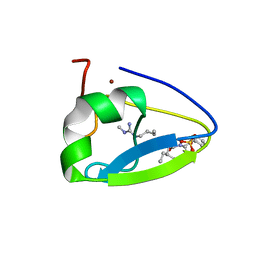
 | | Human UBR-box from UBR2 in complex with HIFS peptide | | 分子名称: | 1,2-ETHANEDIOL, E3 ubiquitin-protein ligase UBR2, HIS-ILE-PHE-SER peptide, ... | | 著者 | Munoz-Escobar, J, Kozlov, G, Gehring, K. | | 登録日 | 2016-09-19 | | 公開日 | 2017-03-22 | | 最終更新日 | 2023-10-04 | | 実験手法 | X-RAY DIFFRACTION (1.55 Å) | | 主引用文献 | Bound Waters Mediate Binding of Diverse Substrates to a Ubiquitin Ligase.
Structure, 25, 2017
|
|
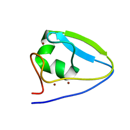
5TDB
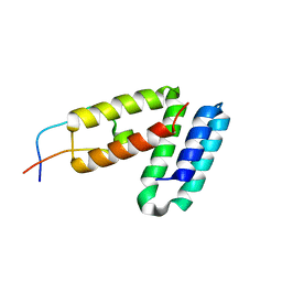
 | | Crystal structure of the human UBR-box domain from UBR2 in complex with asymmetrically double methylated arginine peptide | | 分子名称: | 1,2-ETHANEDIOL, DA2-ILE-PHE-SER peptide, E3 ubiquitin-protein ligase UBR2, ... | | 著者 | Munoz-Escobar, J, Kozlov, G, Gehring, K. | | 登録日 | 2016-09-19 | | 公開日 | 2017-03-22 | | 最終更新日 | 2023-11-15 | | 実験手法 | X-RAY DIFFRACTION (1.101 Å) | | 主引用文献 | Bound Waters Mediate Binding of Diverse Substrates to a Ubiquitin Ligase.
Structure, 25, 2017
|
|
5VMD
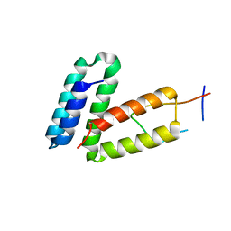
 | | Crystal structure of UBR-box from UBR6 in a domain-swapping conformation | | 分子名称: | 1,2-ETHANEDIOL, F-box only protein 11, ZINC ION | | 著者 | Munoz-Escobar, J, Kozlov, G, Gehring, K. | | 登録日 | 2017-04-27 | | 公開日 | 2017-07-12 | | 最終更新日 | 2024-03-13 | | 実験手法 | X-RAY DIFFRACTION (2.202 Å) | | 主引用文献 | Crystal structure of the UBR-box from UBR6/FBXO11 reveals domain swapping mediated by zinc binding.
Protein Sci., 26, 2017
|
|
5TDC
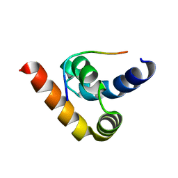
 | | Crystal structure of the human UBR-box domain from UBR1 in complex with monomethylated arginine peptide. | | 分子名称: | E3 ubiquitin-protein ligase UBR1, NMM-ILE-PHE-SER peptide, SULFATE ION, ... | | 著者 | Kozlov, G, Munoz-Escobar, J, Matta-Camacho, E, Gehring, K. | | 登録日 | 2016-09-19 | | 公開日 | 2017-03-22 | | 最終更新日 | 2023-10-04 | | 実験手法 | X-RAY DIFFRACTION (1.607 Å) | | 主引用文献 | Bound Waters Mediate Binding of Diverse Substrates to a Ubiquitin Ligase.
Structure, 25, 2017
|
|
5VRQ
 | | Crystal structure of Legionella pneumophila effector AnkC | | 分子名称: | Ankyrin repeat-containing protein | | 著者 | Kozlov, G, Wong, K, Wang, W, Skubak, P, Munoz-Escobar, J, Liu, Y, Pannu, N.S, Gehring, K, Montreal-Kingston Bacterial Structural Genomics Initiative (BSGI) | | 登録日 | 2017-05-11 | | 公開日 | 2017-11-29 | | 最終更新日 | 2024-03-13 | | 実験手法 | X-RAY DIFFRACTION (3.205 Å) | | 主引用文献 | Ankyrin repeats as a dimerization module.
Biochem. Biophys. Res. Commun., 495, 2018
|
|
5V8Z
 | |
5V90
 | |
3NTW
 | |
5UM3
 | |


