8AFQ
 
 | | Tube assembly of Atg18-PR72AA | | 分子名称: | Autophagy-related protein 18 | | 著者 | Mann, D, Fromm, S, Martinez-Sanchez, A, Gopaldass, N, Mayer, A, Sachse, C. | | 登録日 | 2022-07-18 | | 公開日 | 2023-11-01 | | 最終更新日 | 2024-05-15 | | 実験手法 | ELECTRON MICROSCOPY (3.3 Å) | | 主引用文献 | Atg18 oligomer organization in assembled tubes and on lipid membrane scaffolds.
Nat Commun, 14, 2023
|
|
8AFW
 | | Tube assembly of Atg18-WT | | 分子名称: | Autophagy-related protein 18 | | 著者 | Mann, D, Fromm, S, Martinez-Sanchez, A, Gopaldass, N, Mayer, A, Sachse, C. | | 登録日 | 2022-07-18 | | 公開日 | 2023-11-01 | | 最終更新日 | 2024-05-15 | | 実験手法 | ELECTRON MICROSCOPY (3.8 Å) | | 主引用文献 | Atg18 oligomer organization in assembled tubes and on lipid membrane scaffolds.
Nat Commun, 14, 2023
|
|
8AFY
 | | Subtomogram average of membrane-bound Atg18 oligomers | | 分子名称: | Autophagy-related protein 18 | | 著者 | Mann, D, Fromm, S, Martinez-Sanchez, A, Gopaldass, N, Mayer, A, Sachse, C. | | 登録日 | 2022-07-18 | | 公開日 | 2023-11-01 | | 最終更新日 | 2024-05-15 | | 実験手法 | ELECTRON MICROSCOPY (26 Å) | | 主引用文献 | Atg18 oligomer organization in assembled tubes and on lipid membrane scaffolds.
Nat Commun, 14, 2023
|
|
8AFX
 | | Single particle structure of Atg18-WT | | 分子名称: | Autophagy-related protein 18 | | 著者 | Mann, D, Fromm, S, Martinez-Sanchez, A, Gopaldass, N, Mayer, A, Sachse, C. | | 登録日 | 2022-07-18 | | 公開日 | 2024-01-31 | | 実験手法 | ELECTRON MICROSCOPY (4.8 Å) | | 主引用文献 | Cryo-EM structures of Atg18 oligomers reveal a tilted structural scaffold for Atg2 at the isolation membrane
To Be Published
|
|
6EPC
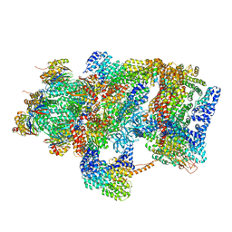 | | Ground state 26S proteasome (GS2) | | 分子名称: | 26S proteasome non-ATPase regulatory subunit 1, 26S proteasome non-ATPase regulatory subunit 11, 26S proteasome non-ATPase regulatory subunit 13, ... | | 著者 | Guo, Q, Lehmer, C, Martinez-Sanchez, A, Rudack, T, Beck, F, Hartmann, H, Hipp, M.S, Hartl, F.U, Edbauer, D, Baumeister, W, Fernandez-Busnadiego, R. | | 登録日 | 2017-10-11 | | 公開日 | 2018-02-07 | | 最終更新日 | 2024-05-15 | | 実験手法 | ELECTRON MICROSCOPY (12.3 Å) | | 主引用文献 | In Situ Structure of Neuronal C9orf72 Poly-GA Aggregates Reveals Proteasome Recruitment.
Cell, 172, 2018
|
|
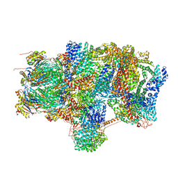
6EPD
 | | Substrate processing state 26S proteasome (SPS1) | | 分子名称: | 26S proteasome non-ATPase regulatory subunit 1, 26S proteasome non-ATPase regulatory subunit 11, 26S proteasome non-ATPase regulatory subunit 13, ... | | 著者 | Guo, Q, Lehmer, C, Martinez-Sanchez, A, Rudack, T, Beck, F, Hartmann, H, Hipp, M.S, Hartl, F.U, Edbauer, D, Baumeister, W, Fernandez-Busnadiego, R. | | 登録日 | 2017-10-11 | | 公開日 | 2018-02-07 | | 最終更新日 | 2024-05-15 | | 実験手法 | ELECTRON MICROSCOPY (15.4 Å) | | 主引用文献 | In Situ Structure of Neuronal C9orf72 Poly-GA Aggregates Reveals Proteasome Recruitment.
Cell, 172, 2018
|
|
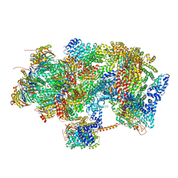
6EPE
 | | Substrate processing state 26S proteasome (SPS2) | | 分子名称: | 26S proteasome non-ATPase regulatory subunit 1, 26S proteasome non-ATPase regulatory subunit 11, 26S proteasome non-ATPase regulatory subunit 13, ... | | 著者 | Guo, Q, Lehmer, C, Martinez-Sanchez, A, Rudack, T, Beck, F, Hartmann, H, Hipp, M.S, Hartl, F.U, Edbauer, D, Baumeister, W, Fernandez-Busnadiego, R. | | 登録日 | 2017-10-11 | | 公開日 | 2018-02-07 | | 最終更新日 | 2024-05-15 | | 実験手法 | ELECTRON MICROSCOPY (12.8 Å) | | 主引用文献 | In Situ Structure of Neuronal C9orf72 Poly-GA Aggregates Reveals Proteasome Recruitment.
Cell, 172, 2018
|
|
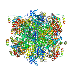
6EPF
 | | Ground state 26S proteasome (GS1) | | 分子名称: | 26S proteasome non-ATPase regulatory subunit 1, 26S proteasome non-ATPase regulatory subunit 11, 26S proteasome non-ATPase regulatory subunit 13, ... | | 著者 | Guo, Q, Lehmer, C, Martinez-Sanchez, A, Rudack, T, Beck, F, Hartmann, H, Hipp, M.S, Hartl, F.U, Edbauer, D, Baumeister, W, Fernandez-Busnadiego, R. | | 登録日 | 2017-10-11 | | 公開日 | 2018-02-07 | | 最終更新日 | 2024-05-15 | | 実験手法 | ELECTRON MICROSCOPY (11.8 Å) | | 主引用文献 | In Situ Structure of Neuronal C9orf72 Poly-GA Aggregates Reveals Proteasome Recruitment.
Cell, 172, 2018
|
|
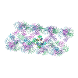
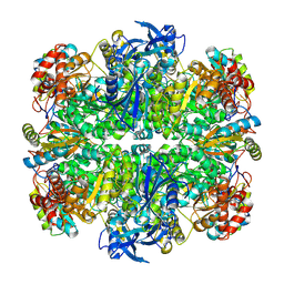
7JFO
 | | EPYC1(49-72)-bound Rubisco | | 分子名称: | LCI5, Ribulose bisphosphate carboxylase large chain, Ribulose bisphosphate carboxylase small chain 2, ... | | 著者 | Matthies, D, Jonikas, M.C, He, S. | | 登録日 | 2020-07-17 | | 公開日 | 2020-11-18 | | 最終更新日 | 2020-12-23 | | 実験手法 | ELECTRON MICROSCOPY (2.13 Å) | | 主引用文献 | The structural basis of Rubisco phase separation in the pyrenoid.
Nat.Plants, 6, 2020
|
|
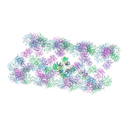
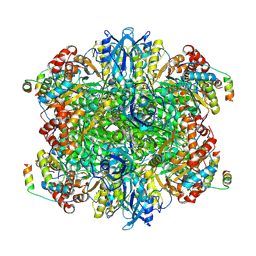
7JN4
 | | Rubisco in the apo state | | 分子名称: | Ribulose bisphosphate carboxylase large chain, Ribulose bisphosphate carboxylase small chain 2, chloroplastic | | 著者 | Matthies, D, Jonikas, M.C, He, S. | | 登録日 | 2020-08-03 | | 公開日 | 2020-11-18 | | 最終更新日 | 2020-12-23 | | 実験手法 | ELECTRON MICROSCOPY (2.68 Å) | | 主引用文献 | The structural basis of Rubisco phase separation in the pyrenoid.
Nat.Plants, 6, 2020
|
|
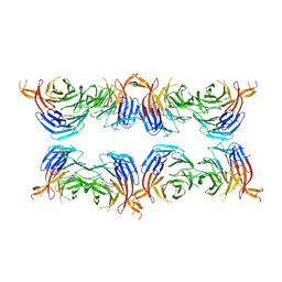
7JSX
 | | EPYC1(106-135) peptide-bound Rubisco | | 分子名称: | EPYC1, Ribulose bisphosphate carboxylase large chain, Ribulose bisphosphate carboxylase small chain 2, ... | | 著者 | Matthies, D, He, S, Jonikas, M.C. | | 登録日 | 2020-08-16 | | 公開日 | 2020-11-18 | | 最終更新日 | 2020-12-23 | | 実験手法 | ELECTRON MICROSCOPY (2.06 Å) | | 主引用文献 | The structural basis of Rubisco phase separation in the pyrenoid.
Nat.Plants, 6, 2020
|
|


