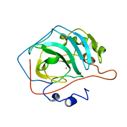
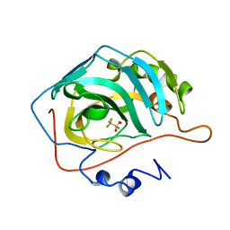
5CAC
 
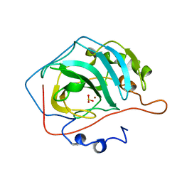 | | REFINED STRUCTURE OF HUMAN CARBONIC ANHYDRASE II AT 2.0 ANGSTROMS RESOLUTION | | 分子名称: | CARBONIC ANHYDRASE FORM C, SULFITE ION, ZINC ION | | 著者 | Lindahl, M, Habash, D, Harrop, S, Helliwell, D.R, Liljas, A. | | 登録日 | 1991-09-05 | | 公開日 | 1994-01-31 | | 最終更新日 | 2024-03-06 | | 実験手法 | X-RAY DIFFRACTION (2.2 Å) | | 主引用文献 | Refined structure of human carbonic anhydrase II at 2.0 A resolution.
Proteins, 4, 1988
|
|
1CA2
 | |
1RZC
 | | X-RAY ANALYSIS OF METAL SUBSTITUTED HUMAN CARBONIC ANHYDRASE II DERIVATIVES | | 分子名称: | CARBONIC ANHYDRASE II, COPPER (II) ION, OXYGEN MOLECULE | | 著者 | Hakansson, K, Wehnert, A, Liljas, A. | | 登録日 | 1993-05-25 | | 公開日 | 1993-10-31 | | 最終更新日 | 2024-02-14 | | 実験手法 | X-RAY DIFFRACTION (1.9 Å) | | 主引用文献 | X-ray analysis of metal-substituted human carbonic anhydrase II derivatives.
Acta Crystallogr.,Sect.D, 50, 1994
|
|
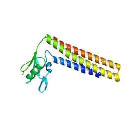
1CTF
 | |
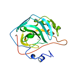
3CA2
 | | CRYSTALLOGRAPHIC STUDIES OF INHIBITOR BINDING SITES IN HUMAN CARBONIC ANHYDRASE II. A PENTACOORDINATED BINDING OF THE SCN-ION TO THE ZINC AT HIGH P*H | | 分子名称: | 3-MERCURI-4-AMINOBENZENESULFONAMIDE, CARBONIC ANHYDRASE II, MERCURY (II) ION, ... | | 著者 | Eriksson, A.E, Jones, T.A, Liljas, A. | | 登録日 | 1989-10-02 | | 公開日 | 1990-01-15 | | 最終更新日 | 2024-02-21 | | 実験手法 | X-RAY DIFFRACTION (2 Å) | | 主引用文献 | Crystallographic studies of inhibitor binding sites in human carbonic anhydrase II: a pentacoordinated binding of the SCN- ion to the zinc at high pH.
Proteins, 4, 1988
|
|
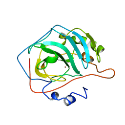
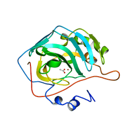
4CAC
 | | REFINED STRUCTURE OF HUMAN CARBONIC ANHYDRASE II AT 2.0 ANGSTROMS RESOLUTION | | 分子名称: | CARBONIC ANHYDRASE FORM C, ZINC ION | | 著者 | Lindahl, M, Habash, D, Harrop, S, Helliwell, D.R, Liljas, A. | | 登録日 | 1991-09-05 | | 公開日 | 1994-01-31 | | 最終更新日 | 2024-02-28 | | 実験手法 | X-RAY DIFFRACTION (2.2 Å) | | 主引用文献 | Refined structure of human carbonic anhydrase II at 2.0 A resolution.
Proteins, 4, 1988
|
|
1CAM
 | | STRUCTURAL ANALYSIS OF THE ZINC HYDROXIDE-THR 199-GLU 106 HYDROGEN BONDING NETWORK IN HUMAN CARBONIC ANHYDRASE II | | 分子名称: | BICARBONATE ION, CARBONIC ANHYDRASE II, ZINC ION | | 著者 | Xue, Y, Liljas, A, Jonsson, B.-H, Lindskog, S. | | 登録日 | 1992-09-17 | | 公開日 | 1993-10-31 | | 最終更新日 | 2024-02-07 | | 実験手法 | X-RAY DIFFRACTION (1.7 Å) | | 主引用文献 | Structural analysis of the zinc hydroxide-Thr-199-Glu-106 hydrogen-bond network in human carbonic anhydrase II.
Proteins, 17, 1993
|
|
1CAZ
 | | WILD-TYPE AND E106Q MUTANT CARBONIC ANHYDRASE COMPLEXED WITH ACETATE | | 分子名称: | ACETIC ACID, CARBONIC ANHYDRASE II, ZINC ION | | 著者 | Hakansson, K, Briand, C, Zaitsev, V, Xue, Y, Liljas, A. | | 登録日 | 1993-02-26 | | 公開日 | 1993-10-31 | | 最終更新日 | 2024-02-07 | | 実験手法 | X-RAY DIFFRACTION (1.9 Å) | | 主引用文献 | Wild-type and E106Q mutant carbonic anhydrase complexed with acetate.
Acta Crystallogr.,Sect.D, 50, 1994
|
|
1CAY
 | | WILD-TYPE AND E106Q MUTANT CARBONIC ANHYDRASE COMPLEXED WITH ACETATE | | 分子名称: | ACETIC ACID, CARBONIC ANHYDRASE II, ZINC ION | | 著者 | Hakansson, K, Briand, C, Zaitsev, V, Xue, Y, Liljas, A. | | 登録日 | 1993-02-26 | | 公開日 | 1993-10-31 | | 最終更新日 | 2024-02-07 | | 実験手法 | X-RAY DIFFRACTION (2.1 Å) | | 主引用文献 | Wild-type and E106Q mutant carbonic anhydrase complexed with acetate.
Acta Crystallogr.,Sect.D, 50, 1994
|
|
1CAL
 | | STRUCTURAL ANALYSIS OF THE ZINC HYDROXIDE-THR 199-GLU 106 HYDROGEN BONDING NETWORK IN HUMAN CARBONIC ANHYDRASE II | | 分子名称: | CARBONIC ANHYDRASE II, ZINC ION | | 著者 | Xue, Y, Liljas, A, Jonsson, B.-H, Lindskog, S. | | 登録日 | 1992-09-17 | | 公開日 | 1993-10-31 | | 最終更新日 | 2024-02-07 | | 実験手法 | X-RAY DIFFRACTION (2.2 Å) | | 主引用文献 | Structural analysis of the zinc hydroxide-Thr-199-Glu-106 hydrogen-bond network in human carbonic anhydrase II.
Proteins, 17, 1993
|
|
1CAI
 | | STRUCTURAL ANALYSIS OF THE ZINC HYDROXIDE-THR 199-GLU 106 HYDROGEN BONDING NETWORK IN HUMAN CARBONIC ANHYDRASE II | | 分子名称: | CARBONIC ANHYDRASE II, SULFATE ION, ZINC ION | | 著者 | Xue, Y, Liljas, A, Jonsson, B.-H, Lindskog, S. | | 登録日 | 1992-09-17 | | 公開日 | 1993-10-31 | | 最終更新日 | 2024-02-07 | | 実験手法 | X-RAY DIFFRACTION (1.8 Å) | | 主引用文献 | Structural analysis of the zinc hydroxide-Thr-199-Glu-106 hydrogen-bond network in human carbonic anhydrase II.
Proteins, 17, 1993
|
|
1BCD
 | |
1DD5
 | | CRYSTAL STRUCTURE OF THERMOTOGA MARITIMA RIBOSOME RECYCLING FACTOR, RRF | | 分子名称: | ACETIC ACID, RIBOSOME RECYCLING FACTOR | | 著者 | Selmer, M, Al-Karadaghi, S, Hirokawa, G, Kaji, A, Liljas, A. | | 登録日 | 1999-11-08 | | 公開日 | 1999-12-22 | | 最終更新日 | 2024-02-07 | | 実験手法 | X-RAY DIFFRACTION (2.55 Å) | | 主引用文献 | Crystal structure of Thermotoga maritima ribosome recycling factor: a tRNA mimic.
Science, 286, 1999
|
|
2BV3
 | | Crystal structure of a mutant elongation factor G trapped with a GTP analogue | | 分子名称: | ELONGATION FACTOR G, MAGNESIUM ION, PHOSPHOAMINOPHOSPHONIC ACID-GUANYLATE ESTER | | 著者 | Hansson, S, Singh, R, Gudkov, A.T, Liljas, A, Logan, D.T. | | 登録日 | 2005-06-22 | | 公開日 | 2005-08-10 | | 最終更新日 | 2023-12-13 | | 実験手法 | X-RAY DIFFRACTION (2.5 Å) | | 主引用文献 | Crystal Structure of a Mutant Elongation Factor G Trapped with a GTP Analogue.
FEBS Lett., 579, 2005
|
|
1DAR
 | | ELONGATION FACTOR G IN COMPLEX WITH GDP | | 分子名称: | ELONGATION FACTOR G, GUANOSINE-5'-DIPHOSPHATE | | 著者 | Al-Karadaghi, S, Aevarsson, A, Garber, M, Zheltonosova, J, Liljas, A. | | 登録日 | 1996-02-15 | | 公開日 | 1996-07-11 | | 最終更新日 | 2024-02-07 | | 実験手法 | X-RAY DIFFRACTION (2.4 Å) | | 主引用文献 | The structure of elongation factor G in complex with GDP: conformational flexibility and nucleotide exchange.
Structure, 4, 1996
|
|
1VID
 | | CATECHOL O-METHYLTRANSFERASE | | 分子名称: | 3,5-DINITROCATECHOL, CATECHOL O-METHYLTRANSFERASE, MAGNESIUM ION, ... | | 著者 | Vidgren, J, Svensson, L.A, Liljas, A. | | 登録日 | 1996-01-05 | | 公開日 | 1996-07-11 | | 最終更新日 | 2024-02-14 | | 実験手法 | X-RAY DIFFRACTION (2 Å) | | 主引用文献 | Crystal structure of catechol O-methyltransferase.
Nature, 368, 1994
|
|
2BM0
 | | Ribosomal elongation factor G (EF-G) Fusidic acid resistant mutant T84A | | 分子名称: | ELONGATION FACTOR G, GUANOSINE-5'-DIPHOSPHATE, MAGNESIUM ION | | 著者 | Hansson, S, Singh, R, Gudkov, A.T, Liljas, A, Logan, D.T. | | 登録日 | 2005-03-09 | | 公開日 | 2005-05-04 | | 最終更新日 | 2023-12-13 | | 実験手法 | X-RAY DIFFRACTION (2.4 Å) | | 主引用文献 | Structural Insights Into Fusidic Acid Resistance and Sensitivity in EF-G
J.Mol.Biol., 348, 2005
|
|
2BM1
 | | Ribosomal elongation factor G (EF-G) Fusidic acid resistant mutant G16V | | 分子名称: | ELONGATION FACTOR G, GUANOSINE-5'-DIPHOSPHATE, MAGNESIUM ION | | 著者 | Hansson, S, Singh, R, Gudkov, A.T, Liljas, A, Logan, D.T. | | 登録日 | 2005-03-09 | | 公開日 | 2005-05-04 | | 最終更新日 | 2023-12-13 | | 実験手法 | X-RAY DIFFRACTION (2.6 Å) | | 主引用文献 | Structural Insights Into Fusidic Acid Resistance and Sensitivity in EF-G
J.Mol.Biol., 348, 2005
|
|
1FNM
 | | STRUCTURE OF THERMUS THERMOPHILUS EF-G H573A | | 分子名称: | ELONGATION FACTOR G, GUANOSINE-5'-DIPHOSPHATE, MAGNESIUM ION | | 著者 | Laurberg, M, Kristensen, O, Martemyanov, K, Gudkov, A.T, Nagaev, I, Hughes, D, Liljas, A. | | 登録日 | 2000-08-22 | | 公開日 | 2000-11-22 | | 最終更新日 | 2024-02-07 | | 実験手法 | X-RAY DIFFRACTION (2.8 Å) | | 主引用文献 | Structure of a mutant EF-G reveals domain III and possibly the fusidic acid binding site.
J.Mol.Biol., 303, 2000
|
|
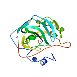
2CA2
 | | CRYSTALLOGRAPHIC STUDIES OF INHIBITOR BINDING SITES IN HUMAN CARBONIC ANHYDRASE II. A PENTACOORDINATED BINDING OF THE SCN-ION TO THE ZINC AT HIGH P*H | | 分子名称: | CARBONIC ANHYDRASE II, MERCURY (II) ION, THIOCYANATE ION, ... | | 著者 | Eriksson, A.E, Kylsten, P.M, Jones, T.A, Liljas, A. | | 登録日 | 1989-02-06 | | 公開日 | 1990-01-15 | | 最終更新日 | 2024-02-14 | | 実験手法 | X-RAY DIFFRACTION (1.9 Å) | | 主引用文献 | Crystallographic studies of inhibitor binding sites in human carbonic anhydrase II: a pentacoordinated binding of the SCN- ion to the zinc at high pH.
Proteins, 4, 1988
|
|
1ELO
 | | ELONGATION FACTOR G WITHOUT NUCLEOTIDE | | 分子名称: | ELONGATION FACTOR G | | 著者 | Aevarsson, A, Brazhnikov, E, Garber, M, Zheltonosova, J, Chirgadze, Yu, Al-Karadaghi, S, Svensson, L.A, Liljas, A. | | 登録日 | 1996-03-13 | | 公開日 | 1996-08-01 | | 最終更新日 | 2024-02-07 | | 実験手法 | X-RAY DIFFRACTION (2.8 Å) | | 主引用文献 | Three-dimensional structure of the ribosomal translocase: elongation factor G from Thermus thermophilus.
EMBO J., 13, 1994
|
|
1T6C
 | | Structural characterization of the Ppx/GppA protein family: crystal structure of the Aquifex aeolicus family member | | 分子名称: | (4S)-2-METHYL-2,4-PENTANEDIOL, CALCIUM ION, CHLORIDE ION, ... | | 著者 | Kristensen, O, Laurberg, M, Liljas, A, Kastrup, J.S, Gajhede, M. | | 登録日 | 2004-05-06 | | 公開日 | 2004-08-03 | | 最終更新日 | 2024-04-03 | | 実験手法 | X-RAY DIFFRACTION (1.53 Å) | | 主引用文献 | Structural characterization of the stringent response related exopolyphosphatase/guanosine pentaphosphate phosphohydrolase protein family
Biochemistry, 43, 2004
|
|
1T6D
 | | MIRAS phasing of the Aquifex aeolicus Ppx/GppA phosphatase: crystal structure of the type II variant | | 分子名称: | 2-AMINO-2-HYDROXYMETHYL-PROPANE-1,3-DIOL, CHLORIDE ION, exopolyphosphatase | | 著者 | Kristensen, O, Laurberg, M, Liljas, A, Kastrup, J.S, Gajhede, M. | | 登録日 | 2004-05-06 | | 公開日 | 2004-08-03 | | 最終更新日 | 2021-11-10 | | 実験手法 | X-RAY DIFFRACTION (2.15 Å) | | 主引用文献 | Structural characterization of the stringent response related exopolyphosphatase/guanosine pentaphosphate phosphohydrolase protein family
Biochemistry, 43, 2004
|
|
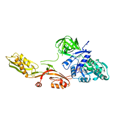
1CJS
 | | CRYSTAL STRUCTURE OF RIBOSOMAL PROTEIN L1 FROM METHANOCOCCUS JANNASCHII | | 分子名称: | 50S RIBOSOMAL PROTEIN L1P | | 著者 | Nevskaya, N, Tishchenko, S, Fedorov, R, Al-Karadaghi, S, Liljas, A, Kraft, A, Piendl, W, Garber, M, Nikonov, S. | | 登録日 | 1999-04-19 | | 公開日 | 2000-05-31 | | 最終更新日 | 2023-12-27 | | 実験手法 | X-RAY DIFFRACTION (2.3 Å) | | 主引用文献 | Archaeal ribosomal protein L1: the structure provides new insights into RNA binding of the L1 protein family.
Structure Fold.Des., 8, 2000
|
|
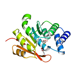
1RZA
 | | X-RAY ANALYSIS OF METAL SUBSTITUTED HUMAN CARBONIC ANHYDRASE II DERIVATIVES | | 分子名称: | CARBONIC ANHYDRASE II, COBALT (II) ION, OXYGEN MOLECULE | | 著者 | Hakansson, K, Wehnert, A, Liljas, A. | | 登録日 | 1993-05-25 | | 公開日 | 1993-10-31 | | 最終更新日 | 2024-02-14 | | 実験手法 | X-RAY DIFFRACTION (1.9 Å) | | 主引用文献 | X-ray analysis of metal-substituted human carbonic anhydrase II derivatives.
Acta Crystallogr.,Sect.D, 50, 1994
|
|


